. 2021 Apr 13;116(1):25.
doi: 10.1007/s00395-021-00865-9.

SKI activates the Hippo pathway via LIMD1 to inhibit cardiac fibroblast activation

Affiliations

SKI activates the Hippo pathway via LIMD1 to inhibit cardiac fibroblast activation

Natalie M Landry et al. Basic Res Cardiol. .

Abstract

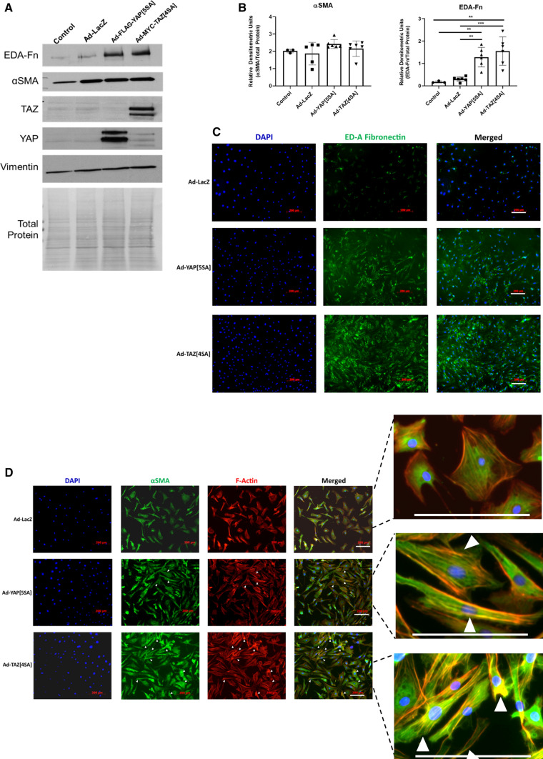
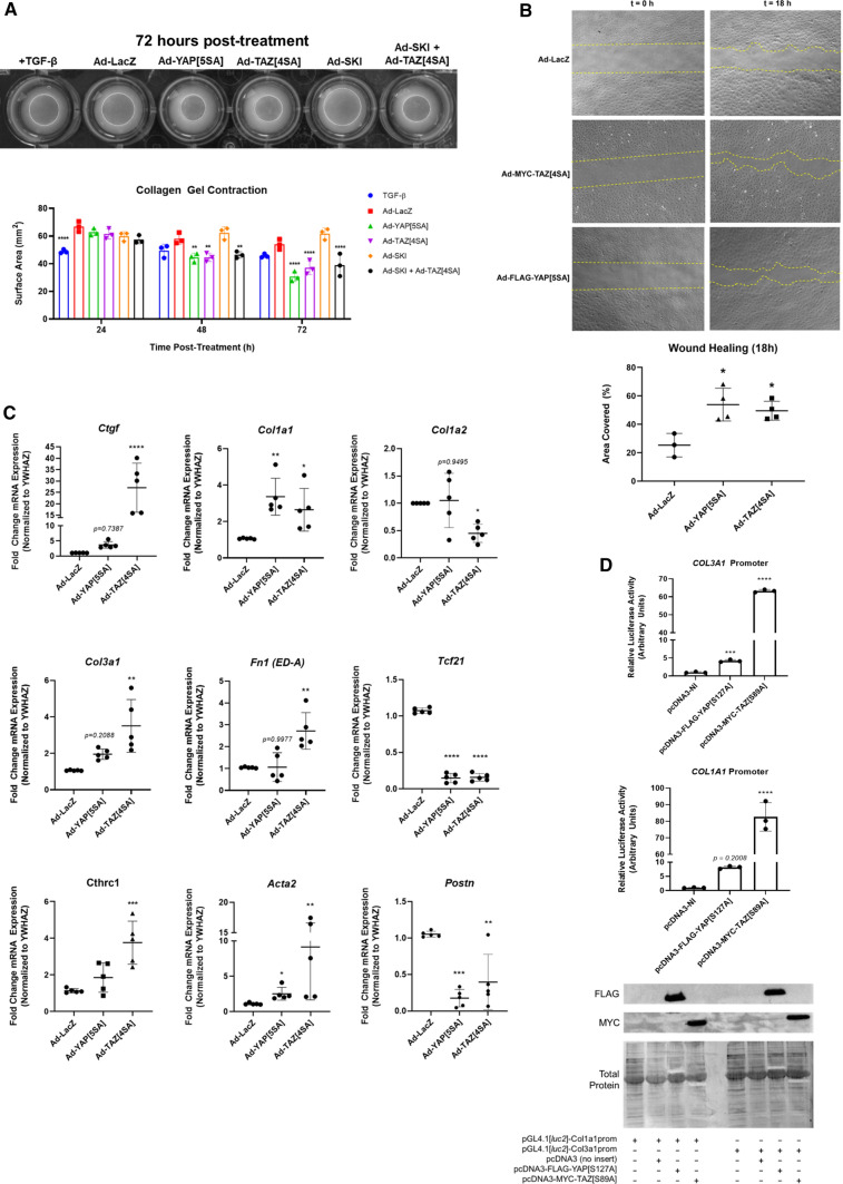
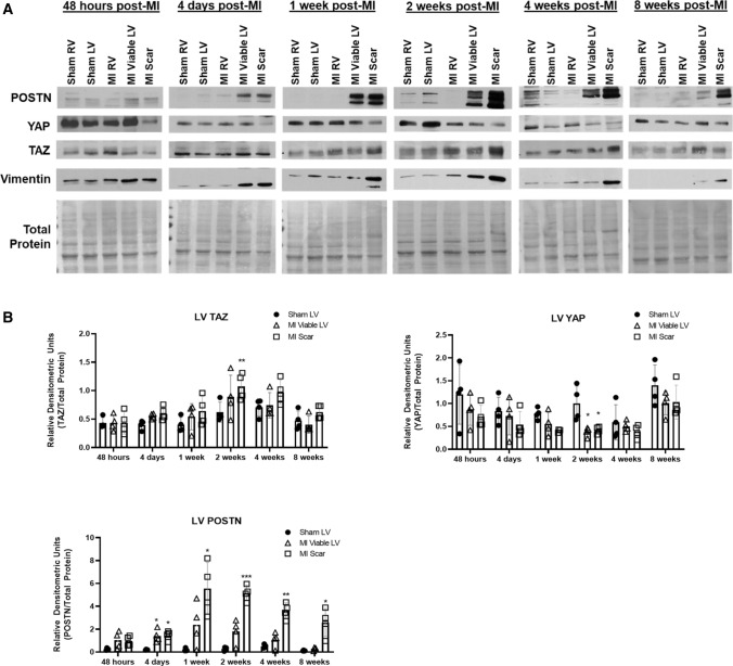
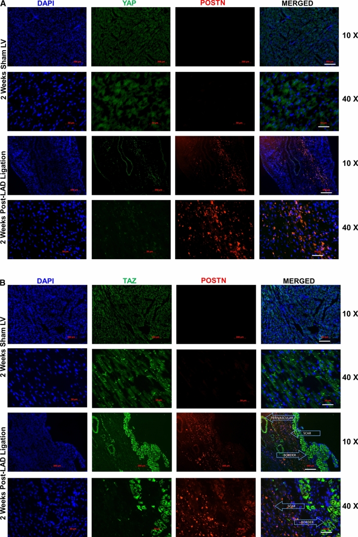
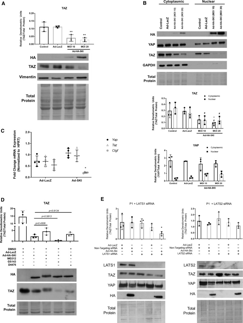
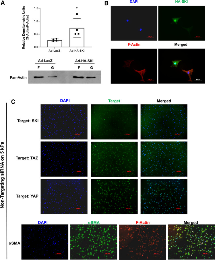
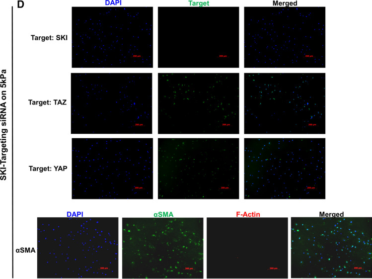
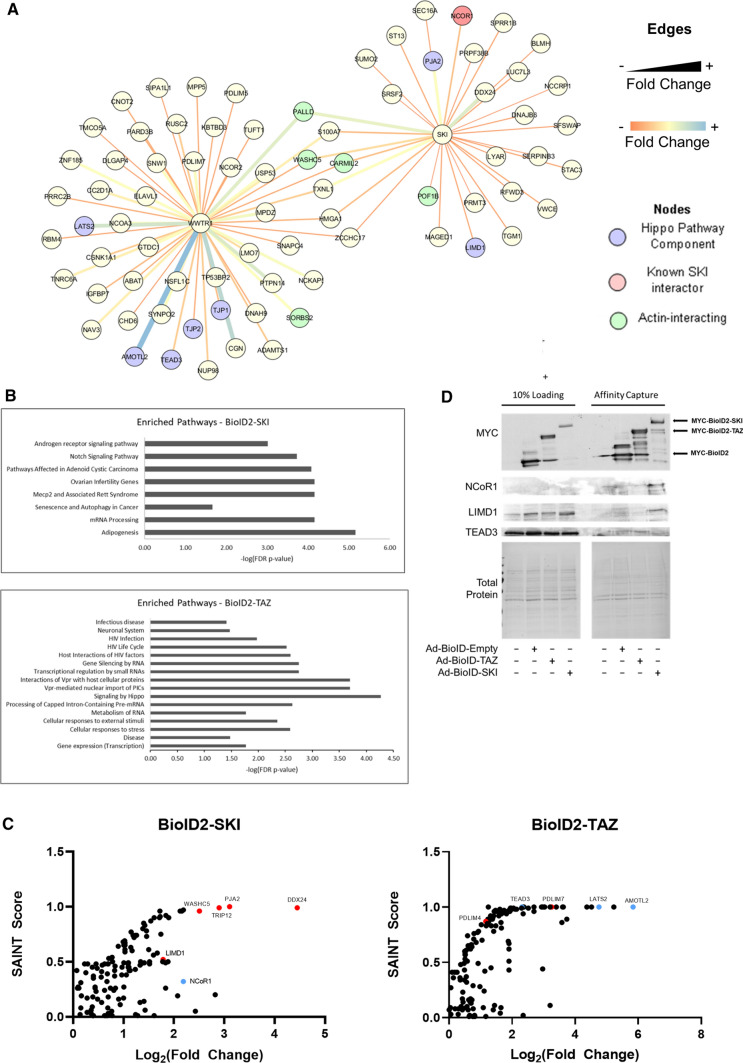
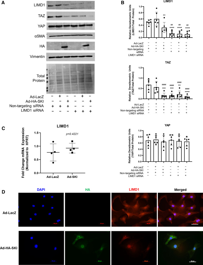
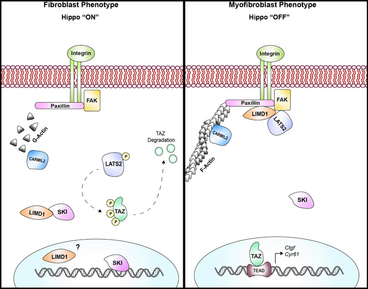
We have previously shown that overexpression of SKI, an endogenous TGF-β1 repressor, deactivates the pro-fibrotic myofibroblast phenotype in the heart. We now show that SKI also functions independently of SMAD/TGF-β signaling, by activating the Hippo tumor-suppressor pathway and inhibiting the Transcriptional co-Activator with PDZ-binding motif (TAZ or WWTR1). The mechanism(s) by which SKI targets TAZ to inhibit cardiac fibroblast activation and fibrogenesis remain undefined. A rat model of post-myocardial infarction was used to examine the expression of TAZ during acute fibrogenesis and chronic heart failure. Results were then corroborated with primary rat cardiac fibroblast cell culture performed both on plastic and on inert elastic substrates, along with the use of siRNA and adenoviral expression vectors for active forms of SKI, YAP, and TAZ. Gene expression was examined by qPCR and luciferase assays, while protein expression was examined by immunoblotting and fluorescence microscopy. Cell phenotype was further assessed by functional assays. Finally, to elucidate SKI's effects on Hippo signaling, the SKI and TAZ interactomes were captured in human cardiac fibroblasts using BioID2 and mass spectrometry. Potential interactors were investigated in vitro to reveal novel mechanisms of action for SKI. In vitro assays on elastic substrates revealed the ability of TAZ to overcome environmental stimuli and induce the activation of hypersynthetic cardiac myofibroblasts. Further cell-based assays demonstrated that SKI causes specific proteasomal degradation of TAZ, but not YAP, and shifts actin cytoskeleton dynamics to inhibit myofibroblast activation. These findings were supported by identifying the bi-phasic expression of TAZ in vivo during post-MI remodeling and fibrosis. BioID2-based interactomics in human cardiac fibroblasts suggest that SKI interacts with actin-modifying proteins and with LIM Domain-containing protein 1 (LIMD1), a negative regulator of Hippo signaling. Furthermore, we found that LATS2 interacts with TAZ, whereas LATS1 does not, and that LATS2 knockdown prevented TAZ downregulation with SKI overexpression. Our findings indicate that SKI's capacity to regulate cardiac fibroblast activation is mediated, in part, by Hippo signaling. We postulate that the interaction between SKI and TAZ in cardiac fibroblasts is arbitrated by LIMD1, an important intermediary in focal adhesion-associated signaling pathways. This study contributes to the understanding of the unique physiology of cardiac fibroblasts, and of the relationship between SKI expression and cell phenotype.

Keywords: Cardiac fibrosis; Extracellular matrix; Fibroblast; Hippo signaling; SKI; TAZ.


Conflict of interest statement

The authors declare that they have no competing interests.

Figures

Fig. 1
Fig. 1
YAP and TAZ induce cardiac myofibroblast marker expression. a Immunoblotting of whole cell lysates from unpassaged (P0) primary rat cardiac fibroblasts cultured on 5 kPa elastic silicone tissue culture surfaces coated with gelatin. Cells were treated with adenoviral constructs overexpressing constitutively active forms of YAP (Ad-FLAG-YAP[5SA]), TAZ (Ad-MYC-TAZ[4SA]), or Ad-LacZ controls for approximately 36 h. b Quantification of densitometric measurements represented in panel A (n = 3 untreated controls; n = 6 biological replicates per test condition). Data are presented at mean ± SD. *P < 0.05 versus untreated and Ad-LacZ infected controls. c P0 primary rat cardiac fibroblasts cultured on 5 kPa elastic silicone coverslips were infected with constitutively active Ad-YAP[5SA], Ad-TAZ[4SA], or Ad-LacZ for 36 h prior to fixation and for indirect immunofluorescence detection of fibronectin extracellular domain splice variant A (ED-A FN; green); nuclei were counterstained with DAPI (blue). Scale bar = 200 µm. d P0 primary rat cardiac fibroblasts treated as described for panel C, with indirect immunofluorescence detection of alpha-Smooth Muscle Actin (αSMA; green) and F-actin (phalloidin staining; red). White arrows indicate cells with greater inclusion of αSMA into F-actin stress fibers. Data shown for C and D are representative of n = 3 biological replicates
Fig. 2
Fig. 2
Cardiac myofibroblast gene expression and physiology are enhanced by TAZ expression. a P0 rat cardiac fibroblasts seeded on two-dimensional collagen matrices were assayed for gel contraction following infection with Ad-YAP[5SA], Ad-TAZ[4SA], Ad-SKI, a combination thereof, or with Ad-LacZ control. Treatment with recombinant human TGF-β1 served as a positive control. Images were captured at 24, 48, and 72 h post-treatment; the image in the upper panel was captured at the 72-h timepoint. The lower panel displays the quantification of the surface area of the top of the collagen matrices, measured in mm2. Data shown are representative of n = 4 biological replicates, where **P < 0.01, ****P < 0.0001 when compared to Ad-LacZ infected controls. b Rat cardiac fibroblasts seeded into inserts with a defined cell-free gap on 5 kPa elastic silicone surfaces were infected with either YAP[5SA], TAZ[4SA], or LacZ-expressing adenoviral constructs at an MOI of 50. Wound healing rate was assessed as percent surface area covered by cells at 18 h post-infection. Data are representative of n = 4 biological replicates, with *P < 0.05 when compared to Ad-LacZ infected controls. c P0 primary rat cardiac fibroblasts cultured on 5 kPa elastic silicone tissue culture surfaces coated with gelatin were transduced with adenoviral constructs overexpressing Hippo effectors, Ad-YAP[5SA] or Ad-TAZ[4SA], or Ad-LacZ control. mRNA was isolated 48 h post-infection, and analyzed by qRT-PCR, with n = 5 biological replicates per condition. Data are reported as mean fold-change with respect to Ad-LacZ infected controls. *P < 0.05, **P < 0.01, ***P < 0.001, ****P < 0.0001 when compared to Ad-LacZ infected controls. d NIH-3T3 fibroblasts were transfected with either an empty pcDNA3-NI (NI, no insert), -YAP[S127A], or -TAZ[S89A] expressing vectors in conjunction with a luciferase reporter–promoter plasmid (pGL4.1[luc2]) containing either the human Collagen 1α1 (COL1A1) or 3α1 (COL3A1) promoter. Luciferase activity was assayed 48 h post-transfection, and normalized to pcDNA3 transfected controls. Data are representative of n = 3 biological replicates (3 technical replicates each), with ***P < 0.001, ****P < 0.00001 when compared to pcDNA3-transfected controls. All data (A-D) are reported as mean ± SD
Fig. 3
Fig. 3
YAP and TAZ expression during post-MI fibrogenesis, in vivo. a Immunoblotting of whole tissue lysate from male Sprague–Dawley rats subject to left anterior descending (LAD) coronary artery ligation or sham operation. Hearts were excised at various timepoints, spanning 48 h to 8-week post-ligation, and tissue from left (LV) and right (RV) ventricles were isolated for analysis. b Quantification of densitometric measurements of data shown in A. Data are representative of experiments originating from n = 4 to 6 animals per timepoint, and are reported as the mean ± SD. *P < 0.05, **P < 0.01, ***P < 0.001, when compared to that tissue’s (RV or LV) corresponding sham animals
Fig. 4
Fig. 4
TAZ expression is increased in the surviving noninfarcted myocardium adjacent to the infarct scar and in the infarct scar following MI. Indirect immunofluorescence of LV scar at 2-week post-MI (LAD ligation) or sham operation. Sections were probed for YAP (A panels, green), or TAZ (B panels, green) and extracellular periostin (POSTN—red) for identification of areas containing remodeling tissues. Perivascular medial tissues and infarct scar are indicated with arrows. Periostin staining was apparent in the infarct scar in 2-week post-LAD ligation group. TAZ staining was observed in the Z-line regions of myocytes of noninfarcted control heart sections, and in the perivascular space, surviving myocytes of the border zone myocardium and in the infarct scar. Nuclei were counterstained with DAPI (blue). Scale bars = 200 µm at 10X magnification, and 50 µm at 40X magnification. Images are representative of n = 3 biological replicates
Fig. 5
Fig. 5
SKI induces proteasomal degradation of TAZ, but not YAP. First-passage (P1) primary rat cardiac myofibroblast were cultured on stiff plastic surfaces and infected with SKI overexpressing adenovirus (Ad-HA-SKI) at a low and high MOI (10 and 20, respectively) for 36 h prior to harvesting. a Whole cell lysates were probed by immunoblotting. Data are representative of n = 3 biological replicates, where **P < 0.01 when compared to non-treated and Ad-LacZ infected controls. b Immunoblotting of nuclear and cytoplasmic subcellular fractions, showing an enrichment of nuclear-localized SKI. Data are representative of n = 3 biological replicates, where *P < 0.05 when compared to Ad-LacZ infected controls and #P < 0.05 when compared to untreated controls. c Gene expression was assayed by qRT-PCR, specifically targeting Yap and Taz, as well as their genetic target, Ctgf. Data are representative of n = 4 biological replicates, where *P < 0.05 when compared to Ad-LacZ infected controls for the given genetic target. d P1 rat cardiac fibroblasts were pre-treated with either MG132 (1 µM), GS143 (1 µM), or D4476 (500 nM) for 3 h prior to infection with either Ad-HA-SKI or Ad-LacZ control for 24 h. Data are representative of n = 3 biological replicates. e Prior to infection with SKI-expressing adenovirus, P1 rat cardiac myofibroblasts where transfected with siRNA targeting Lats1 and Lats2 kinases for 24 h. Cells were subsequently harvested after 36 h of viral infection. Data are representative of n = 3 biological replicates for each condition, where *P < 0.05 compared to cells treated with non-targeting siRNA and Ad-LacZ. All data are displayed (A-E) as the mean ± SD
Fig. 6
Fig. 6
F-Actin polymerization is modulated by SKI expression. First-passage (P1) primary rat cardiac myofibroblasts were infected with SKI-expressing adenovirus (Ad-HA-SKI) or LacZ-expressing control (Ad-LacZ) for 36 h prior to harvesting and isolation of F-actin and soluble G-actin. a Equal volumes of each F- or G-actin isolate from one culture dish was separated by SDS-PAGE and immunoblotted with pan-actin antibody. Data are reported as the ratio of G-actin to F-actin in a given sample. *P < 0.05 when compared to Ad-LacZ infected control. b HA-SKI overexpressing P1 rat cardiac myofibroblasts were cultured on glass coverslips for 48 h prior to fixation. Cells were probed by indirect immunofluorescence for HA-SKI (green) and F-actin (red), with nuclei counterstained with DAPI (blue). Scale bar = 50 µm. Data shown are representative of n = 3 biological replicates, with 2 technical replicates each. c, d Unpassaged primary cardiac fibroblasts were cultured on 5 kPa compressible silicone surfaces and treated with 50 nM siRNA pools targeting Ski (D.) or non-targeting control pools (C.) for 48 h in serum-free F10 culture medium. Cultures were then treated with 10 ng/mL recombinant human TGF-β1 for another 24 h. Cells were then fixed and probed for YAP, TAZ, or αSMA (green) and F-actin (phalloidin; red), with DAPI nuclear counterstaining (blue). Images shown are representative of n = 3 biological replicates, with 2 technical replicates for each
Fig. 6
Fig. 6
F-Actin polymerization is modulated by SKI expression. First-passage (P1) primary rat cardiac myofibroblasts were infected with SKI-expressing adenovirus (Ad-HA-SKI) or LacZ-expressing control (Ad-LacZ) for 36 h prior to harvesting and isolation of F-actin and soluble G-actin. a Equal volumes of each F- or G-actin isolate from one culture dish was separated by SDS-PAGE and immunoblotted with pan-actin antibody. Data are reported as the ratio of G-actin to F-actin in a given sample. *P < 0.05 when compared to Ad-LacZ infected control. b HA-SKI overexpressing P1 rat cardiac myofibroblasts were cultured on glass coverslips for 48 h prior to fixation. Cells were probed by indirect immunofluorescence for HA-SKI (green) and F-actin (red), with nuclei counterstained with DAPI (blue). Scale bar = 50 µm. Data shown are representative of n = 3 biological replicates, with 2 technical replicates each. c, d Unpassaged primary cardiac fibroblasts were cultured on 5 kPa compressible silicone surfaces and treated with 50 nM siRNA pools targeting Ski (D.) or non-targeting control pools (C.) for 48 h in serum-free F10 culture medium. Cultures were then treated with 10 ng/mL recombinant human TGF-β1 for another 24 h. Cells were then fixed and probed for YAP, TAZ, or αSMA (green) and F-actin (phalloidin; red), with DAPI nuclear counterstaining (blue). Images shown are representative of n = 3 biological replicates, with 2 technical replicates for each
Fig. 7
Fig. 7
SKI and TAZ interactomes overlap in primary human cardiac fibroblasts. Primary human cardiac fibroblasts were infected with adenovirus constructs overexpressing MYC-BioID2 fusion proteins (TAZ or SKI) or empty MYC-BioID2 for 24 h. Cell cultures were then supplemented with 20 µM biotin, and incubated for another 24 h prior to harvesting. Untreated controls were also included to exclude endogenously biotinylated proteins. a Graphical representation of the TAZ (WWTR1) and SKI interactomes in human cardiac fibroblasts. Edge thickness and color is representative of the fold-change enrichment of the prey obtained by affinity capture. Hippo pathway components are highlighted in violet, while known SKI interactors are highlighted in red. b Pathway enrichment analysis for both SKI and TAZ interactomes. c Plotting of SAINT scores versus log2 fold-change enrichment of potential interactors. A SAINT score closer to 1 indicates greater likelihood of interaction. Select known interactors are indicated in blue, while novel interactors are indicated in red. d Immunoblotting was used to confirm novel interaction between SKI and LIMD1. Data shown are representative of n = 4 biological replicates
Fig. 8
Fig. 8
TAZ expression is regulated by LIMD1 in cardiac myofibroblasts. a Immunoblotting of whole cell lysates from activated (P1) primary rat cardiac myofibroblasts transfected with siRNA targeting Limd1 24 h prior to infection with Ad-HA-SKI or Ad-LacZ control. A non-targeting siRNA pool functioned as a control. b Quantification of data shown in A, with n = 6 biological replicates. **P < 0.01, ****P < 0.001 when compared to Ad-LacZ infected controls; ##P < 0.01, ###P < 0.001, ####P < 0.0001 when compared to cells only treated with non-targeting siRNA pool. c, d P1 primary rat cardiac myofibroblasts were cultured on stiff plastic (C) or glass (D) surfaces and infected with SKI-expressing adenovirus (Ad-HA-SKI) for 36 h. mRNA was isolated and (C) qRT-PCR of Limd1 was performed on n = 4 biological replicates. Fixed cells (D) were probed for LIMD1 (red) and HA-SKI (green) by indirect immunofluorescence, with nuclei counterstained with DAPI (blue). Scale bar = 50 µm. Images are representative of n = 3 biological replicates, with 2 technical replicates each. Data shown in B and C are reported as the mean ± SD
Fig. 9
Fig. 9
Model of SKI-mediated regulation of Hippo signaling and cardiac fibroblast activation. When SKI is localized in the cytoplasm, LIMD1 can freely associate and inhibit the function of LATS2 kinase, thus allowing TAZ-dependent, pro-fibrotic signaling to occur. Conversely, when SKI is functioning in the nucleus, it inhibits LIMD1 which, in turn, de-represses LATS2 kinase. The result is the phosphorylation and proteasomal degradation of TAZ, and the inhibition of the activated myofibroblast phenotype

Similar articles

Cited by

References

    1. Anorga S, Overstreet JM, Falke LL, Tang J, Goldschmeding RG, Higgins PJ, Samarakoon R. Deregulation of Hippo-TAZ pathway during renal injury confers a fibrotic maladaptive phenotype. FASEB J. 2018;32:2644–2657. doi: 10.1096/fj.201700722R. - DOI - PMC - PubMed
    1. Bai SW, Herrera-Abreu MT, Rohn JL, Racine V, Tajadura V, Suryavanshi N, Bechtel S, Wiemann S, Baum B, Ridley AJ. Identification and characterization of a set of conserved and new regulators of cytoskeletal organization, cell morphology and migration. BMC Biol. 2011;9:54. doi: 10.1186/1741-7007-9-54. - DOI - PMC - PubMed
    1. Bertero T, Cottrill KA, Lu Y, Haeger CM, Dieffenbach P, Annis S, Hale A, Bhat B, Kaimal V, Zhang YY, Graham BB, Kumar R, Saggar R, Saggar R, Wallace WD, Ross DJ, Black SM, Fratz S, Fineman JR, Vargas SO, Haley KJ, Waxman AB, Chau BN, Fredenburgh LE, Chan SY. Matrix Remodeling Promotes Pulmonary Hypertension through Feedback Mechanoactivation of the YAP/TAZ-miR-130/301 Circuit. Cell Rep. 2015;13:1016–1032. doi: 10.1016/j.celrep.2015.09.049. - DOI - PMC - PubMed
    1. Bhandary B, Meng Q, James J, Osinska H, Gulick J, Valiente-Alandi I, Sargent MA, Bhuiyan MS, Blaxall BC, Molkentin JD, Robbins J. Cardiac fibrosis in proteotoxic cardiac disease is dependent upon myofibroblast TGF -beta signaling. JAHA. 2018;7:e010013. doi: 10.1161/JAHA.118.010013. - DOI - PMC - PubMed
    1. Bonnon C, Atanasoski S. c-Ski in health and disease. Cell Tissue Res. 2012;347:51–64. doi: 10.1007/s00441-011-1180-z. - DOI - PubMed

Publication types

MeSH terms

Substances