. 2021 May 24;6(10):e145994.
doi: 10.1172/jci.insight.145994.

Base editing repairs an SGCA mutation in human primary muscle stem cells

Affiliations

Base editing repairs an SGCA mutation in human primary muscle stem cells

Helena Escobar et al. JCI Insight. .

Abstract

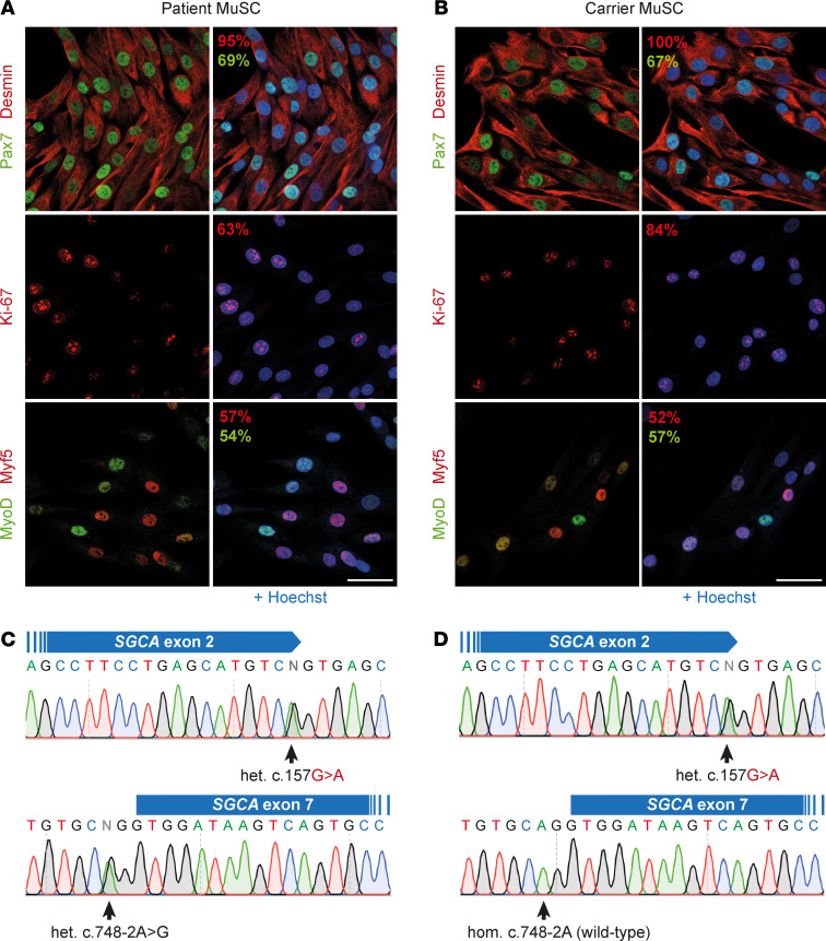
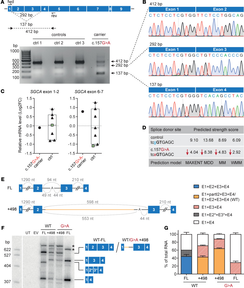
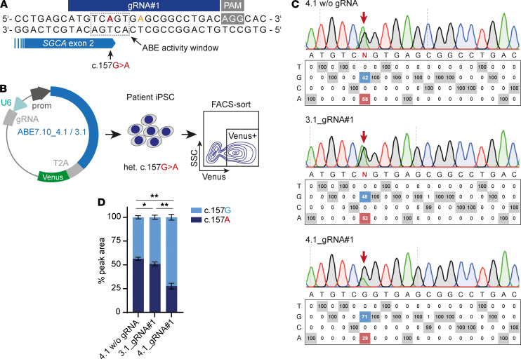
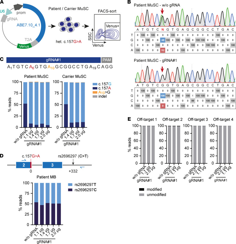
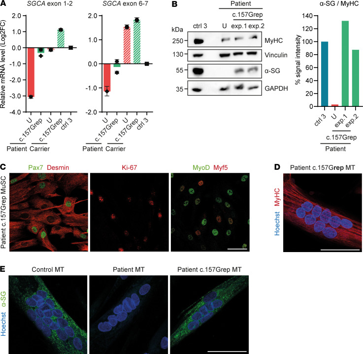
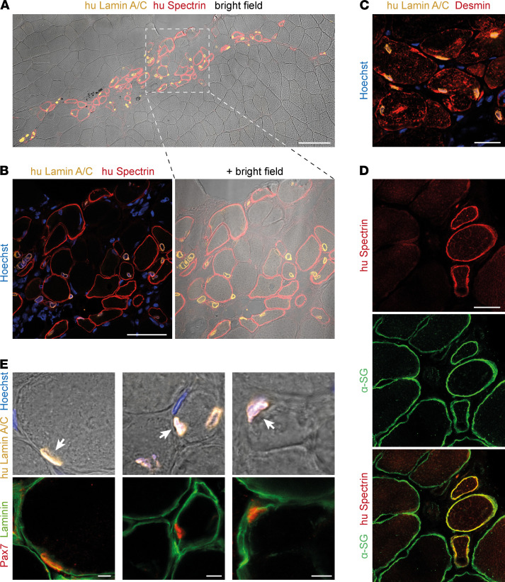
Skeletal muscle can regenerate from muscle stem cells and their myogenic precursor cell progeny, myoblasts. However, precise gene editing in human muscle stem cells for autologous cell replacement therapies of untreatable genetic muscle diseases has not yet been reported. Loss-of-function mutations in SGCA, encoding α-sarcoglycan, cause limb-girdle muscular dystrophy 2D/R3, an early-onset, severe, and rapidly progressive form of muscular dystrophy affecting both male and female patients. Patients suffer from muscle degeneration and atrophy affecting the limbs, respiratory muscles, and heart. We isolated human muscle stem cells from 2 donors, with the common SGCA c.157G>A mutation affecting the last coding nucleotide of exon 2. We found that c.157G>A is an exonic splicing mutation that induces skipping of 2 coregulated exons. Using adenine base editing, we corrected the mutation in the cells from both donors with > 90% efficiency, thereby rescuing the splicing defect and α-sarcoglycan expression. Base-edited patient cells regenerated muscle and contributed to the Pax7+ satellite cell compartment in vivo in mouse xenografts. Here, we provide the first evidence to our knowledge that autologous gene-repaired human muscle stem cells can be harnessed for cell replacement therapies of muscular dystrophies.

Keywords: Human stem cells; Monogenic diseases; Skeletal muscle; Stem cells; Therapeutics.


Conflict of interest statement

Conflict of interest: SS is an inventor on a pending patent application (2016/030371), regarding technology for primary human MuSC isolation and manufacturing. SS and HE are coinventors on a pending patent application (European Patent Office 21 160 696.7) relevant to this publication.

Figures

Figure 1
Figure 1. Characterization of primary MuSC from 2 donors with a heterozygous SGCA c.157G>A mutation.
(A and B) Immunostaining for the satellite cell marker Pax7; the myogenic markers Desmin, MyoD, and Myf5; and the proliferation marker Ki-67 in patient (A) and carrier (B) primary MuSC cultures. The percentage of cells expressing each marker is displayed on the images in the corresponding colors. Nuclei were counterstained with Hoechst. At least 100 nuclei were counted per sample, and staining was done before cryopreservation and after thawing. (C) SGCA sequence analysis from the patient shows the compound heterozygous mutation in exon 2 (c.157G>A) and the splice acceptor of exon 7 (c.748-2A>G). (D) SGCA sequence analysis from the carrier shows the compound heterozygous mutations in exon 2 (c.157G>A) and a homozygous WT exon 7 sequence. Scale bars: 50 μm.
Figure 2
Figure 2. SGCA c.157G>A is an exonic splicing mutation.
(A) RT-PCR analysis of SGCA mRNA in muscle tissue from controls and the c.157G>A carrier. Primer binding sites and expected band sizes are displayed in the panel above. Control 3 is a heterozygous SGCA c.748-2A>G carrier with a WT SGCA exon 2 sequence. The RT-PCR was performed 3 times. (B) Sequencing of the bands from A shows skipping of exon 2 or exon 2+3 in the c.157G>A carrier. (C) qPCR analysis of SGCA mRNA in muscle tissue from controls and the c.157G>A carrier using primers against the exon 1–2 or exon 6–7 boundaries. Control 3 is represented by a green square. The qPCR was performed in technical triplicates. Values were normalized to GAPDH and relativized to the mean of controls. (D) Strength scores of the SGCA exon 2 splice donor for the WT and the mutant sequence predicted by MaxEntScan:score5ss. (E) Minigene construct schemes. FL, full-length SGCA exon 1–4 (blue boxes) with the intermediate introns (black lines); +498, a 498 bp-long low complexity intronic sequence from the human HBB gene (yellow) was inserted to extend intron 2. The size of each intron is indicated above in gray. For intron 2, the size before and after the branch point is indicated below in gray. UT, untransfected; EV, empty vector; WT, WT SGCA; G>A, c.157G>A mutation. (F) Minigene splicing patterns in HEK293T cells analyzed by low-cycle RT-PCR with a 32P-labeled forward primer; products were separated by denaturing PAGE. The splice isoforms identified are shown on the right (identity confirmed by Sanger sequencing). *, intron-retention isoforms; 2*, truncated exon 2 (40 nt); 3*, truncated exon 3 (–70 nt). (G) Splice isoform quantification from F using Phosphorimager analysis. Quantified values are presented as mean ± SD (n = 3). E, exon; I, intron.
Figure 3
Figure 3. ABE corrects the SGCA c.157G>A mutation in patient-derived iPSC.
(A) c.157G>A is located in the center of the ABE activity window for gRNA#1. (B) Experimental design. A plasmid encoding ABE7.10_4.1 or ABE7.10_3.1 was transfected into patient iPSC. Venus+ cells were selected via FACS, and the bulk sorted population was analyzed. (C) EditR analysis of nucleotide rates at each protospacer position in patient iPSC transfected with ABE7.10_4.1 and _3.1 in combination with gRNA#1. iPSC transfected with ABE7.10_4.1 without gRNA are shown as control. (D) c.157G/A nucleotide rates from EditR analysis. Quantified values are presented as mean ± SD (n = 5). Statistical analysis of the difference in c.157G nucleotide rates (light blue) between the columns was performed using the Mann-Whitney U test. Two-tailed P values were calculated. *P < 0.02; **P < 0.01.
Figure 4
Figure 4. ABE repairs the SGCA c.157G>A mutation in patient and carrier primary MuSC without detectable off-target editing.
(A) Experimental design. Primary MuSC from the patient and the carrier were transfected with ABE7.10_4.1/gRNA#1 or without gRNA. FACS was performed to enrich for Venus+ cells, which were subsequently analyzed. (B) EditR analysis of nucleotide rates at each protospacer in patient MuSC transfected with ABE7.10_4.1/gRNA#1. (C) Percentage of reads containing c.157G/A, bystander editing of A10, and indels in patient and carrier MuSC transfected with a range of ABE7.10_4.1/gRNA#1 vector concentrations. (D) A SNP located 332 bp downstream of the mutation and heterozygous in the patient was included in the amplicon to rule out allele detection bias. The plot shows the percentage of reads aligned to each allele. (E) Amplicon sequencing analysis of the 4 predicted exonic off-target sites. Amplicon sequencing data were analyzed using CRISPResso2.
Figure 5
Figure 5. SGCA c.157Grep patient MuSC express α-sarcoglycan, and they are viable and myogenic.
(A) qPCR analysis of SGCA mRNA in SGCA c.157Grep compared with unedited (U) patient and carrier myotubes. Values are normalized to GAPDH and relativized to control 3. SGCA mRNA values were additionally normalized to MYH2 to correct for differences in the differentiation stage of the cells, which may affect promoter activity. Each data point represents the mean ± SD for the n = 3 technical replicates of each biological sample. (B) Western blot analysis of α-sarcoglycan protein in SGCA c.157Grep patient myotubes compared with unedited (U) patient and control 3 myotubes. Myosin heavy chain (MyHC) was used as a differentiation marker. Vinculin and GAPDH were used as loading controls. The intensity of α-sarcoglycan Western blot bands was quantified using ImageJ and normalized to Vinculin and MyHC. (C) SGCA c.157Grep patient MuSC were immunostained for Pax7, Desmin, MyoD, Myf5, and Ki-67. The analysis was performed for all SGCA c.157Grep patient cell populations, and a quantification is shown in Supplemental Table 1. (D) SGCA c.157Grep MuSC readily fused into multinucleated myotubes expressing MyHC. The analysis was performed for all SGCA c.157Grep patient cell populations. (E) α-Sarcoglycan immunostaining in control myotubes, as well as unedited and SGCA c.157Grep patient myotubes. The analysis was performed for all SGCA c.157Grep patient cell populations. MT, myotubes. Scale bars: 50 μm.
Figure 6
Figure 6. SGCA c.157Grep patient MuSC regenerate muscle in vivo.
(A) SGCA c.157Grep patient MuSC were injected into preirradiated anterior tibial muscles of immunocompromised NSG mice. Grafted muscles were collected for analysis after 19 days. (A and B) Grafted muscles were immunostained with antibodies that specifically recognize human Lamin A/C and human Spectrin, labeling donor nuclei and donor-derived myofibers, respectively. In total, 31–87 human nuclei and 33–72 human myofibers were found per section (n = 2). (C) Grafted muscles were immunostained with antibodies against human Lamin A/C and Desmin. (D) Grafted muscles were immunostained with antibodies against human Spectrin and α-sarcoglycan. (E) Grafted muscles were immunostained with antibodies against human Lamin A/C, Pax7, and Laminin to identify satellite cells of human origin located in the stem cell niche under the basal lamina (n = 2). Human Pax7+ satellite cells are indicated by arrows. Nuclei were counterstained with Hoechst. Scale bars: 100 μm (A), 50 μm (B), 20 μm (C and D), 5 μm (E).

References

    1. Liu W, et al. Estimating prevalence for limb-girdle muscular dystrophy based on public sequencing databases. Genet Med. 2019;21(11):2512–2520. doi: 10.1038/s41436-019-0544-8. - DOI - PubMed
    1. Roberds SL, et al. Primary structure and muscle-specific expression of the 50-kDa dystrophin-associated glycoprotein (adhalin) J Biol Chem. 1993;268(32):23739–23742. doi: 10.1016/S0021-9258(20)80440-2. - DOI - PubMed
    1. McNally EM, et al. Human adhalin is alternatively spliced and the gene is located on chromosome 17q21. Proc Natl Acad Sci U S A. 1994;91(21):9690–9694. doi: 10.1073/pnas.91.21.9690. - DOI - PMC - PubMed
    1. Carrié A, et al. Mutational diversity and hot spots in the alpha-sarcoglycan gene in autosomal recessive muscular dystrophy (LGMD2D) J Med Genet. 1997;34(6):470–475. doi: 10.1136/jmg.34.6.470. - DOI - PMC - PubMed
    1. Alonso-Pérez J, et al. New genotype-phenotype correlations in a large European cohort of patients with sarcoglycanopathy. Brain. 2020;143(9):2696–2708. doi: 10.1093/brain/awaa228. - DOI - PubMed

Publication types