Clinical Trial
. 2021 May 17;131(10):e142014.
doi: 10.1172/JCI142014.

Tissue-resident-like CD4+ T cells secreting IL-17 control Mycobacterium tuberculosis in the human lung

Affiliations
Clinical Trial

Tissue-resident-like CD4+ T cells secreting IL-17 control Mycobacterium tuberculosis in the human lung

Paul Ogongo et al. J Clin Invest. .

Abstract

T cell immunity is essential for the control of tuberculosis (TB), an important disease of the lung, and is generally studied in humans using peripheral blood cells. Mounting evidence, however, indicates that tissue-resident memory T cells (Trms) are superior at controlling many pathogens, including Mycobacterium tuberculosis (M. tuberculosis), and can be quite different from those in circulation. Using freshly resected lung tissue, from individuals with active or previous TB, we identified distinct CD4+ and CD8+ Trm-like clusters within TB-diseased lung tissue that were functional and enriched for IL-17-producing cells. M. tuberculosis-specific CD4+ T cells producing TNF-α, IL-2, and IL-17 were highly expanded in the lung compared with matched blood samples, in which IL-17+ cells were largely absent. Strikingly, the frequency of M. tuberculosis-specific lung T cells making IL-17, but not other cytokines, inversely correlated with the plasma IL-1β levels, suggesting a potential link with disease severity. Using a human granuloma model, we showed the addition of either exogenous IL-17 or IL-2 enhanced immune control of M. tuberculosis and was associated with increased NO production. Taken together, these data support an important role for M. tuberculosis-specific Trm-like, IL-17-producing cells in the immune control of M. tuberculosis in the human lung.

Keywords: Bacterial infections; Immunology; Infectious disease; T cells.


Conflict of interest statement

Conflict of interest: MF, BHL, and A Nardin are shareholders and/or employees of ImmunoScape Pte Ltd. A Nardin is a board director of ImmunoScape Pte Ltd.

Figures

Figure 1
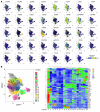
Figure 1. Human lung tissue contains populations of Trm-like T cells.
(A) Cumulative staining of lung CD4+ T cells from 12 biological replicates, defined as having either active TB or previous TB, by CyTOF high-dimensional phenotyping based on uniform manifold approximation and projection (UMAP) plotted as UMAP1 (x axis) versus UMAP2 (y axis) for each cell type. CRTH2, chemoattractant receptor–homologous molecule expressed on receptor on Th2 cells; ITGB7, integrin β7; PD-1, programmed cell death 1; TIGIT, T cell immunoreceptor with immunoglobulin and ITIM domain. (B) PhenoGraph clustering (left) identified 18 clusters (clusters 1–18) depicted on the heatmap of staining intensity of T cell markers (right).
Figure 2
Figure 2. Expression pattern of surface markers in CyTOF panel significantly differentially expressed on CD4+ T cells in lung homogenate according to coexpression of CD69 and CD103.
Markers not significantly expressed presented in Supplemental Figure 1A. Significance test applied stated within each individual plot.
Figure 3
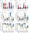
Figure 3. Lung Trm-like T cells are functional and predominantly effector memory.
(A) Frequencies of CD69+ CD4+ and CD8+ T cells isolated from lung tissues from participants with active TB (red), participants with previous TB (dark blue), or cancer controls (dark green). (B) Frequencies of CD103+ CD4+ and CD8+ T cells isolated from lung tissues from participants with active TB (red), participants with previous TB (dark blue), or cancer controls (dark green). (C) Frequencies of CD4+ (left) and CD8+ T cells (right) expressing naive, central memory (CM), effector memory (EM), and terminally differentiated effector memory T cells (TEMRA) phenotypes in blood (yellow) and lungs (blue) from participants with active/previous TB. (D) Frequencies of TNF-α–, IL-2–, IFN-γ–, and IL-17–producing CD4+ and CD8+ T cells from blood (yellow) and lungs (blue) of participants with active/previous TB. Significance calculated by Mann-Whitney test. Asterisk denotes P values that remained significant after stringent Bonferroni’s correction for multiple comparisons.
Figure 4
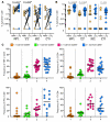
Figure 4. HIV severely depletes cytokine-producing T cells from the lungs of TB-infected study participants.
(A) Frequencies of TNF-α–, IL-2–, IFN-γ–, and IL-17–producing CD4+ T cells from blood (yellow) and lungs (blue) of participants with (squares) and without (circles) HIV coinfection. (B) Tissue-resident phenotypes of TNF-α–, IL-2–, IFN-γ–, and IL-17–producing CD4+ T cells from lungs from participants with (squares) and without (circles) HIV coinfections, where 1 (orange) = CD103+CD69, 2 (green) = CD103CD69+, 3 (pink) = CD103+CD69+, and 4 (blue) = CD103CD69. Significance calculated by Mann-Whitney test.
Figure 5
Figure 5. A portion of lung T cells are TB specific and produce cytokines in response to M. tuberculosis peptides.
(A) Frequencies of TB-specific TNF-α–, IL-2–, IFN-γ–, and IL-17–producing CD4+ T cells in paired blood (yellow) and lung (blue) samples from the same participant. Significance by Wilcoxon’s matched pairs signed-rank test. (B) Frequencies of TB-specific TNF-α–, IL-2–, IFN-γ–, and IL-17–producing CD4+ T cells in blood (yellow) and lung (blue) samples from participants with active/previous TB. Significance by Mann-Whitney test. Asterisk denotes P values that remained significant after stringent Bonferroni’s correction for multiple comparisons. (C) Tissue-resident phenotypes of TNF-α–, IL-2–, IFN-γ–, and IL-17–producing CD4+ T cells where 1 (orange) = CD103+CD69, 2 (green) = CD103CD69+, 3 (pink) = CD103+CD69+, and 4 (blue) = CD103CD69. Significance calculated by Kruskal-Wallis test, although none was found.
Figure 6
Figure 6. IL-17–producing M. tuberculosis–specific CD4+ T cells in lung homogenate inversely correlate with systemic markers of inflammation.
(AD) Correlations between plasma IL-1β and TNF-α–, IL-2–, IFN-γ–, and IL-17–producing CD4+ T cells in lung tissue from participants with active TB (red) or previous TB (dark blue). Spearman’s correlation with P value reported for 2-tailed analysis.
Figure 7
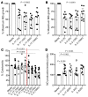
Figure 7. Exogenous IL-17 and IL-2 decrease M. tuberculosis growth in 3D culture system.
(A and B) Addition of IL-17 and IL-2 to culture media decreases M. tuberculosis growth in granuloma-like 3D human cell culture system. Data from 3 separate experiments using PBMCs from 3 healthy controls conducted in triplicate. Concentrations shown in parentheses (ng/mL). Impact of exogenous IL-17 and IL-2 on (C) cell viability in above experiments as measured by concentration of lactate dehydrogenase (LDH) in culture supernatant on day 7 and (D) production of NO. Statistical differences tested by Kruskal-Wallis (A and B), with Dunn’s correction for multiple comparisons (C and D).

References

    1. Harding E. WHO global progress report on tuberculosis elimination. Lancet Respir Med. 2020;8(1):19. doi: 10.1016/S2213-2600(19)30418-7. - DOI - PubMed
    1. Young D, Dye C. The development and impact of tuberculosis vaccines. Cell. 2006;124(4):683–687. doi: 10.1016/j.cell.2006.02.013. - DOI - PubMed
    1. Cohen A, et al. The global prevalence of latent tuberculosis: a systematic review and meta-analysis. Eur Respir J. 2019;54(3):1900655. doi: 10.1183/13993003.00655-2019. - DOI - PubMed
    1. Jeyanathan M, et al. New tuberculosis vaccine strategies: taking aim at un-natural immunity. Trends Immunol. 2018;39(5):419–433. doi: 10.1016/j.it.2018.01.006. - DOI - PubMed
    1. Orme IM. A new unifying theory of the pathogenesis of tuberculosis. Tuberculosis (Edinb) 2014;94(1):8–14. doi: 10.1016/j.tube.2013.07.004. - DOI - PMC - PubMed

Publication types