. 2021 Sep 16;28(9):1298-1309.e7.
doi: 10.1016/j.chembiol.2021.03.012. Epub 2021 Apr 12.

Protein acylation by saturated very long chain fatty acids and endocytosis are involved in necroptosis

Affiliations

Protein acylation by saturated very long chain fatty acids and endocytosis are involved in necroptosis

Apoorva J Pradhan et al. Cell Chem Biol. .

Abstract

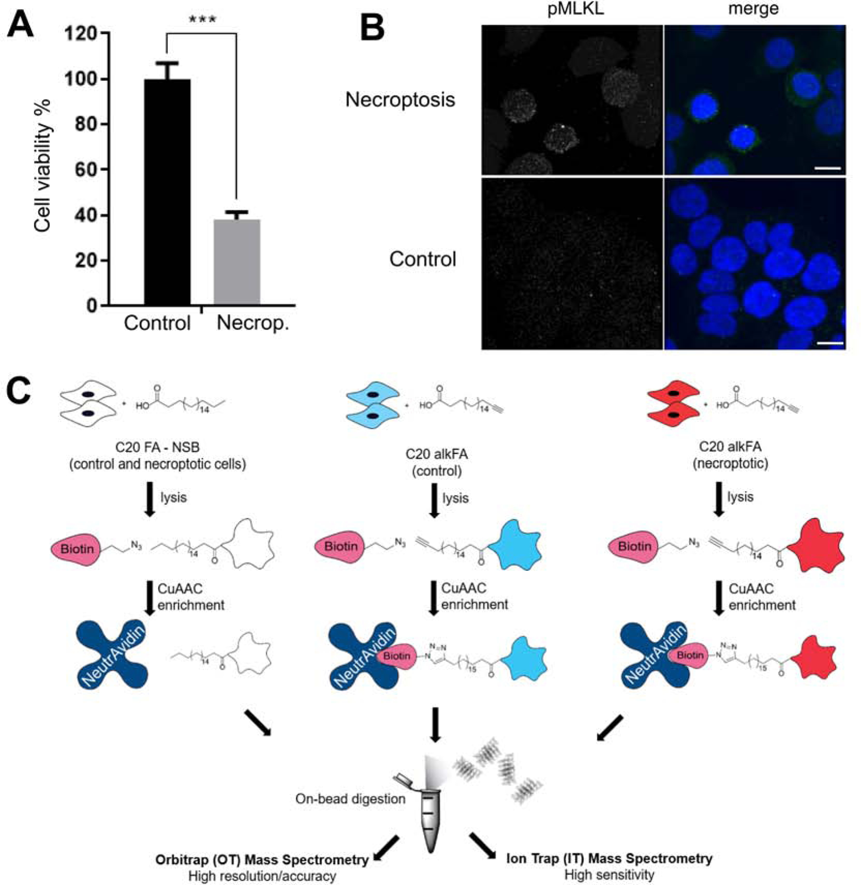
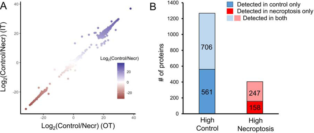
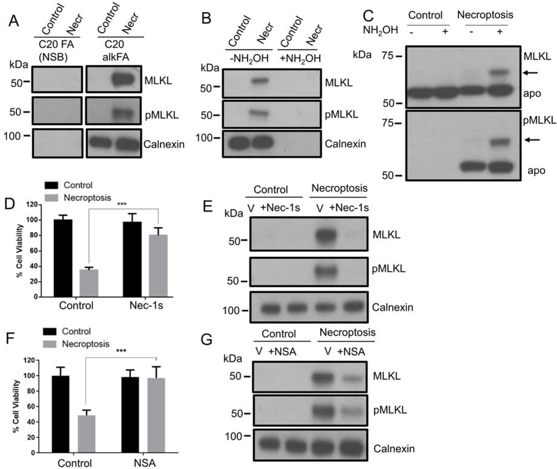
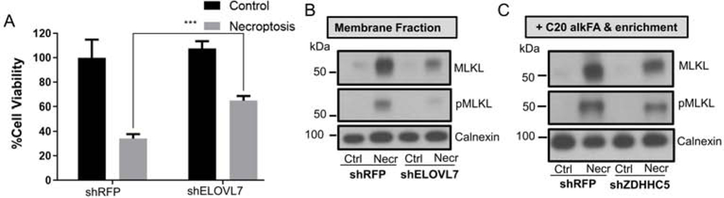
Necroptosis is a form of cell death characterized by receptor-interacting protein kinase activity and plasma membrane permeabilization via mixed-lineage kinase-like protein (MLKL). This permeabilization is responsible for the inflammatory properties of necroptosis. We previously showed that very long chain fatty acids (VLCFAs) are functionally involved in necroptosis, potentially through protein fatty acylation. Here, we define the scope of protein acylation by saturated VLCFAs during necroptosis. We show that MLKL and phosphoMLKL, key for membrane permeabilization, are exclusively acylated during necroptosis. Reducing the levels of VLCFAs decreases their membrane recruitment, suggesting that acylation by VLCFAs contributes to their membrane localization. Acylation of phosphoMLKL occurs downstream of phosphorylation and oligomerization and appears to be, in part, mediated by ZDHHC5 (a palmitoyl transferase). We also show that disruption of endosomal trafficking increases cell viability during necroptosis, possibly by preventing recruitment, or removal, of phosphoMLKL from the plasma membrane.

Keywords: VLCFA; ZDHHC5; endocytosis; lipids; necroptosis; programmed cell death; protein acylation; vesicular trafficking.


Conflict of interest statement

Declaration of interests The authors declare no competing interest.

Figures

Figure 1.
Figure 1.. Induction of necroptosis and workflow of C20 fatty acid acylation and proteomics.
(A) Cell viability decreases as HT-29 cells undergo necroptosis. Necroptosis is induced with BV6/zVAD-FMK/TNF-α treatment. Data represent mean ± 1 SD; n=5. *** represents p < 0.001. (B) Cellular distribution of pMLKL during necroptosis. Sample 3D project images of pMLKL immunostaining in necroptotic (top) or control (bottom) cells show an increase in pMLKL staining, mainly concentrated at the plasma membrane. Merge images include DAPI nuclear staining (blue) and pMLKL (green). Scalebar represents 10 μm. (C) HT-29 cells were treated with endogenous C20:0 FA or C20:0 alkFA for 3h to account for non-specific and specific interactions respectively. Control and necroptotic cells were then membrane fractionated, subjected to CuAAC and installed with a biotin reporter. Biotinylated proteins were captured on neutravidin and subjected to on-bead trypsin digestion followed by quantitative proteomics. See also Figure S1.
Figure 2.
Figure 2.. Quantitative analysis of acylated proteome in necroptosis.
(A) High correlation between the mean intensities of acylated proteins detected in control and necroptotic cells in Orbitrap Lumos (x axis) and Ion trap (y axis) platforms. Strong correlation between difference in mean intensities indicates biological variation rather than technical variation among the proteins detected in control and necroptotic cells. (B) Bar plot shows the total number of proteins (1672) undergoing acylation in either control, necroptotic or both conditions. Blue and red bars represent the number of proteins with high abundance in control or necroptotic cells, respectively. A higher number of proteins appear acylated in only control (561) as compared to only necroptosis (158). Overall, fewer proteins undergo acylation in necroptosis (405) as compared to control (1267). See also Figure S2 and Table S1.
Figure 3.
Figure 3.. MLKL and pMLKL are acylated downstream of RIPK1 activation.
(A) Western blot analysis of proteins from C20 alkFA treated control and necroptosis cells. After cell lysis and Biotin-azide attachment via CuAAC, acylated proteins are enriched on neutravidin resin. The samples are blotted for MLKL and pMLKL and Calnexin. Calnexin is used as a loading control. Equal amounts of protein are used for enrichment of each condition (see Materials and Methods for details). We note that the treatments with C20 FA and C20 alkFA and the western blot analysis were carried out during the same experiment. Uncropped western blot image in shown in Figure S3F. (B) Acylation of MLKL and pMLKL with C20 alkFA show NH2OH sensitivity. Control and necroptotic cells treated C20 alkFA were membrane fractionated and subjected to CuAAC with Biotin-azide. The proteins were treated with 0.3 M NH2OH for 30 min, enriched using neutravidin beads, then blotted for MLKL and pMLKL and Calnexin. Calnexin is used as a positive control for NH2OH sensitivity. We note that NH2OH treatment does not cause protein degradation (Figure S3A). Loss of signal with NH2OH treatment indicates that MLKL and pMLKL acylation occurs primarily through S-linkages. (C) HT-29 cells were lysed and total cell lysates were subjected to APE with NEM, NH2OH, and mPEG-Mal, and compared with negative controls. The samples are blotted for MLKL and pMLKL. (D) 1 μM Nec-1s and (F) 1 μM NSA reduce necroptotic cell death. Percent cell viability of control, Nec-1s or NSA-treated and necroptotic cells are shown. Necroptosis is induced with BV6/zVAD-FMK/TNF-α treatment. Data represent mean ± 1 SD; n=5. *** represents p < 0.001. (E and G) Western blot analysis of C20 alkFA treated control and necroptotic cells in the presence of Nec-1s and NSA. Control and necroptotic cells treated with C20 alkFA and Nec-1s/NSA were membrane fractionated and subjected to CuAAC with Biotin-azide and enrichment using neutravidin beads. The proteins on the beads were blotted for MLKL and pMLKL. Equal amounts of protein are used for enrichment of each condition. Calnexin is used as loading control (see Materials and Methods for details). Nec-1s (E) and NSA (G) treatment significantly reduce VLCFAcylation of MLKL and pMLKL (see Figure S3B and D for quantification of band intensities). See also Figure S3.
Figure 4.
Figure 4.. Membrane recruitment of MLKL and pMLKL in shELOVL7 and shZDHHC5 cells during necroptosis.
(A) ELOVL7 knockdown reduces necroptotic cell death. Percent cell viability of shRFP (used as control) and shELOVL7 transduced cells treated with BV6/zVAD-FMK/TNF-α to induce necroptosis compared to vehicle-treated control cells. Data represent mean ± 1 SD; n=5. *** represents p < 0.001 (B) Western blot analysis of membrane fraction from untreated and necroptotic shRFP (used as control) and shELOVL7 cells. Cell lysates were fractionated by ultracentrifugation and membrane fractions analyzed. Decrease in MLKL and pMLKL were observed in necroptotic shELOVL7 as compared to necroptotic control cells suggesting that membrane recruitment is reduced in shELOVL7 cells (see Figure S4A for quantification of band intensities). (D) Control and necroptotic shRFP and shZDHHC5 cells treated with C20 alkFA were membrane fractionated and subjected to CuAAC with Biotin-azide and enrichment using neutravidin beads. Equal amounts of protein were used for enrichment of each condition (see Materials and Methods for details). The proteins on the beads were blotted for MLKL, pMLKL and calnexin. An approximate 50% decrease in the enriched pMLKL was observed as compared to necroptotic control cells suggesting a role for ZDHHC5 in VLCFAcylation during necroptosis. (see Figure S4B for quantification of band intensities). See also Figure S4.
Figure 5.
Figure 5.. Inhibition of endocytic pathway specifically ameliorate necroptotic cell death.
(A) Pathway enrichment analysis of proteins that show differential fatty acylation during necroptosis. 8 pathways significantly enriched (corrected p value < 0.05). See also Table S3. (B) Small molecule inhibitor targeting endocytic pathway rescues necroptotic cell death, but not apoptotic cell death, in HT-29 cells. HT-29 cells were pretreated with 100 μM dynasore for 2 hours. Vehicle-treated control cells were treated with DMSO. For induction of necroptosis, cells were treated with BV6/zVAD-FMK/TNF-α. For induction of apoptosis, cells were treated with BV6/TNF-α. The percent cell viability in the presences of inhibitor during necroptosis was compared to the vehicle-treated control cells. Data represent mean ± 1 SD; n ≥ 3. ** represents p < 0.01, *** represents p < 0.001. (C-D) Western blot analysis of membrane fraction (C) and whole lysate (D) of vehicle-treated cells and dynasore treated cells with or without necroptosis. Cell lysates are fractionated by ultracentrifugation and membrane fractions are analyzed. The decrease in pMLKL observed in necroptosis when dynasore is present suggesting endocytosis maybe involved in necroptosis.

References

    1. Akimov SA, Volynsky PE, Galimzyanov TR, Kuzmin PI, Pavlov KV & Batishchev OV (2017). Pore formation in lipid membrane I: Continuous reversible trajectory from intact bilayer through hydrophobic defect to transversal pore. Sci. Rep 7, 12152. - PMC - PubMed
    1. Blanc M, David FPA & Van Der Goot FG (2019). SwissPalm 2: Protein S-Palmitoylation Database. Methods Mol. Biol 2009, 203–214. - PubMed
    1. Breusegem SY & Seaman MNJ (2014). Genome-wide RNAi screen reveals a role for multipass membrane proteins in endosome-to-golgi retrieval. Cell Rep 9, 1931–1945. - PMC - PubMed
    1. Bunney TD & Katan M (2010). Phosphoinositide signalling in cancer: beyond PI3K and PTEN. Nat. Rev. Cancer 10, 342–52. - PubMed
    1. Cai Z, Jitkaew S, Zhao J, Chiang HC, Choksi S, Liu J, Ward Y, Wu LG & Liu ZG (2014). Plasma membrane translocation of trimerized MLKL protein is required for TNF-induced necroptosis. Nat. Cell Biol 16, 55–65. - PMC - PubMed

Publication types