. 2021 May 7;49(8):4705-4724.
doi: 10.1093/nar/gkab242.

Global RNA profiles show target selectivity and physiological effects of peptide-delivered antisense antibiotics

Affiliations

Global RNA profiles show target selectivity and physiological effects of peptide-delivered antisense antibiotics

Linda Popella et al. Nucleic Acids Res. .

Abstract

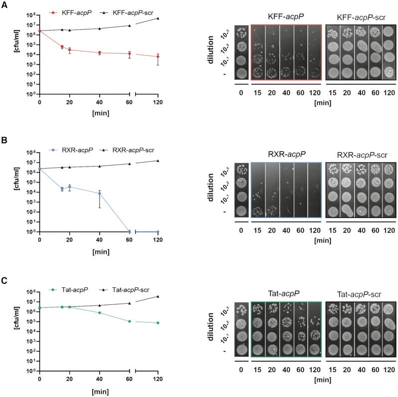
Antisense peptide nucleic acids (PNAs) inhibiting mRNAs of essential genes provide a straight-forward way to repurpose our knowledge of bacterial regulatory RNAs for development of programmable species-specific antibiotics. While there is ample proof of PNA efficacy, their target selectivity and impact on bacterial physiology are poorly understood. Moreover, while antibacterial PNAs are typically designed to block mRNA translation, effects on target mRNA levels are not well-investigated. Here, we pioneer the use of global RNA-seq analysis to decipher PNA activity in a transcriptome-wide manner. We find that PNA-based antisense oligomer conjugates robustly decrease mRNA levels of the widely-used target gene, acpP, in Salmonella enterica, with limited off-target effects. Systematic analysis of several different PNA-carrier peptides attached not only shows different bactericidal efficiency, but also activation of stress pathways. In particular, KFF-, RXR- and Tat-PNA conjugates especially induce the PhoP/Q response, whereas the latter two additionally trigger several distinct pathways. We show that constitutive activation of the PhoP/Q response can lead to Tat-PNA resistance, illustrating the utility of RNA-seq for understanding PNA antibacterial activity. In sum, our study establishes an experimental framework for the design and assessment of PNA antimicrobials in the long-term quest to use these for precision editing of microbiota.


Figures

Figure 1.
Figure 1.
(A) Schematic overview of the herein applied strategy. Antisense peptide nucleic acid (PNA)-coupling to cell penetrating peptides (CPP) facilitates its delivery into the bacterial cell, e.g. Salmonella. PNA sequences targeting either the Shine-Dalgarno (SD) or the translational start codon (AUG) possess the strongest inhibitory efficacy in preventing ribosomal binding (30 S subunit) and thus translation. Strong bactericidal effects are elicited when the PNA is targeted against an essential gene. (B) Structure of the acpP-fabF transcription unit including a snapshot of the acpP mRNA sequence in Salmonella. The sequence of the herein used antisense PNA as well as its target sequence in acpP is shaded in cyan. The translational start codon is highlighted in bold type, the Shine-Dalgarno sequence is underlined. The sequence and charge (https://pepcalc.com/) of the three most potent peptides used in the present study—KFF, RXR and Tat—as well as of the additionally tested peptides—ANT, SAP and SAPE—are highlighted in gray. The indicated peptide charges correspond to peptide conjugated to PNA.
Figure 2.
Figure 2.
Growth kinetics and MIC determination of wild-type (wt) Salmonella enterica serovar Typhimurium strain SL1344 (∼106 cfu/ml) in the presence of varying concentrations of the following PPNA, PNA and peptide constructs. (A) KFF-acpP, KFF-acpP-scrambled (scr) or KFF (10–0.3 μM), (B) RXR-acpP, RXR-acpP-scr or RXR (10–0.3 μM), (C) Tat-acpP, Tat-acpP-scr or Tat (40–1.25 μM), and (D) acpP (40–0.6 μM). Growth curves are depicted as OD600 (y-axis) over time (indicated in hours, x-axis). The experiment was performed three times. Error bars indicate standard error of the mean.
Figure 3.
Figure 3.
Kinetics of bactericidal effects for (A) 5 μM KFF-acpP versus 5 μM KFF-acpP-scr, (B) 5 μM RXR-acpP versus 5 μM RXR-acpP-scr and (C) 10 μM Tat-acpP versus 10 μM Tat-acpP-scr against Salmonella wt (∼106 cfu/ml). After the indicated time points post treatment (15, 20, 40, 60, 120 min), aliquots of each sample were harvested to determine the number of viable cells over time (cfu/ml per min; left panels). Additional aliquots were subjected to spot assays on LB agar plates, including the indicated dilutions (right panels). The experiment was performed three times. Error bars indicate standard error of the mean.
Figure 4.
Figure 4.
An optimal RNA isolation protocol for the analysis of PNA-mediated transcriptomic effects. (A, B) Seven different RNA isolation protocols (#1–7, described in Materials and Methods) were applied to analyze their capacity to enrich for all size classes of total RNA. In brief, 109 cfu were pelleted for the isolation of RNA, which was subsequently analyzed using (A) a denaturing polyacrylamide gel (1 μg/lane) and (B) northern blotting (10 μg/lane). (A) Prominent RNAs, such as ribosomal 23S/16S or 5S, and tRNA, are indicated on the right. (B) Transcript levels of two sRNAs, SroC and SdhX (SdhX1, SdhX2), and the loading control 5S were detected using sequence specific radioactive-labeled probes. The experiment was performed two times independently. (C, D) A Salmonella culture (OD600 0.5) was diluted to 106 cfu/ml and cells were treated with the following conditions for 15 min: 1) water – untreated control, 2) 5 μM KFF-acpP, 3) 5 μM KFF-acpP-scrambled (scr), 4) 5 μM KFF peptide, 5) 5 μM RXR-acpP, 6) 5 μM RXR-acpP-scr, 7) 5 μM RXR peptide, 8) 10 μM Tat-acpP, 9) 10 μM Tat-acpP-scr or 10) 10 μM Tat peptide. After isolating RNA using (C) protocol #4 or (D) protocol #3, a total of 60–100 ng RNA was subjected to denaturing PAGE. After staining with SybrGold, ribosomal 23S/16S or 5S were visualized. Asterisks indicate the appearance of a smear in the range of short RNAs. The experiments were performed each three times independently. (E) A Salmonella culture (OD600 0.5) was diluted to 106 cfu/ml and cells were treated with the following constructs for 5, 10 or 15 min: water (control) or 5 μM of either KFF-acpP, KFF-acpP-scrambled or KFF peptide, as indicated. After isolating RNA using protocol #3, a total of 100–150 ng RNA was subjected to denaturing PAGE, followed by northern blotting. Transcript levels of acpP or the loading control 5S were detected using sequence specific DIG-labeled probes. Results show exemplary data from one out of two biological replicates.
Figure 5.
Figure 5.
Transcriptomic profiling of Salmonella in response to treatment with peptide–PNA conjugates. (A) Experimental workflow showing the different conditions used for the analyses. ‘w/o’ denotes the untreated water control. Parts of the image have been created with BioRender.com. (B) Principal component analysis (PCA) of all 10 conditions including three independent biological replicates, after TMM normalization. Clusters 1, 2 and 3 were added manually after creating the plot.
Figure 6.
Figure 6.
Transcriptomic responses of Salmonella upon PNA, PPNA or peptide treatment for 15 min. Volcano plots show calculated changes in Salmonella gene expression as false discovery rate (FDR)-adjusted P-value (–log10, y-axis) and fold change (log2, x-axis). The following conditions are shown: (A) KFF-acpP, KFF-acpP-scrambled or KFF peptide versus untreated (water) control, (B) RXR-acpP, RXR-acpP-scrambled or RXR peptide versus untreated (water) control, (C) Tat-acpP, Tat-acpP-scrambled or Tat peptide versus untreated (water) control. (A–C) Significantly differentially regulated genes are characterized by an absolute fold change >2 (down-regulated log2 < –1, up-regulated log2 > 1; vertical dashed line) and an FDR-adjusted P-value < 0.001 (–log10 > 3, horizontal dashed line). Significantly down-regulated genes are highlighted in blue, whereas up-regulated genes are highlighted in red. The top-3 differentially expressed transcripts, showing the strongest up- and down-regulation, are specified.
Figure 7.
Figure 7.
Heatmap showing differentially expressed sRNAs upon the indicated treatment conditions. Each comparison includes triplicate RNA-seq samples for the indicated conditions. The coloring indicates log2 fold change (FC) of the selected samples, while red and blue denote up- and down-regulation, respectively. Asterisks (*) show significantly differentially regulated sRNAs are characterized by an absolute fold change >2 (down-regulated log2 < –1, up-regulated log2 > 1; vertical dashed line) and an false discovery rate (FDR) adjusted P-value < 0.001.
Figure 8.
Figure 8.
Analysis of gene enrichment classified according to annotated KEGG pathways and known regulons (marked in green, (18). Pathway and regulon analysis reveals a broad transcriptomic response upon RXR- and Tat-PNA exposure, but mostly PmrAB, PhoP/Q and CAMP-resistance-restricted gene enrichment post KFF-PNA or KFF treatment. Statistically significant (P-valueadj < 0.05) gene sets are marked with an asterisk (*). Color indicates median log2 fold change (FC) of all genes belonging to the gene set, while red denotes up- and blue down-regulation. Each column represents a single KEGG pathway or manually created regulon.
Figure 9.
Figure 9.
PNA-mediated growth inhibition is independent of established factors for regulation by endogenous small RNAs. Growth kinetics for MIC determination of 105 cfu/ml of the indicated Salmonella strains (A–D) in the presence of varying concentrations of KFF-acpP, KFF-acpP-scrambled (scr) or KFF peptide in serial dilutions from 10 μM to 0.3 μM. Data represent the average of three biological replicates, error bars indicate standard error of the mean (SEM).

Similar articles

Cited by

References

    1. Kole R., Krainer A.R., Altman S.. RNA therapeutics: beyond RNA interference and antisense oligonucleotides. Nat. Rev. Drug Discov. 2012; 11:125–140. - PMC - PubMed
    1. Pifer R., Greenberg D.E.. Antisense antibacterial compounds. Transl. Res. J. Lab. Clin. Med. 2020; 223:89–106. - PubMed
    1. Vogel J. An RNA biology perspective on species-specific programmable RNA antibiotics. Mol. Microbiol. 2020; 113:550–559. - PubMed
    1. Barkowsky G., Lemster A.-L., Pappesch R., Jacob A., Krüger S., Schröder A., Kreikemeyer B., Patenge N.. Influence of different cell-penetrating peptides on the antimicrobial efficiency of PNAs in Streptococcus pyogenes. Mol. Ther. Nucleic Acids. 2019; 18:444–454. - PMC - PubMed
    1. Geller B.L., Li L., Martinez F., Sully E., Sturge C.R., Daly S.M., Pybus C., Greenberg D.E.. Morpholino oligomers tested in vitro, in biofilm and in vivo against multidrug-resistant Klebsiella pneumoniae. J. Antimicrob. Chemother. 2018; 73:1611–1619. - PMC - PubMed

Publication types

MeSH terms