. 2021 Apr;17(4):20210073.
doi: 10.1098/rsbl.2021.0073. Epub 2021 Apr 14.

Age and social experience induced plasticity across brain regions of the paper wasp Polistes fuscatus

Affiliations

Age and social experience induced plasticity across brain regions of the paper wasp Polistes fuscatus

Christopher M Jernigan et al. Biol Lett. 2021 Apr.

Abstract

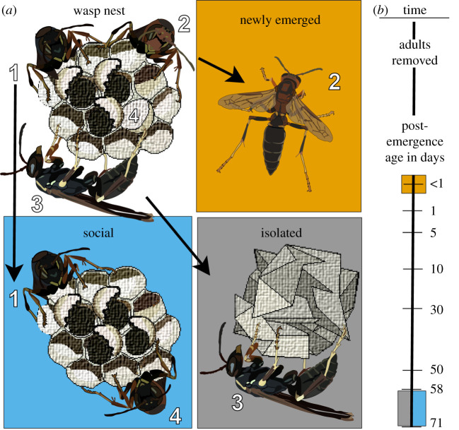
Developmental studies of brain volumes can reveal which portions of neural circuits are sensitive to environmental inputs. In social insects, differences in relative investment across brain regions emerge as behavioural repertoires change during ontogeny or as a result of experience. Here, we test the effects of maturation and social experience on morphological brain development in Polistes fuscatus paper wasps, focusing on brain regions involved in visual and olfactory processing. We find that mature wasps regardless of social experience have relatively larger brains than newly emerged wasps and this difference is driven by changes to mushroom body calyx and visual regions but not olfactory processing neuropils. Notably, social wasps invest more in the anterior optic tubercle (AOT), a visual glomerulus involved in colour and object processing in other taxa, relative to other visual integration centres the mushroom body calyces compared with aged socially naive wasps. Differences in developmental plasticity between visual and olfactory neuropil volumes are discussed in light of behavioural maturation in paper wasps, especially as it relates to social recognition. Previous research has shown that P. fuscatus need social experience to develop specialized visual processing of faces, which is used to individually recognize conspecifics. The present study suggests that the AOT is a candidate brain region that could mediate facial processing in this species.

Keywords: Polistes fuscatus; anterior optic tubercle; neuroplasticity; paper wasp; social experience.


Figures

Figure 1.
Figure 1.
Development and social rearing protocol. Nests with capped pupa were collected and newly emerged adults were split into one of three treatments. Numbers denote individual wasp labels as they are placed into treatments from a small pool of nests. (a) Depiction of treatment groups. Newly Emerged: adults less than 24 h of age (top right, orange), Social: mature wasps socially housed on the nest with siblings (bottom left, blue) and Isolated: mature isolated individuals (bottom right, grey). (b) Timeline of experiment in days. Coloured blocks denote age at the time of dissection and corresponding social treatment groups in (a).
Figure 2.
Figure 2.
Impact of social experience on subregions of the wasp brain. (a) Anti-synapsin immunofluorescent image of one hemisphere of the wasp brain, with white lines highlighting brain regions that were reconstructed (AOT in more peripheral slices); scale bar 250 µm. (b) Reconstruction rendering of the quantified regions from each brain. (c) Total measured brain volume in μm3 across treatments. (d) Brain region volume differences across treatment in μm3: MBcx (left), Lo (centre left), AL (centre right) and AOT (right). Visual representations of corresponding brain regions are placed in the bottom right corner of each plot. (e) Relative brain volume investment across treatments, AOT to MBcx (left), lip (centre left), collar (centre right) and basal ring (right). (f) Proposed trade-off of central brain investment as P. fuscatus age, with and without social interaction. Box plot fills denote treatment: Isolated (I, grey), Newly Emerged (NE, orange), Social (S, blue). Letters denote significance via ANCOVA with body size covariate and Tukey HSD post hoc comparisons (all statistical comparisons and values are provided in electronic supplementary material, tables S1 and S2). Sample sizes: Newly Emerged N = 12, Isolated N = 14, Social N = 14. Me, medulla; Lo, lobula; lip, lip of mushroom body calyx; co, collar of mushroom body calyx; br, basal ring of mushroom body calyx; AL, antennal lobe; MBcx, mushroom body calyx; AOT, anterior optic tubercle.

References

    1. Gronenberg W, Hölldobler B. 1999. Morphologic representation of visual and antennal information in the ant brain. J. Comp. Neurol. 412, 229-240. (10.1002/(SICI)1096-9861(19990920)412:2<229::AID-CNE4>3.0.CO;2-E). - DOI - PubMed
    1. O'Donnell S, Clifford MR, DeLeon S, Papa C, Zahedi N, Bulova SJ. 2013. Brain size and visual environment predict species differences in paper wasp sensory processing brain regions (Hymenoptera: Vespidae, Polistinae). Brain Behav. Evol. 82, 177-184. (10.1159/000354968) - DOI - PubMed
    1. Stöckl A, Heinze S, Charalabidis A, el Jundi B, Warrant E, Kelber A. 2016. Differential investment in visual and olfactory brain areas reflects behavioural choices in hawk moths. Scient. Rep. 6, 26041. (10.1038/srep26041) - DOI - PMC - PubMed
    1. Sheehan ZBV, Kamhi JF, Seid MA, Narendra A. 2019. Differential investment in brain regions for a diurnal and nocturnal lifestyle in Australian Myrmecia ants. J. Comp. Neurol. 527, 1261-1277. (10.1002/cne.24617) - DOI - PubMed
    1. Barth M, Hirsch HVB, Meinertzhagen IA, Heisenberg M. 1997. Experience-dependent developmental plasticity in the optic lobe of Drosophila melanogaster. J. Neurosci. 17, 1493. (10.1523/JNEUROSCI.17-04-01493.1997) - DOI - PMC - PubMed

Publication types