. 2021 Apr 13;16(1):253.
doi: 10.1186/s13018-021-02391-9.

Circ_0116061 regulated the proliferation, apoptosis, and inflammation of osteoarthritis chondrocytes through regulating the miR-200b-3p/SMURF2 axis

Affiliations

Circ_0116061 regulated the proliferation, apoptosis, and inflammation of osteoarthritis chondrocytes through regulating the miR-200b-3p/SMURF2 axis

Wei Zheng et al. J Orthop Surg Res. .

Abstract

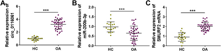
Background: Circular RNA (circRNA) has been shown to be associated with osteoarthritis (OA) progression. Circ_0116061 has been found to be highly expressed in OA cartilage tissues, but its role and mechanism in OA progression remain unclear.

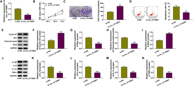
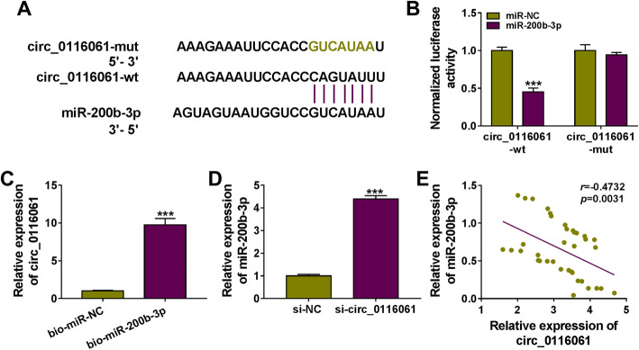
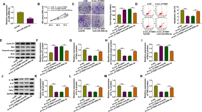
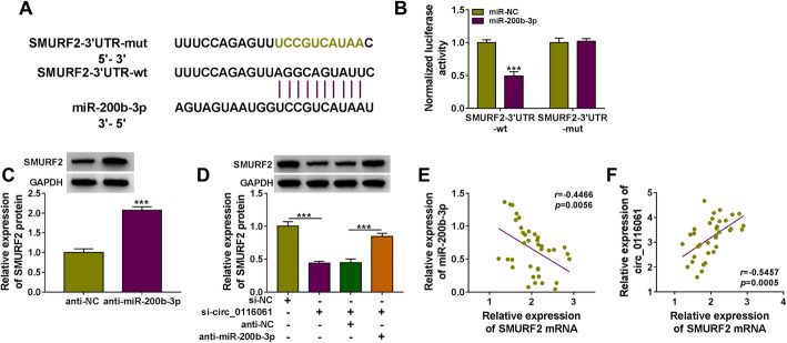
Methods: Expression levels of circ_0116061, microRNA (miR)-200b-5p, and Smad ubiquitin regulatory factor 2 (SMURF2) were detected using quantitative real-time PCR. The proliferation and apoptosis of cells were measured using cell counting kit 8 (CCK8) assay, colony formation assay, and flow cytometry. Furthermore, the protein levels of proliferation-related marker, apoptosis-related markers, inflammatory factors, and SMURF2 were tested using western blot (WB) analysis. In addition, the interaction between miR-200b-3p and circ_0116061 or SMURF2 was examined using dual-luciferase reporter assay and biotin-labeled RNA pull-down assay.

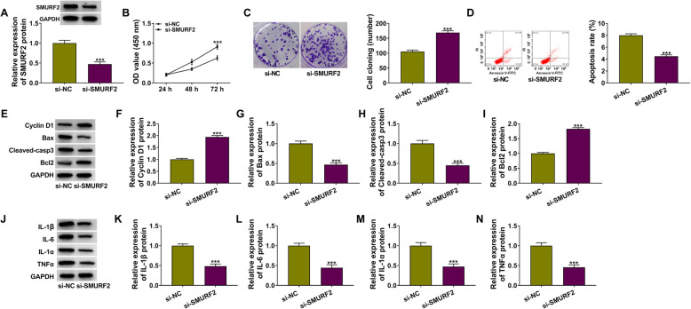
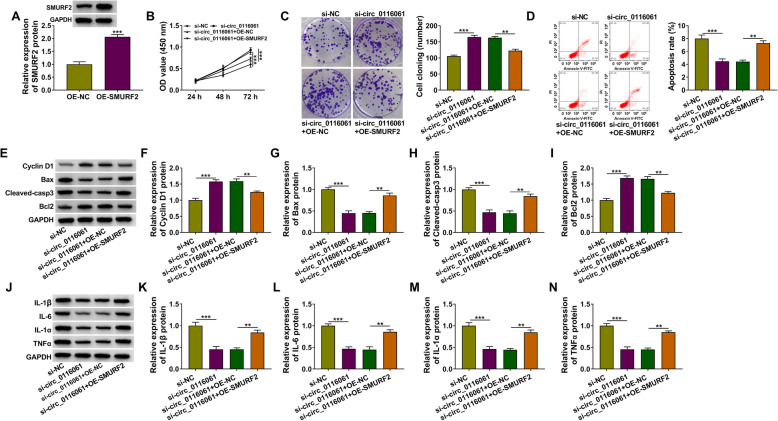
Results: Circ_0116061 and SMURF2 were highly expressed, and miR-200b-3p was lowly expressed in OA cartilage tissues. Knockdown of circ_0116061 could promote the proliferation and inhibit the apoptosis and inflammation of OA chondrocytes. MiR-200b-3p could be sponged by circ_0116061, and its inhibitor could reverse the regulation of circ_0116061 silencing on the biological functions of OA chondrocytes. SMURF2 was a target of miR-200b-3p, and its expression was positively regulated by circ_0116061. Silencing of SMURF2 also could enhance the proliferation and suppress the apoptosis and inflammation of OA chondrocytes. Furthermore, the regulation of circ_0116061 silencing on the biological functions of OA chondrocytes also could be reversed by SMURF2 overexpression.

Conclusion: Our data showed that circ_0116061 might regulate the miR-200b-3p/SMURF2 axis to promote the progression of OA.

Keywords: Chondrocytes; Circ_0116061; MiR-200b-3p; Osteoarthritis; SMURF2.


Conflict of interest statement

The authors declare that they have no competing interests.

Figures

Fig. 1
Fig. 1
The expression of circ_0116061, miR-200b-3p, and SMURF2 in cartilage tissues. The expression levels of circ_0116061 (a), miR-200b-3p (b), and SMURF2 (c) in the cartilage tissues of healthy controls and OA patients were detected by qRT-PCR. ***P < 0.001
Fig. 2
Fig. 2
Silencing of circ_0116061 regulated the biological functions of OA chondrocytes. OA chondrocytes were transfected with si-NC or si-circ_0116061. a The expression of circ_0116061 was detected by qRT-PCR. CCK8 assay (b), colony formation assay (c), and flow cytometry (d) were used to test cell viability, the cell cloning number, and apoptosis rate. en The protein levels of Cyclin D1, Bcl2, Bax, Cleaved-casp3, IL-1β, IL-6, IL-1α, and TNFα were determined by WB analysis. ***P < 0.001
Fig. 3
Fig. 3
Circ_0116061 sponged miR-200b-3p. a The binding sites between miR-200b-3p and circ_0116061 were shown. Dual-luciferase reporter assay (b) and biotin-labeled RNA pull-down assay (c) were performed to measure the interaction between miR-200b-3p and circ_0116061. d After transfecting with si-NC or si-circ_0116061 into OA chondrocytes, the expression of miR-200b-3p was detected by qRT-PCR. e Pearson correlation analysis was used to analyze the correlation between miR-200b-3p and circ_0116061 in OA cartilage tissues. ***P < 0.001
Fig. 4
Fig. 4
Effects of circ_0116061 silencing and miR-477 inhibitor on the biological functions of OA chondrocytes. a The transfection efficiency of anti-miR-200b-3p was evaluated by detecting miR-200b-3p expression using qRT-PCR. bn OA chondrocytes were transfected with si-NC, si-circ_0116061, si-circ_0116061 + anti-NC or si-circ_0116061 + anti-miR-200b-3p. Cell viability, the cell cloning number, and apoptosis rate were determined using CCK8 assay (b), colony formation assay (c), and flow cytometry (d). en WB analysis was used to examine the protein levels of Cyclin D1, Bcl2, Bax, Cleaved-casp3, IL-1β, IL-6, IL-1α, and TNFα. **P < 0.01, ***P < 0.001
Fig. 5
Fig. 5
miR-200b-3p targeted SMURF2. a The binding sites between SMURF2 3′UTR and miR-200b-3p were presented. b The interaction between SMURF2 and miR-200b-3p was confirmed by a dual-luciferase reporter assay. c After transfecting with anti-NC or anti-miR-200b-3p into OA chondrocytes, the protein expression of SMURF2 was examined using WB analysis. d OA chondrocytes were transfected with si-NC, si-circ_0116061, si-circ_0116061 + anti-NC, or si-circ_0116061 + anti-miR-200b-3p. The protein expression of SMURF2 was determined by WB analysis. ef The correlation between SMURF2 and miR-200b-3p or circ_0116061 was determined using Pearson correlation analysis. ***P < 0.001
Fig. 6
Fig. 6
SMURF2 knockdown regulated the biological functions of OA chondrocytes. OA chondrocytes were transfected with si-NC or si-SMURF2. a WB analysis was performed to test the protein expression of SMURF2. CCK8 assay (b), colony formation assay (c), and flow cytometry (d) were used to determine the cell viability, the cell cloning number, and apoptosis rate. en WB analysis was employed to test the protein levels of Cyclin D1, Bcl2, Bax, Cleaved-casp3, IL-1β, IL-6, IL-1α, and TNFα. ***P < 0.001
Fig. 7
Fig. 7
Effects of circ_0116061 silencing and SMURF2 overexpression on the biological functions of OA chondrocytes. a OA chondrocytes were transfected with OE-NC or OE-SMURF2. WB analysis was used to measure the protein expression of SMURF2. bn OA chondrocytes were transfected with si-NC, si-circ_0116061, si-circ_0116061 + OE-NC, or si-circ_0116061 + OE-SMURF2. Cell viability, the cell cloning number, and apoptosis rate were measured by CCK8 assay (b), colony formation assay (c), and flow cytometry (d). en The protein levels of Cyclin D1, Bcl2, Bax, Cleaved-casp3, IL-1β, IL-6, IL-1α, and TNFα were assessed using WB analysis. **P < 0.01, ***P < 0.001

Similar articles

Cited by

References

    1. Pereira D, Ramos E, Branco J. Osteoarthritis. Acta Medica Port. 2015;28(1):99–106. doi: 10.20344/amp.5477. - DOI - PubMed
    1. Taruc-Uy RL, Lynch SA. Diagnosis and treatment of osteoarthritis. Prim Care. 2013;40(4):821–836. doi: 10.1016/j.pop.2013.08.003. - DOI - PubMed
    1. Wenham CY, Conaghan PG. The role of synovitis in osteoarthritis. Ther Adv Musculoskelet Dis. 2010;2(6):349–359. doi: 10.1177/1759720X10378373. - DOI - PMC - PubMed
    1. Loeser RF, Goldring SR, Scanzello CR, Goldring MB. Osteoarthritis: a disease of the joint as an organ. Arthritis Rheum. 2012;64(6):1697–1707. doi: 10.1002/art.34453. - DOI - PMC - PubMed
    1. Belluzzi E, Macchi V, Fontanella CG, Carniel EL, Olivotto E, Filardo G, Sarasin G, Porzionato A, Granzotto M, Pozzuoli A, et al. Infrapatellar fat pad gene expression and protein production in patients with and without osteoarthritis. Int J Mol Sci. 2020;21(17):6016. doi: 10.3390/ijms21176016. - DOI - PMC - PubMed

MeSH terms