. 2021 Apr 13;12(2):e00065-21.
doi: 10.1128/mBio.00065-21.

SARS-CoV-2 ORF6 Disrupts Bidirectional Nucleocytoplasmic Transport through Interactions with Rae1 and Nup98

Affiliations

SARS-CoV-2 ORF6 Disrupts Bidirectional Nucleocytoplasmic Transport through Interactions with Rae1 and Nup98

Amin Addetia et al. mBio. .

Abstract

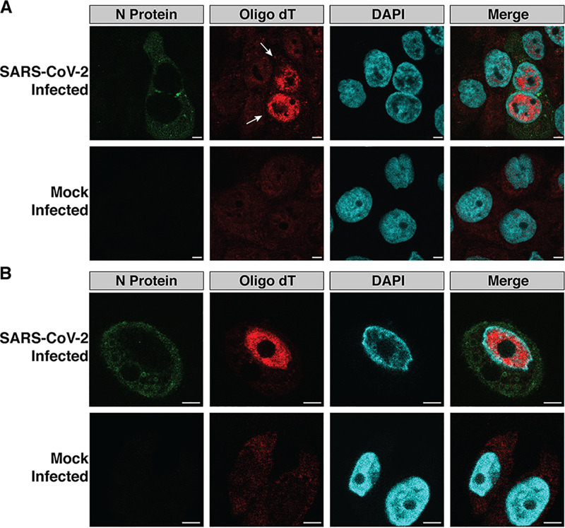
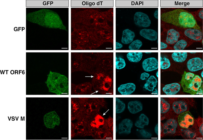
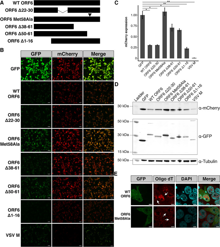
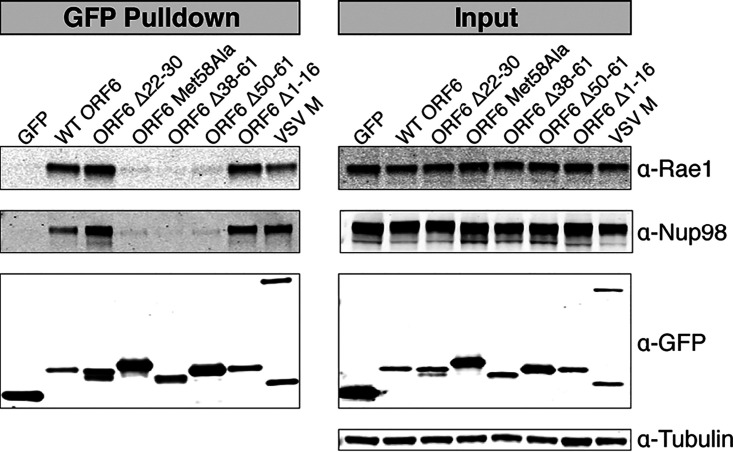
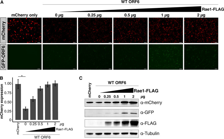
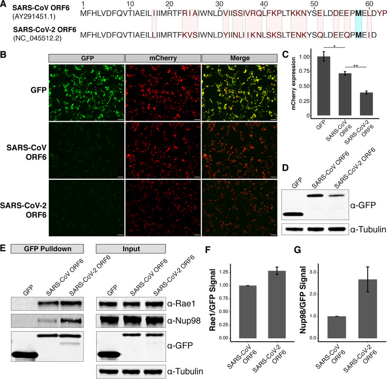
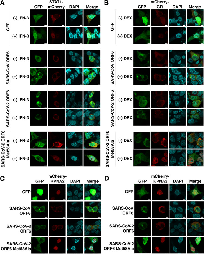
RNA viruses that replicate in the cytoplasm often disrupt nucleocytoplasmic transport to preferentially translate their own transcripts and prevent host antiviral responses. The Sarbecovirus accessory protein ORF6 has previously been shown to be a major inhibitor of interferon production in both severe acute respiratory syndrome coronavirus (SARS-CoV) and severe acute respiratory syndrome coronavirus 2 (SARS-CoV-2). Here, we show SARS-CoV-2-infected cells display an elevated level of nuclear mRNA accumulation compared to mock-infected cells. We demonstrate that ORF6 is responsible for this nuclear imprisonment of host mRNA, and using a cotransfected reporter assay, we show this nuclear retention of mRNA blocks expression of newly transcribed mRNAs. ORF6's nuclear entrapment of host mRNA is associated with its ability to copurify with the mRNA export factors, Rae1 and Nup98. These protein-protein interactions map to the C terminus of ORF6 and can be abolished by a single amino acid mutation in Met58. Overexpression of Rae1 restores reporter expression in the presence of SARS-CoV-2 ORF6. SARS-CoV ORF6 also interacts with Rae1 and Nup98. However, SARS-CoV-2 ORF6 more strongly copurifies with Rae1 and Nup98 and results in significantly reduced expression of reporter proteins compared to SARS-CoV ORF6, a potential mechanism for the delayed symptom onset and presymptomatic transmission uniquely associated with the SARS-CoV-2 pandemic. We also show that both SARS-CoV and SARS-CoV-2 ORF6 block nuclear import of a broad range of host proteins. Together, these data support a model in which ORF6 clogs the nuclear pore through its interactions with Rae1 and Nup98 to prevent both nuclear import and export, rendering host cells incapable of responding to SARS-CoV-2 infection.IMPORTANCE SARS-CoV-2, the causative agent of coronavirus disease 2019 (COVID-19), is an RNA virus with a large genome that encodes multiple accessory proteins. While these accessory proteins are not required for growth in vitro, they can contribute to the pathogenicity of the virus. We demonstrate that SARS-CoV-2-infected cells accumulate poly(A) mRNA in the nucleus, which is attributed to the accessory protein ORF6. Nuclear entrapment of mRNA and reduced expression of newly transcribed reporter proteins are associated with ORF6's interactions with the mRNA export proteins Rae1 and Nup98. SARS-CoV ORF6 also shows the same interactions with Rae1 and Nup98. However, SARS-CoV-2 ORF6 more strongly represses reporter expression and copurifies with Rae1 and Nup98 compared to SARS-CoV ORF6. Both SARS-CoV ORF6 and SARS-CoV-2 ORF6 block nuclear import of a wide range of host factors through interactions with Rae1 and Nup98. Together, our results suggest ORF6's disruption of nucleocytoplasmic transport prevents infected cells from responding to the invading virus.

Keywords: Nup98; ORF6; RNA virus; Rae1; SARS-CoV-2; VSV M; nucleocytoplasmic transport; virology.


Figures

FIG 1
FIG 1
SARS-CoV-2-infected cells accumulate mRNA in the nuclei. (A and B) Calu3 (A) and HBEC3-ACE2 (B) cells were infected with SARS-CoV-2. Twenty-four hours postinfection, the cells were stained with an oligo(dT)(30) probe conjugated to an Alexa Fluor 594 fluorophore. Poly(A) mRNA was localized to the nuclei in infected cells (white arrows). mRNA was more evenly distributed throughout the nuclei and cytoplasm in mock and uninfected cells. Bars, 5 μm.
FIG 2
FIG 2
The SARS-CoV-2 accessory protein ORF6 is responsible for the nuclear mRNA accumulation phenotype observed in SARS-CoV-2-infected cells. 293T cells were transiently transfected with GFP, GFP-SARS-CoV-2 ORF6, or GFP-VSV M. Staining for poly(A) mRNA was conducted 24 h posttransfection. Cells expressing SARS-CoV-2 ORF6 or VSV M (white arrows) displayed an accumulation of mRNA in the nuclei, while those transfected with GFP displayed mRNA localization patterns identical to those of untransfected cells. Bars, 5 μm.
FIG 3
FIG 3
ORF6 of SARS-CoV-2 results in reduced mCherry reporter protein expression in 293T cells. (A) Schematic representation of ORF6 constructs used in this study. (B) 293T cells were transiently transfected with GFP-tagged constructs and mCherry and visualized 48 h after transfection. All images were taken with identical fluorescence gain settings. Bars, 100 μm. (C) The fluorescent intensities for three fields per cotransfection were measured with ImageJ and displayed as means ± standard errors (error bars). Wild-type (WT) ORF6 caused a significant reduction in mCherry expression. ORF6 constructs with deletions or a single amino acid substitution (Met58Ala) in the C terminus showed a smaller reduction in mCherry expression than WT ORF6. Cell lysates were collected for each of the conditions. (D) Western blotting confirmed the role of the C terminus of ORF6 in reducing protein expression in transfected cells. (E) Poly(A) mRNA staining of transiently transfected 293T cells revealed ORF6 Met58Ala-expressing cells do not accumulate mRNA in the nuclei, consistent with the results from the mCherry cotransfection assay. Bars, 5 μm. *, P < 0.05; **, P < 0.01.
FIG 4
FIG 4
Affinity purification of GFP-tagged constructs. 293T cells were transiently transfected with GFP-tagged constructs. Forty-eight hours after transfection, the GFP-tagged proteins were rapidly captured using an anti-GFP resin. Western blotting revealed that ORF6 interacts with the mRNA nuclear export factor Rae1 and the nuclear pore complex protein Nup98. ORF6 constructs with C-terminal deletions or a substitution did not pull down Rae1 or Nup98.
FIG 5
FIG 5
Overexpression of Rae1 rescues mCherry expression in cells transfected with ORF6. (A) 293T cells were cotransfected with equal amounts GFP-ORF6 and mCherry and an increasing amount of Rae1. Bars, 100 μm. (B) Expression of the fluorescent reporters was visualized and quantified 48 h after transfection. In the presence of ORF6, mCherry expression was restored in a dose-dependent manner. (C) Western blotting confirmed that mCherry expression was rescued in a dose-dependent manner with increasing Rae1-FLAG. *, P < 0.05.
FIG 6
FIG 6
SARS-CoV-2 ORF6 represses reporter expression and copurifies with relatively more Rae1-Nup98 than SARS-CoV ORF6. (A) Comparison between the amino acid sequences of ORF6 of SARS-CoV and ORF6 of SARS-CoV-2. Residues differing between the two viruses are highlighted in red. The residue (Met58) implicated in binding the Rae1·Nup98 complex is highlighted in blue. (B) 293T cells were transiently transfected with GFP-tagged constructs and mCherry and visualized 24 h after transfection. Bars, 100 μm. (C) Cells transfected with SARS-CoV-2 ORF6 showed significantly reduced mCherry expression compared to those transfected with SARS-CoV ORF6. (D) Western blotting showed decreased expression of SARS-CoV-2 ORF6 compared to SARS-CoV ORF6 in 293T cells. (E) Affinity purification of GFP-tagged constructs demonstrates both ORF6 of SARS-CoV and ORF6 of SARS-CoV-2 interact with Rae1 and Nup98. (F and G) Densitometry shows SARS-CoV-2 ORF6 copurifies with relatively more Rae1 (F) and Nup98 (G) compared to SARS-CoV ORF6. *, P < 0.05; **, P < 0.01.
FIG 7
FIG 7
SARS-CoV ORF6 and SARS-CoV-2 ORF6 inhibit nuclear import of a broad range of host factors. (A) 293T cells were cotransfected with equal amounts of GFP-tagged constructs and STAT1 with an mCherry tag. STAT1 localized to the nuclei following IFN-β stimulation in GFP- and SARS-CoV-2 ORF6 Met58Ala-expressing cells but remained in the cytoplasm in SARS-CoV ORF6- and SARS-CoV-2 ORF6-expressing cells. (B) 293T cells were cotransfected with GFP-tagged constructs and mCherry-tagged glucocorticoid receptor (GR). The glucocorticoid receptor was translocated into the nuclei following dexamethasone (DEX) stimulation in GFP- and SARS-CoV-2 ORF6 Met58Ala-expressing cells yet remained in the cytoplasm in SARS-CoV ORF6- and SARS-CoV-2-expressing cells. (C and D) The importins KPNA2 (C) and KPNA3 (D) displayed distinct localization patterns based on the GFP-tagged constructs that was transfected with the mCherry-tagged importin. In GFP- and SARS-CoV-2 ORF6 Met58Ala-expressing cells, the importins localized to the nuclei, while in SARS-CoV ORF6- and SARS-CoV-2 ORF6-expressing cells, the importins were localized to the cytoplasm. Bars, 5 μm.

References

    1. Zhu N, Zhang D, Wang W, Li X, Yang B, Song J, Zhao X, Huang B, Shi W, Lu R, Niu P, Zhan F, Ma X, Wang D, Xu W, Wu G, Gao GF, Tan W, China Novel Coronavirus Investigating and Research Team . 2020. A novel coronavirus from patients with pneumonia in China, 2019. N Engl J Med 382:727–733. doi: 10.1056/NEJMoa2001017. - DOI - PMC - PubMed
    1. Narayanan K, Huang C, Makino S. 2008. SARS coronavirus accessory proteins. Virus Res 133:113–121. doi: 10.1016/j.virusres.2007.10.009. - DOI - PMC - PubMed
    1. Frieman M, Yount B, Heise M, Kopecky-Bromberg SA, Palese P, Baric RS. 2007. Severe acute respiratory syndrome coronavirus ORF6 antagonizes STAT1 function by sequestering nuclear import factors on the rough endoplasmic reticulum/Golgi membrane. J Virol 81:9812–9824. doi: 10.1128/JVI.01012-07. - DOI - PMC - PubMed
    1. Gordon DE, Jang GM, Bouhaddou M, Xu J, Obernier K, White KM, O’Meara MJ, Rezelj VV, Guo JZ, Swaney DL, Tummino TA, Hüttenhain R, Kaake RM, Richards AL, Tutuncuoglu B, Foussard H, Batra J, Haas K, Modak M, Kim M, Haas P, Polacco BJ, Braberg H, Fabius JM, Eckhardt M, Soucheray M, Bennett MJ, Cakir M, McGregor MJ, Li Q, Meyer B, Roesch F, Vallet T, Mac Kain A, Miorin L, Moreno E, Naing ZZC, Zhou Y, Peng S, Shi Y, Zhang Z, Shen W, Kirby IT, Melnyk JE, Chorba JS, Lou K, Dai SA, Barrio-Hernandez I, Memon D, Hernandez-Armenta C, Lyu J. et al. 2020. A SARS-CoV-2 protein interaction map reveals targets for drug repurposing. Nature 583:459–468. doi: 10.1038/s41586-020-2286-9. - DOI - PMC - PubMed
    1. Pritchard CE, Fornerod M, Kasper LH, van Deursen JM. 1999. RAE1 is a shuttling mRNA export factor that binds to a GLEBS-like NUP98 motif at the nuclear pore complex through multiple domains. J Cell Biol 145:237–254. doi: 10.1083/jcb.145.2.237. - DOI - PMC - PubMed

Publication types

MeSH terms

LinkOut - more resources