. 2021 Apr 13;12(2):e00296-21.
doi: 10.1128/mBio.00296-21.

Dynamics of the Bacillus subtilis Min System

Affiliations

Dynamics of the Bacillus subtilis Min System

Helge Feddersen et al. mBio. .

Abstract

Division site selection is a vital process to ensure generation of viable offspring. In many rod-shaped bacteria, a dynamic protein system, termed the Min system, acts as a central regulator of division site placement. The Min system is best studied in Escherichia coli, where it shows a remarkable oscillation from pole to pole with a time-averaged density minimum at midcell. Several components of the Min system are conserved in the Gram-positive model organism Bacillus subtilis However, in B. subtilis, it is commonly believed that the system forms a stationary bipolar gradient from the cell poles to midcell. Here, we show that the Min system of B. subtilis localizes dynamically to active sites of division, often organized in clusters. We provide physical modeling using measured diffusion constants that describe the observed enrichment of the Min system at the septum. Mathematical modeling suggests that the observed localization pattern of Min proteins corresponds to a dynamic equilibrium state. Our data provide evidence for the importance of ongoing septation for the Min dynamics, consistent with a major role of the Min system in controlling active division sites but not cell pole areas.IMPORTANCE The molecular mechanisms that help to place the division septum in bacteria is of fundamental importance to ensure cell proliferation and maintenance of cell shape and size. The Min protein system, found in many rod-shaped bacteria, is thought to play a major role in division site selection. It was assumed that there are strong differences in the functioning and in the dynamics of the Min system in E. coli and B. subtilis Most previous attempts to address Min protein dynamics in B. subtilis have been hampered by the use of overexpression constructs. Here, functional fusions to Min proteins have been constructed by allelic exchange and state-of-the-art imaging techniques allowed to unravel an unexpected fast dynamic behavior of the B. subtilis Min system. Our data show that the molecular mechanisms leading to Min protein dynamics are not fundamentally different in E. coli and B. subtilis.

Keywords: B. subtilis; FRAP; Min system; PALM; cell division; protein patterns; reaction diffusion equations; super resolution microscopy.


Figures

FIG 1
FIG 1
FRAP experiments in growing B. subtilis cells reveal Min protein dynamics. (a) Representative microscopy images of msfGFP-MinD (BHF017), MinJ-msfGFP (BHF007), and DivIVA-GFP (1803) before bleaching of the indicated spot with a 488-nm laser pulse, directly after bleaching, and after recovery of fluorescence. Scale bars, 2 μm. (b) Representation of the normalized fluorescence recovery in the green channel over time. T1/2 = time when fluorescence recovery reaches half height of total recovery; the shown value corresponds to the displayed cell, indicated on the graph with a dashed square. The red line represents measured values of the displayed cell, and the black line represents the fitted values. Values were obtained as described in Materials and Methods (equations 1 to 3).
FIG 2
FIG 2
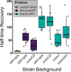
B. subtilis Min proteins form dynamic complexes. Shown are median half-time recovery values, indicated by the black bar inside each box. Each box represents a different strain; see also Table 3 for mean values. Every dot represents a single FRAP experiment (n ≥ 8).
FIG 3
FIG 3
Model and simulation of the Min system in B. subtilis. (a) The geometry sensing protein DivIVA (green) preferentially localizes to regions of highest negative curvature and stabilizes MinJ (purple) to these regions. Membrane-bound DivIVA acts as a template for MinD recruitment of cytosolic MinD-ATP (orange) facilitated through MinJ. MinD-ATP binds to the membrane with a rate, kD, and recruits cytosolic MinD-ATP with a (space-dependent) recruitment rate, kdD, to the membrane. Membrane-bound MinD is stabilized by MinJ-DivIVA complexes, which is reflected in a space-dependent detachment rate, kdet. After detachment, MinD is in a hydrolyzed state, MinD-ADP, and can rebind to the membrane only after nucleotide exchange with a rate λ. (b) MinD binds to flat membrane regions as well and recruits MinD-ATP from the cytosol. Binding to flat regions is, however, less favored, due to the lower concentration of MinJ-DivIVA complexes. (c) Simulation of the reaction-diffusion model in a 3D rod-shaped cell; shown is the membrane-bound MinD density distribution. As the initial condition, we take the steady-state distribution of the scenario where DivIVA is localized at the poles (left figure). At simulation start, we assume that MinD is losing its affinity to the poles by making the recruitment and detachment rate uniform on the entire cell membrane (this is, for example, the case at the onset of septum formation). From left to right, the time evolution of membrane-bound MinD is shown, where the far-right side shows the final steady-state density distribution. We find that polar localization of MinD becomes unstable and that the proteins preferentially localize at the cell center. (d) To test whether MinD can be localized at midplane through MinJ-DivIVA complexes after septum formation, we took the same initial condition as described for panel c and enhanced recruitment and decreased detachment near midcell. We find that MinD can sharply localize at the septum.
FIG 4
FIG 4
PALM imaging of strains expressing Dendra2-MinD, MinJ-mNeonGreen, and DivIVA-PAmCherry. Representative PALM images of Dendra2-MinD (BHF011), MinJ-mNeonGreen (JB40), and DivIVA-PAmCherry (JB37) expressing cells at different divisional states are shown. Upon formation of a division site, DivIVA, MinJ, and MinD partially relocalize from the poles to the division septum, where they reside after successful cytokinesis. Samples were fixed prior to imaging; every image represents a different cell. Scale bar, 500 nm.
FIG 5
FIG 5
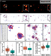
PALM imaging and representative cluster analysis of Dendra2-MinD and DivIVA-PAmCherry. (a) Representative PALM image of Dendra2-MinD (BHF011) in a cell in a late division state. Scale bar, 500 nm. (b) Cluster analysis of the same PALM data as shown in panel a with three highlighted regions (i, ii, and iii). Cluster analysis was performed in R using the OPTICS algorithm from the DBSCAN package. Every point indicates a single event and thus a Dendra2-MinD/DivIVA-PAmCherry protein, and precision is indicated by color and size of the circle. (c) Box plot of the number of clusters of Dendra2-MinD and DivIVA-PAmCherry per cell (MinD, ncells = 48; DivIVA, ncells = 37). (d) Box plot of the number of proteins per cluster; no jitter is shown due to the high sample number (Dendra2-MinD, nclusters = 1,171; DivIVA-PAmCherry, nclusters = 586). (e) Box plot of fraction of clusters localized at poles and septa per cell (MinD, ncells = 48; DivIVA, ncells = 37). Outliers in box plots are indicated by a red outline.

Similar articles

Cited by

References

    1. Bi EF, Lutkenhaus J. 1991. FtsZ ring structure associated with division in Escherichia coli. Nature 354:161–164. doi:10.1038/354161a0. - DOI - PubMed
    1. Margolin W. 2005. FtsZ and the division of prokaryotic cells and organelles. Nat Rev Mol Cell Biol 6:862–871. doi:10.1038/nrm1745. - DOI - PMC - PubMed
    1. Rowlett VW, Margolin W. 2015. The Min system and other nucleoid-independent regulators of Z ring positioning. Front Microbiol 6:478. doi:10.3389/fmicb.2015.00478. - DOI - PMC - PubMed
    1. Shih YL, Rothfield L. 2006. The bacterial cytoskeleton. Microbiol Mol Biol Rev 70:729–754. doi:10.1128/MMBR.00017-06. - DOI - PMC - PubMed
    1. Lutkenhaus J, Pichoff S, Du S. 2012. Bacterial cytokinesis: from Z ring to divisome. Cytoskeleton (Hoboken) 69:778–790. doi:10.1002/cm.21054. - DOI - PMC - PubMed

Publication types

MeSH terms

LinkOut - more resources