. 2021 Apr 13;12(2):e00620-21.
doi: 10.1128/mBio.00620-21.

The Small t Antigen of JC Virus Antagonizes RIG-I-Mediated Innate Immunity by Inhibiting TRIM25's RNA Binding Ability

Affiliations

The Small t Antigen of JC Virus Antagonizes RIG-I-Mediated Innate Immunity by Inhibiting TRIM25's RNA Binding Ability

Cindy Chiang et al. mBio. .

Abstract

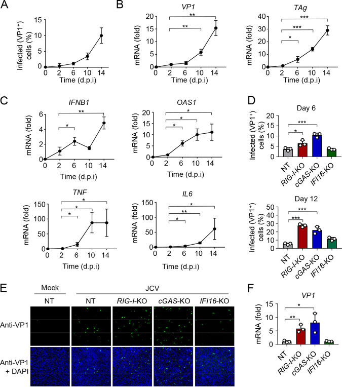
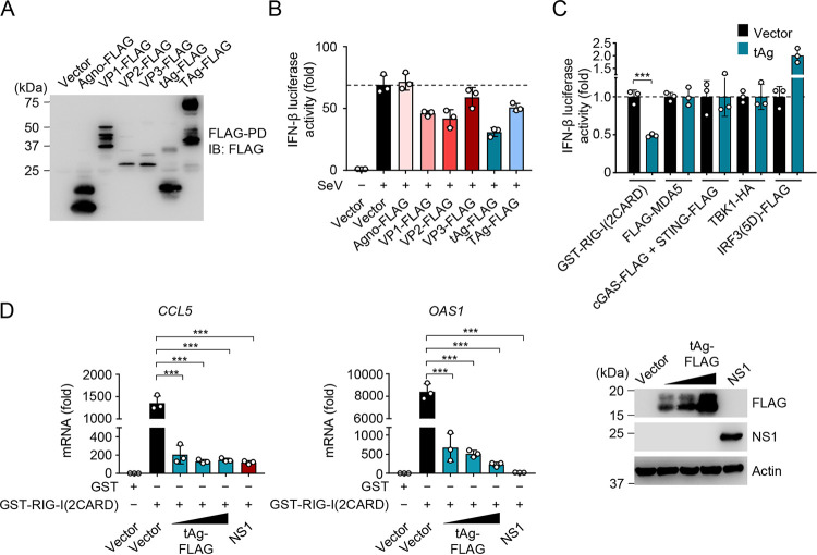
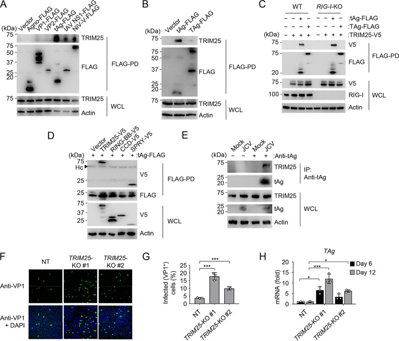
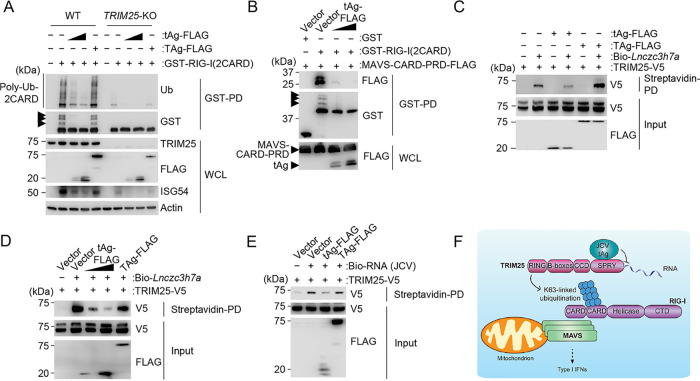
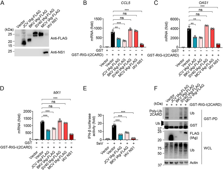
JC polyomavirus (JCV), a DNA virus that leads to persistent infection in humans, is the causative agent of progressive multifocal leukoencephalopathy, a lethal brain disease that affects immunocompromised individuals. Almost nothing is currently known about how JCV infection is controlled by the innate immune response and, further, whether JCV has evolved mechanisms to antagonize antiviral immunity. Here, we show that the innate immune sensors retinoic acid-inducible gene I (RIG-I) and cGMP-AMP synthase (cGAS) control JCV replication in human astrocytes. We further identify that the small t antigen (tAg) of JCV functions as an interferon (IFN) antagonist by suppressing RIG-I-mediated signal transduction. JCV tAg interacts with the E3 ubiquitin ligase TRIM25, thereby preventing its ability to bind RNA and to induce the K63-linked ubiquitination of RIG-I, which is known to facilitate RIG-I-mediated cytokine responses. Antagonism of RIG-I K63-linked ubiquitination and antiviral signaling is also conserved in the tAg of the related polyomavirus BK virus (BKV). These findings highlight how JCV and BKV manipulate a key innate surveillance pathway, which may stimulate research into designing novel therapies.IMPORTANCE The innate immune response is the first line of defense against viral pathogens, and in turn, many viruses have evolved strategies to evade detection by the host's innate immune surveillance machinery. Investigation of the interplay between viruses and the innate immune response provides valuable insight into potential therapeutic targets against viral infectious diseases. JC polyomavirus (JCV) is associated with a lifelong, persistent infection that can cause a rare neurodegenerative disease, called progressive multifocal leukoencephalopathy, in individuals that are immunosuppressed. The molecular mechanisms of JCV infection and persistence are not well understood, and very little is currently known about the relevance of innate immunity for the control of JCV replication. Here, we define the intracellular innate immune sensors responsible for controlling JCV infection and also demonstrate a novel mechanism by which a JCV-encoded protein acts as an antagonist of the type I interferon-mediated innate immune response.

Keywords: JC polyomavirus; RIG-I; innate immunity; small t antigen; type I interferon response; viral immune evasion.


Figures

FIG 1
FIG 1
RIG-I and cGAS control JCV replication in human glial astrocyte cells. (A) Frequency of JCV-positive SVGA cells that were infected with JCV (multiplicity of infection [MOI], 0.2) for the indicated times, determined by immunostaining with anti-VP1 and FACS analysis. (B) qRT-PCR analysis of JCV VP1 (left) and TAg (right) transcripts in SVGA cells that were infected with JCV (MOI, 0.2) for the indicated times. (C) qRT-PCR analysis of IFNB1, OAS1, TNF, and IL6 transcripts in SVGA cells that were infected with JCV (MOI, 0.2) for the indicated times. (D) JCV replication in CRISPR SVGA nontargeting control (NT) cells, or RIG-I-KO, cGAS-KO, or IFI16-KO SVGA cells that were infected with JCV (MOI, 0.2) for 6 (top) or 12 (bottom) days, assessed by immunostaining with anti-VP1 and FACS analysis. (E) JCV infection in CRISPR SVGA NT or KO cells that were infected with JCV (MOI, 0.2) for 6 days, determined by immunostaining with anti-VP1 (green) and confocal microscopy analysis. Mock-infected SVGA NT cells served as an infection specificity control. DAPI was used to stain cell nuclei (blue). (F) qRT-PCR analysis of JCV VP1 transcripts in CRISPR SVGA NT or indicated KO cells that were infected with JCV (MOI, 0.2) for 12 days. Data are means ± standard deviations (SD) (n = 3) and are representative of at least two independent experiments. *, P < 0.05; **, P < 0.005; ***, P < 0.001 (Student's t test in panels A, B, C, D, and F). d.p.i, days postinfection.
FIG 2
FIG 2
JCV tAg antagonizes RIG-I-mediated innate immune signaling. (A) Representative protein expression of the indicated FLAG-tagged JCV proteins in HEK 293T cells that were transfected for 48 h to express those proteins, determined by FLAG pulldown assay (FLAG-PD) and IB with anti-FLAG. (B) IFN-β luciferase activity in HEK 293T cells that were transfected for 40 h with an empty vector or the indicated FLAG-tagged JCV proteins and then infected with SeV (5 HAU/ml) (which specifically activates RIG-I) for 20 h or left uninfected. Luciferase activity was normalized to values for cotransfected β-galactosidase, and values are presented relative to those for uninfected cells that expressed the empty vector, set to 1. (C) IFN-β luciferase activity induced by transfection of constitutively active GST-RIG-I(2CARD), FLAG-MDA5, cGAS-FLAG and STING-FLAG, TBK1-HA, or the constitutively active mutant of IRF3 [IRF3(5D)-FLAG] with or without cotransfected JCV tAg. Luciferase values were normalized to values for cotransfected β-galactosidase and are presented relative to values for vector-transfected cells, set to 1. (D, left and middle panels) qRT-PCR analysis of CCL5 or OAS1 transcripts in HEK 293T cells that were transfected for 40 h with GST or GST-RIG-I(2CARD), together with an empty vector, increasing amounts of FLAG-tagged JCV tAg or IAV NS1 (positive control). (Right) Representative IB confirming protein expression of FLAG-tagged JCV tAg or IAV NS1, determined by IB with anti-FLAG or anti-NS1. IB with antiactin served as a loading control. Data are means ± SD (n = 3) and are representative of at least two independent experiments. **, P < 0.005; ***, P < 0.001 (Student's t test in panels B, C, and D). ns, statistically not significant.
FIG 3
FIG 3
JCV tAg interacts with TRIM25, an E3 ligase that controls JCV infection. (A) Binding of endogenous TRIM25 in HEK 293T cells that were transfected for 48 h with an empty vector, the indicated FLAG-tagged JCV proteins, or the IAV NS1 or NiV V protein (positive controls), determined by FLAG-PD and IB with anti-TRIM25 and anti-FLAG. WCLs were probed by IB with anti-TRIM25 and antiactin (loading control). (B) Binding of endogenous TRIM25 to JCV tAg in HEK 293T cells that were transiently transfected for 48 h with the empty vector or FLAG-tagged JCV tAg or TAg (control), determined by FLAG-PD and IB with anti-TRIM25 and anti-FLAG. WCLs were probed by IB with anti-TRIM25 and antiactin (loading control). (C) Binding of JCV tAg to TRIM25-V5 in WT or RIG-I-KO HEK 293T cells that were transfected for 48 h with the vector or TRIM25-V5 together with the empty vector or FLAG-tagged JCV tAg or TAg (control), determined by FLAG-PD and IB with anti-V5 and anti-FLAG. WCLs were probed by IB with anti-V5, anti-RIG-I, and antiactin (loading control). (D) TRIM25-tAg binding assay of HEK 293T cells that were transfected for 48 h with FLAG-tagged JCV tAg together with the empty vector, the V5-tagged TRIM25 full-length (TRIM25-V5) construct, or the indicated TRIM25 truncation constructs, determined by FLAG-PD and IB with anti-V5 and anti-FLAG. WCLs were probed by IB with anti-V5 and antiactin (loading control). RING-BB, RING-finger and B-box domains; CCD, coiled-coil domain; SPRY, SPla/ryanodine receptor (SPRY)/B30.2 domain; Hc, antibody heavy chain. (E) Binding of endogenous TRIM25 to JCV tAg in SVGA cells that were infected with JCV (MOI, 0.2) for 8 days, determined by IP with anti-tAg and IB with anti-TRIM25 and anti-tAg. WCLs were probed by IB with anti-TRIM25, anti-tAg, and antiactin (loading control). Uninfected cells (mock) served as a control. (F) JCV infection in SVGA NT or TRIM25-KO cells (nonclonal populations #1 and #2) that were infected with JCV (MOI, 0.2) for 6 days, determined by immunostaining with anti-VP1 (green) and confocal microscopy analysis. DAPI was used to stain cell nuclei (blue). (G) Frequency of JCV infection in CRISPR SVGA NT or TRIM25-KO cells (nonclonal cell populations #1 and #2) that were infected with JCV (MOI, 0.2) for 6 days, determined by immunostaining with anti-VP1 and FACS analysis. (H) qRT-PCR analysis of JCV TAg transcripts in CRISPR SVGA NT or TRIM25-KO cells (nonclonal cell populations #1 and #2) infected with JCV (MOI, 0.2) for 6 or 12 days. Data are means ± SD (n = 3) and are representative of at least two independent experiments. *, P < 0.05; ***, P < 0.001 (Student's t test in panels G and H).
FIG 4
FIG 4
JCV tAg inhibits RNA binding by TRIM25 and blocks the K63-linked ubiquitination of RIG-I. (A) RIG-I CARD ubiquitination in WT or TRIM25-KO HEK 293T cells that were transfected for 48 h with GST-RIG-I(2CARD) together with an empty vector or FLAG-tagged tAg or TAg, determined by GST-PD and IB with antiubiquitin (anti-Ub) and anti-GST. WCLs were probed by IB with anti-FLAG, anti-TRIM25, anti-ISG54, and antiactin (loading control). Arrows indicate the ubiquitinated forms of GST-RIG-I(2CARD). (B) Binding of GST-RIG-I(2CARD) or GST (negative control) to FLAG-tagged MAVS-CARD-PRD (proline-rich domain) in the presence or absence of FLAG-tagged JCV tAg in HEK 293T cells that were transfected for 48 h to express those proteins, determined by GST-PD and IB with anti-FLAG and anti-GST. WCLs were probed with anti-FLAG to confirm the expression of MAVS-CARD-PRD and tAg. Arrows indicate the ubiquitinated forms of GST-RIG-I(2CARD). (C) In vitro binding of biotinylated Lnczc3h7a (Bio-Lnczc3h7a) to TRIM25-V5 expressed in HEK 293T cells that were cotransfected with either the empty vector or FLAG-tagged JCV tAg or TAg, determined by streptavidin-pulldown assay (streptavidin-PD) and IB with anti-V5. The protein abundances for TRIM25 and JCV tAg and TAg were determined in the input samples by IB with anti-V5 and anti-FLAG, respectively. (D) In vitro binding of biotinylated Lnczc3h7a (Bio-Lnczc3h7a) to TRIM25-V5 expressed in HEK 293T cells that were cotransfected with either the empty vector, increasing amounts of FLAG-tagged JCV tAg, or FLAG-tagged JCV TAg, determined by streptavidin-PD and IB with anti-V5. The protein abundances for TRIM25 and the JCV proteins tAg and TAg were determined in the input samples by IB with anti-V5 and anti-FLAG, respectively. (E) In vitro binding of biotinylated total RNA purified from JCV-infected SVGA cells to TRIM25-V5 expressed in HEK 293T cells that were cotransfected with either the empty vector or FLAG-tagged JCV tAg or TAg, determined by streptavidin-PD and IB with anti-V5. The protein abundances for TRIM25 and the JCV proteins tAg and TAg were determined in the input samples by IB with anti-V5 and anti-FLAG, respectively. (F) Proposed model for the mechanism by which JCV tAg inhibits TRIM25-mediated RIG-I signaling. Data are representative of at least two independent experiments.
FIG 5
FIG 5
Antagonism of K63-polyubiquitin-dependent RIG-I signaling is also conserved in BKV tAg, but not SV40 or MCV tAg. (A) Representative protein expression of the indicated FLAG-tagged polyomavirus tAgs or untagged IAV NS1 protein in HEK 293T cells that were transiently transfected for 48 h to express those proteins, determined by either FLAG-PD and IB with anti-FLAG, or by IB with anti-NS1. (B) qRT-PCR analysis of CCL5 transcripts in HEK 293T cells that were transiently transfected for 40 h with either GST-RIG-I(2CARD) or GST (negative control) together with the vector, the indicated FLAG-tagged polyomavirus tAgs, or IAV NS1 (positive control). (C) qRT-PCR analysis of OAS1 transcripts in HEK 293T cells that were transiently transfected for 40 h with either GST-RIG-I(2CARD) or GST (negative control) together with the vector, the indicated FLAG-tagged polyomavirus tAgs, or IAV NS1 (positive control). (D) qRT-PCR analysis of MX1 transcripts in HEK 293T cells that were transiently transfected for 40 h with either GST-RIG-I(2CARD) or GST (negative control) together with the vector, the indicated FLAG-tagged polyomavirus tAgs, or IAV NS1 (positive control). (E) IFN-β luciferase activity in HEK 293T cells that were transfected for 40 h with the empty vector, FLAG-tagged JCV tAg or BKV tAg, or IAV NS1 (positive control), and then infected with SeV (10 HAU/ml) for 16 h or left uninfected. Luciferase activity was normalized to values for cotransfected β-galactosidase and are presented relative to values for uninfected cells that expressed the empty vector, set to 1. (F) RIG-I CARD ubiquitination in HEK 293T cells that were transfected for 48 h with GST-RIG-I(2CARD) together with the empty vector or the indicated FLAG-tagged polyomavirus tAgs, determined by GST-PD and IB with anti-Ub and anti-GST. WCLs were further probed by IB with anti-FLAG, anti-Ub, and antiactin (loading control). Arrows indicate the ubiquitinated forms of GST-RIG-I(2CARD). Data are means ± SD (n = 3) and are representative of at least two independent experiments. **, P < 0.005; ***, P < 0.001 (Student's t test in panels B, C, D, and E). ns, statistically not significant.

Similar articles

Cited by

References

    1. DeCaprio JA, Garcea RL. 2013. A cornucopia of human polyomaviruses. Nat Rev Microbiol 11:264–276. doi:10.1038/nrmicro2992. - DOI - PMC - PubMed
    1. Frisque RJ, Bream GL, Cannella MT. 1984. Human polyomavirus JC virus genome. J Virol 51:458–469. doi:10.1128/JVI.51.2.458-469.1984. - DOI - PMC - PubMed
    1. Beltrami S, Gordon J. 2014. Immune surveillance and response to JC virus infection and PML. J Neurovirol 20:137–149. doi:10.1007/s13365-013-0222-6. - DOI - PMC - PubMed
    1. Haley SA, Atwood WJ. 2017. Progressive multifocal leukoencephalopathy: endemic viruses and lethal brain disease. Annu Rev Virol 4:349–367. doi:10.1146/annurev-virology-101416-041439. - DOI - PubMed
    1. Wei G, Liu CK, Atwood WJ. 2000. JC virus binds to primary human glial cells, tonsillar stromal cells, and B-lymphocytes, but not to T lymphocytes. J Neurovirol 6:127–136. doi:10.3109/13550280009013156. - DOI - PubMed

Publication types

MeSH terms

LinkOut - more resources