

Anatomic, functional, and radiographic review of the ligaments of the craniocervical junction
- PMID: 33850375
- PMCID: PMC8035576
- DOI: 10.4103/jcvjs.JCVJS_209_20
Anatomic, functional, and radiographic review of the ligaments of the craniocervical junction
Abstract
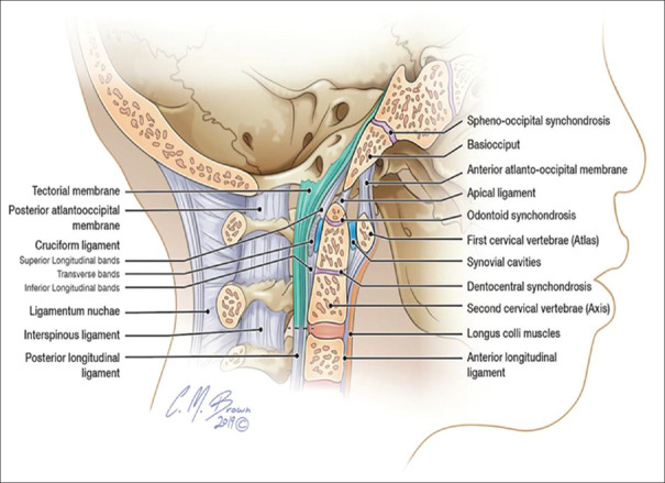
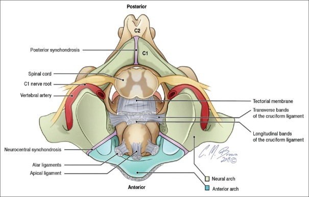
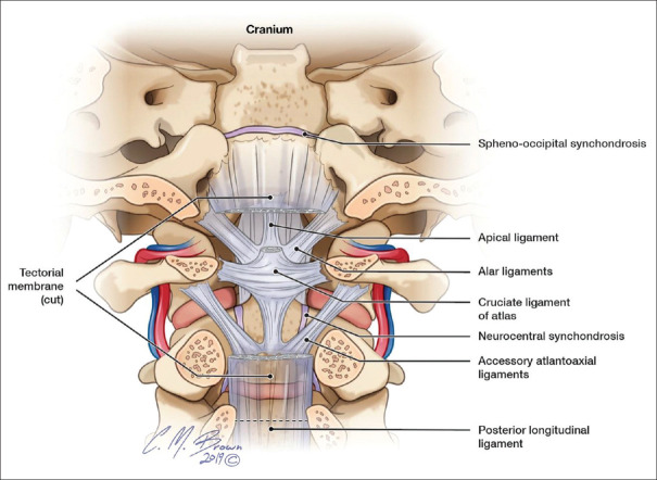
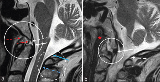
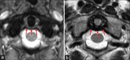
The craniocervical junction (CCJ) is a complex and unique osteoligamentous structure that balances maximum stability and protection of vital neurovascular anatomy with ample mobility and range of motion. With the increasing utilization and improved resolution of cervical magnetic resonance imaging, craniocervical injury is being more accurately defined as a spectrum of injury that ranges in severity from overt craniocervical disassociation to isolated injuries of one more of the craniocervical ligaments, which may also lead to craniocervical instability. Thus, it is vital for the radiologist and neurosurgeon to have a thorough understanding of the imaging anatomy and function of the CCJ.
Keywords: Craniocervical junction; magnetic resonance imaging; trauma.
Copyright: © 2021 Journal of Craniovertebral Junction and Spine.
Conflict of interest statement
There are no conflicts of interest.
Figures
References
-
- Pang D, Nemzek WR, Zovickian J. Atlanto-occipital dislocation--part 2: The clinical use of (occipital) condyle-C1 interval, comparison with other diagnostic methods, and the manifestation, management, and outcome of atlanto-occipital dislocation in children. Neurosurgery. 2007;61:995–1015. - PubMed
-
- Gire JD, Roberto RF, Bobinski M, Klineberg EO, Johnson BD. The utility and accuracy of computed tomography in the diagnosis of occipitocervical dissociation. Spine J. 2013;13:510–19. - PubMed
-
- Kwong Y, Rao N, Latief K. Craniometric measurements in the assessment of craniovertebral settling: Are they still relevant in the age of cross-sectional imaging? AJR Am J Roentgenol. 2011;196:W421–5. - PubMed
Publication types
LinkOut - more resources
Full Text Sources
Other Literature Sources