

Fibromatosis with aggressive demeanor: Benign impersonator of malignancy
- PMID: 33850503
- PMCID: PMC8034791
- DOI: 10.4103/wjnm.WJNM_55_20
Fibromatosis with aggressive demeanor: Benign impersonator of malignancy
Abstract
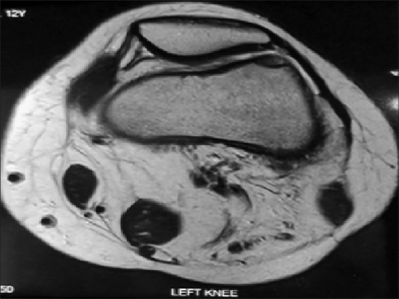
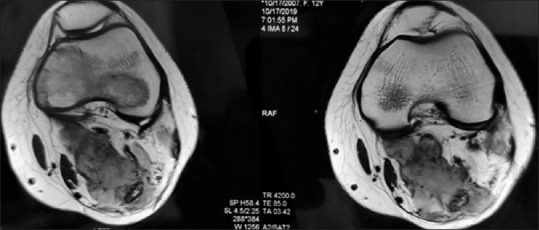
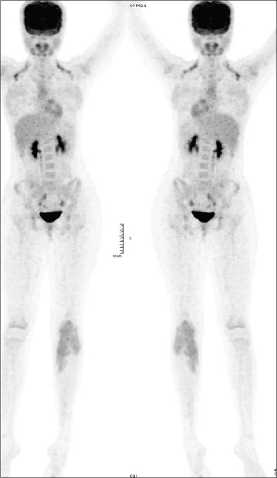
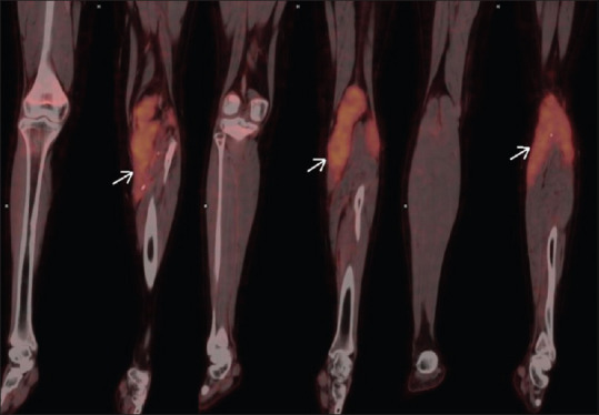
Fibromatosis or desmoid fibromatosis is a rare benign neoplasm and develops commonly in the abdominal wall, abdominal cavity, or extra-abdominal sites. The mainstay of treatment is surgery. Chemotherapy and radiotherapy are preferred in cases of inoperable/relapse or a multifocal disease. Hereby, we report a case of fibromatosis arising in the left popliteal fossa, proven by histopathology and immunohistochemistry. Local excision of the mass was performed. The patient was asymptomatic for 6 months, after which she complained of difficulty in walking. Clinical evaluation elicited recurrence in the surgical bed. In spite of the surgical excision with tumor-free margins, recurrence was seen within a span of 6 months. 18F-fluorodeoxyglucose (FDG) positron emission tomography-computed tomography (PET/CT) was done to rule out multifocal disease and to define the extent of relapse. Although magnetic resonance imaging provides an excellent soft-tissue resolution to delineate the disease, 18F-FDG PET/CT is an important and supplementary tool which aids in the management of fibromatosis.
Keywords: Desmoid tumor; fibromatosis; fluorodeoxyglucose positron emission tomography-computed tomography in fibromatosis; recurrent fibromatosis.
Copyright: © 2020 World Journal of Nuclear Medicine.
Conflict of interest statement
There are no conflicts of interest.
Figures
Similar articles
-
More advantages in detecting bone and soft tissue metastases from prostate cancer using 18F-PSMA PET/CT.Hell J Nucl Med. 2019 Jan-Apr;22(1):6-9. doi: 10.1967/s002449910952. Epub 2019 Mar 7. Hell J Nucl Med. 2019. PMID: 30843003
-
Uptake characteristics of fluorodeoxyglucose (FDG) in deep fibromatosis and abdominal desmoids: potential clinical role of FDG-PET in the management.Br J Radiol. 2007 Sep;80(957):750-6. doi: 10.1259/bjr/53719785. Epub 2007 Aug 20. Br J Radiol. 2007. PMID: 17709361
-
Extra-abdominal fibromatosis (desmoid tumor): a rare tumor of the lower extremity arising from the popliteal fossa.Case Rep Vasc Med. 2011;2011:184906. doi: 10.1155/2011/184906. Epub 2011 Aug 21. Case Rep Vasc Med. 2011. PMID: 22937461 Free PMC article.
-
A case of desmoid-type fibromatosis arising after thoracotomy for lung cancer with a review of the english and Japanese literature.Ann Thorac Cardiovasc Surg. 2014;20 Suppl:465-9. doi: 10.5761/atcs.cr.12.02149. Epub 2013 Apr 5. Ann Thorac Cardiovasc Surg. 2014. PMID: 23558226 Review.
-
Rib and pericardium invaded huge abdominal mass in young woman: A case report with literature review.Medicine (Baltimore). 2022 Sep 2;101(35):e30371. doi: 10.1097/MD.0000000000030371. Medicine (Baltimore). 2022. PMID: 36107577 Free PMC article. Review.
Cited by
-
Annotating the Role of 18F-FDG PET/CT in Fibromatoses: A Benign Masquerader of Malignancies-Is It Really an Advantageous Tool?Nucl Med Mol Imaging. 2024 May;58(3):140-146. doi: 10.1007/s13139-024-00846-5. Epub 2024 Feb 14. Nucl Med Mol Imaging. 2024. PMID: 38633285 Free PMC article.
References
-
- Gourgiotis S, Gemenetzis G, Villias C. Pancreatic desmoid tumour: Extremely rare presentation in an elderly patient. Hell J Surg. 2014;86:378–81.
-
- Basu S, Nair N, Banavali S. Uptake characteristics of fluorodeoxyglucose (FDG) in deep fibromatosis and abdominal desmoids: Potential clinical role of FDG-PET in the management. Br J Radiol. 2007;80:750–6. - PubMed
-
- Alshammari A, Ashkanani R, Alabsi S, Ghanem M. Gardner syndrome with extra and intra-abdominal desmoid tumors and adrenal involvement: PET/CT findings. Case Rep Mol Imaging Radionucl Ther. 2015;24(Suppl 1):38–41.
Publication types
LinkOut - more resources
Full Text Sources
Other Literature Sources