

A coarse-to-fine deep learning framework for optic disc segmentation in fundus images
- PMID: 33850515
- PMCID: PMC8041100
- DOI: 10.1016/j.bspc.2019.01.022
A coarse-to-fine deep learning framework for optic disc segmentation in fundus images
Abstract
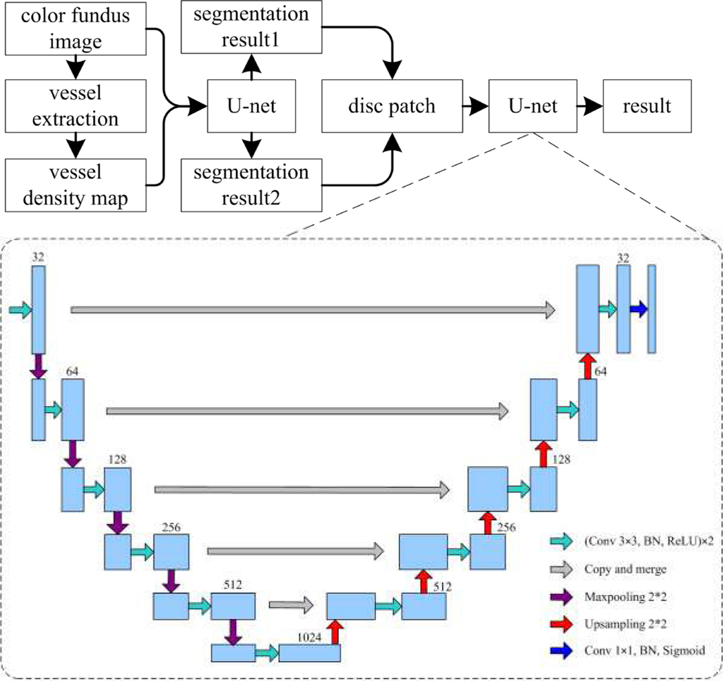
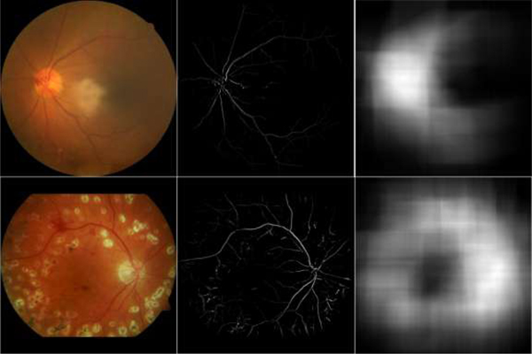
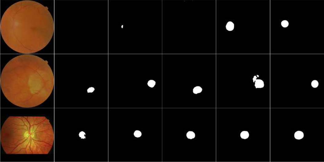
Accurate segmentation of the optic disc (OD) depicted on color fundus images may aid in the early detection and quantitative diagnosis of retinal diseases, such as glaucoma and optic atrophy. In this study, we proposed a coarse-to-fine deep learning framework on the basis of a classical convolutional neural network (CNN), known as the U-net model, to accurately identify the optic disc. This network was trained separately on color fundus images and their grayscale vessel density maps, leading to two different segmentation results from the entire image. We combined the results using an overlap strategy to identify a local image patch (disc candidate region), which was then fed into the U-net model for further segmentation. Our experiments demonstrated that the developed framework achieved an average intersection over union (IoU) and a dice similarity coefficient (DSC) of 89.1% and 93.9%, respectively, based on 2,978 test images from our collected dataset and six public datasets, as compared to 87.4% and 92.5% obtained by only using the sole U-net model. The comparison with available approaches demonstrated a reliable and relatively high performance of the proposed deep learning framework in automated OD segmentation.
Keywords: Color fundus images; Convolutional neural networks; Image segmentation; Optic disc; U-net model.
Figures


References
-
- Singh A, Dutta M, Sarathi M, Uher V, Burget R, Image processing based automatic diagnosis of glaucoma using wavelet features of segmented optic disc from fundus image, Computer Methods and Programs in Biomedicine, 124 (2016) 108–120 - PubMed
-
- Yin F, Liu J, Ong S, et al. , Model-based optic nerve head segmentation on retinal fundus images, Conference of the IEEE Engineering in Medicine and Biology Society (EMBS), 2626–2629, (2011) - PubMed
-
- Zhang D, Yi Y, Shang X, Peng Y, Optic disc localization by projection with vessel distribution and appearance characteristics, International Conference on Pattern Recognition (ICPR), 3176–3179, (2012).
Grants and funding
LinkOut - more resources
Full Text Sources