

Effect of Physiological Noise on Thoracolumbar Spinal Cord Functional Magnetic Resonance Imaging in 3T Magnetic Field
- PMID: 33850611
- PMCID: PMC8019845
- DOI: 10.32598/bcn.11.6.1395.1
Effect of Physiological Noise on Thoracolumbar Spinal Cord Functional Magnetic Resonance Imaging in 3T Magnetic Field
Abstract
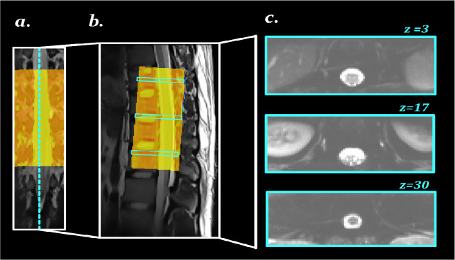
Introduction: Functional Magnetic Resonance Imaging (fMRI) methods have been used to study sensorimotor processing in the spinal cord. However, these techniques confront unwanted noises to the measured signal from the physiological fluctuations. In the spinal cord imaging, most of the challenges are consequences of cardiac and respiratory movement artifacts that are considered as significant sources of noise, especially in the thoracolumbar region. In this study, we investigated the effect of each source of physiological noise and their contribution to the outcome of the analysis of the blood-oxygen-level-dependent signal in the human thoracolumbar spinal cord.
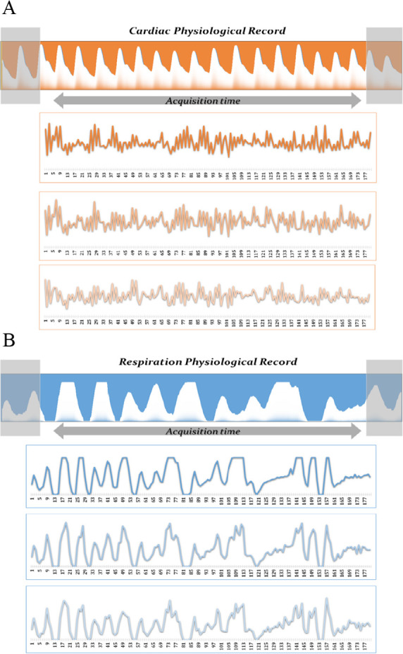
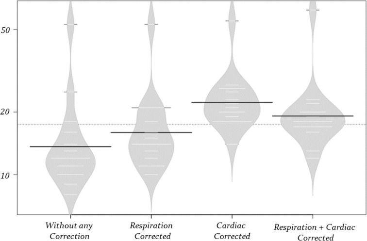
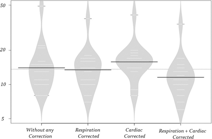
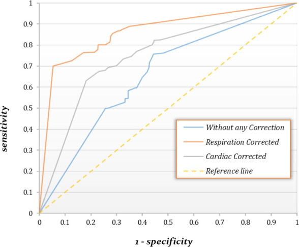
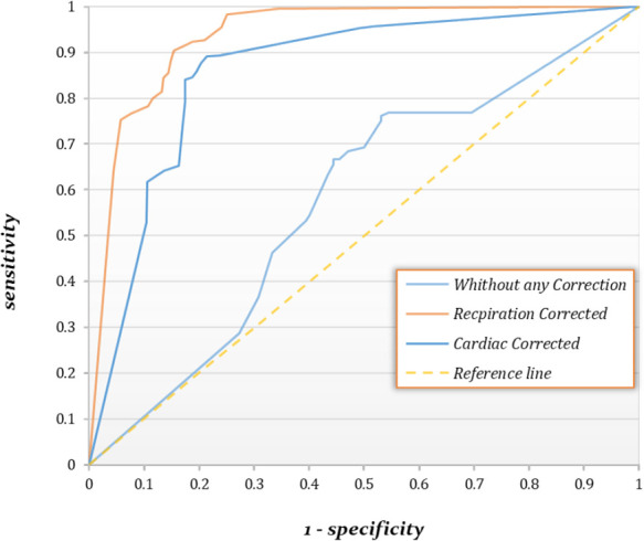
Methods: Fifteen young healthy male volunteers participated in the study, and pain stimuli were delivered on the L5 dermatome between the two malleoli. Respiratory and cardiac signals were recorded during the imaging session, and the generated respiration and cardiac regressors were included in the general linear model for quantification of the effect of each of them on the task-analysis results. The sum of active voxels of the clusters was calculated in the spinal cord in three correction states (respiration correction only, cardiac correction only, and respiration and cardiac noise corrections) and analyzed with analysis of variance statistical test and receiver operating characteristic curve.
Results: The results illustrated that cardiac noise correction had an effective role in increasing the active voxels (Mean±SD = 23.46±9.46) compared to other noise correction methods. Cardiac effects were higher than other physiological noise sources.
Conclusion: In summary, our results indicate great respiration effects on the lumbar and thoracolumbar spinal cord fMRI, and its contribution to the heartbeat effect can be a significant variable in the individual fMRI data analysis. Displacement of the spinal cord and the effects of this noise in the thoracolumbar and lumbar spinal cord fMRI results are significant and cannot be ignored.
Keywords: Functional Magnetic Resonance Imaging (fMRI); General linear model; Imaging; Physiological noise; Spinal cord.
Copyright© 2020 Iranian Neuroscience Society.
Conflict of interest statement
Conflict of interest All authors confirm that they have no conflicts of interest.
Figures
Similar articles
-
Assessment of physiological noise modelling methods for functional imaging of the spinal cord.Neuroimage. 2012 Apr 2;60(2):1538-49. doi: 10.1016/j.neuroimage.2011.11.077. Epub 2011 Dec 8. Neuroimage. 2012. PMID: 22178812
-
Confirmation of resting-state BOLD fluctuations in the human brainstem and spinal cord after identification and removal of physiological noise.Magn Reson Med. 2017 Dec;78(6):2149-2156. doi: 10.1002/mrm.26606. Epub 2017 Jan 11. Magn Reson Med. 2017. PMID: 28074492
-
Reduction of physiological noise with independent component analysis improves the detection of nociceptive responses with fMRI of the human spinal cord.Neuroimage. 2012 Oct 15;63(1):245-52. doi: 10.1016/j.neuroimage.2012.06.057. Epub 2012 Jul 6. Neuroimage. 2012. PMID: 22776463
-
On the impact of physiological noise in spinal cord functional MRI.J Magn Reson Imaging. 2014 Oct;40(4):770-7. doi: 10.1002/jmri.24467. Epub 2013 Nov 7. J Magn Reson Imaging. 2014. PMID: 24925698 Review.
-
The effect of physiological noise in phase functional magnetic resonance imaging: from blood oxygen level-dependent effects to direct detection of neuronal currents.Magn Reson Imaging. 2008 Sep;26(7):1026-40. doi: 10.1016/j.mri.2008.01.010. Epub 2008 May 13. Magn Reson Imaging. 2008. PMID: 18479875 Review.
Cited by
-
Detection of resting-state functional connectivity in the lumbar spinal cord with 3T MRI.Sci Rep. 2023 Oct 24;13(1):18189. doi: 10.1038/s41598-023-45302-0. Sci Rep. 2023. PMID: 37875563 Free PMC article.
-
DeepRetroMoCo: deep neural network-based retrospective motion correction algorithm for spinal cord functional MRI.Front Psychiatry. 2024 Jun 28;15:1323109. doi: 10.3389/fpsyt.2024.1323109. eCollection 2024. Front Psychiatry. 2024. PMID: 39006826 Free PMC article.
-
Employing artificial bee and ant colony optimization in machine learning techniques as a cognitive neuroscience tool.Sci Rep. 2025 Mar 24;15(1):10172. doi: 10.1038/s41598-025-94642-6. Sci Rep. 2025. PMID: 40128279 Free PMC article.
References
-
- Agosta F., Valsasina P., Absinta M., Sala S., Caputo D., Filippi M. (2009). Primary progressive multiple sclerosis: Tactile-associated functional MR activity in the cervical spinal cord. Radiology, 253(1), 209–15. [DOI:10.1148/radiol.2532090187] [PMID https://www.ncbi.nlm.nih.gov/pubmed/19703852] - DOI - PubMed
-
- Agosta F., Valsasina P., Caputo D., Stroman P. W., Filippi M. (2008). Tactile-associated recruitment of the cervical cord is altered in patients with multiple sclerosis. NeuroImage, 39(4), 1542–8. [DOI:10.1016/j.neuroimage.2007.10.048] [PMID https://www.ncbi.nlm.nih.gov/pubmed/18061484] - DOI - PubMed
-
- Agosta F., Valsasina P., Rocca M., Caputo D., Sala S., Judica E., et al. (2008). Evidence for enhanced functional activity of cervical cord in relapsing multiple sclerosis. Magnetic Resonance in Medicine, 59(5), 1035–42. [DOI:10.1002/mrm.21595] [PMID https://www.ncbi.nlm.nih.gov/pubmed/18429010] - DOI - PubMed
-
- Alexander M. S., Kozyrev N., Bosma R. L., Figley C. R., Richards J. S., Stroman P. W. (2016). fMRI localization of spinal cord processing underlying female sexual arousal. Journal of Sex & Marital Therapy, 42(1), 36–47. [DOI:10.1080/0092623X.2015.1010674] [PMID https://www.ncbi.nlm.nih.gov/pubmed/25635474] - DOI - PubMed
LinkOut - more resources
Full Text Sources