

Effect of hormone replacement therapy on bone formation quality and mineralization regulation mechanisms in early postmenopausal women
- PMID: 33850974
- PMCID: PMC8022851
- DOI: 10.1016/j.bonr.2021.101055
Effect of hormone replacement therapy on bone formation quality and mineralization regulation mechanisms in early postmenopausal women
Abstract
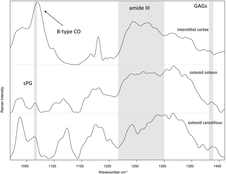
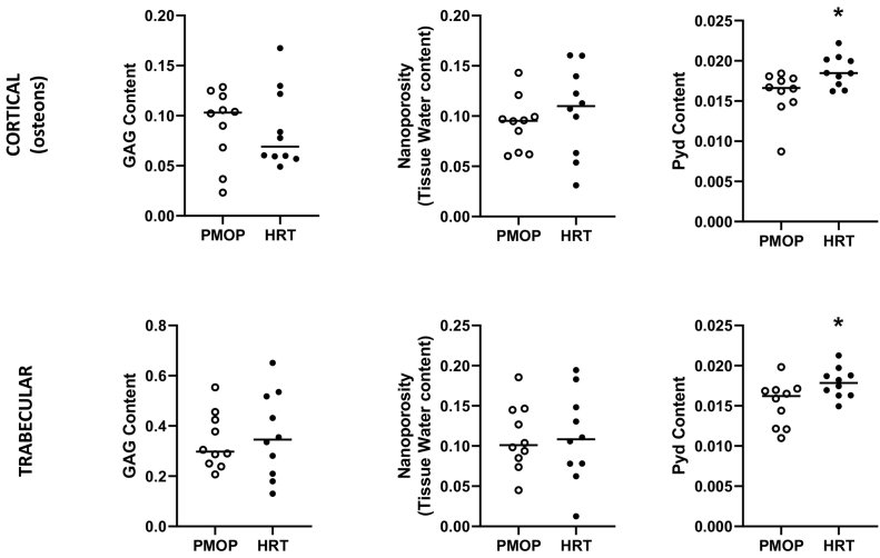
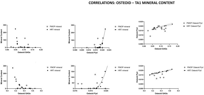
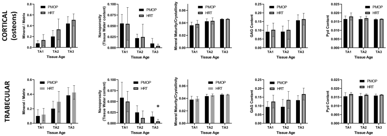
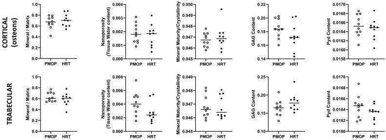
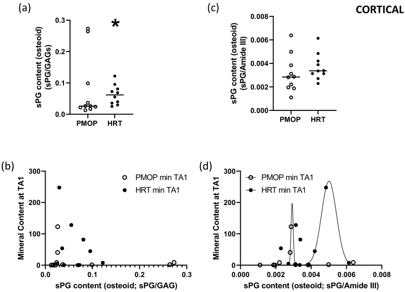
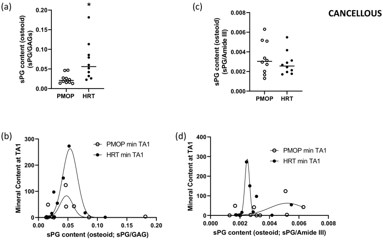
Post-menopausal osteoporosis is characterized by a negative imbalance between bone formation and bone resorption resulting in a net bone loss, increasing the risk of fracture. One of the earliest interventions to protect against this was hormonal replacement therapy (HRT). Bone strength depends on both the amount and quality of bone, the latter including compositional / material and structural properties. Bone compositional / material properties are greatly dependent on both patient-, and tissue-age. Raman spectroscopy is an analytical tool ideally suited for the determination of bone compositional / material properties as a function of tissue age as it is capable of analyzing areas ~1 × 1 μm2 in tetracycline labeled bone forming areas. Using such analysis of humeri from an ovariectomized primate animal model, we reported that loss of estrogen results in alteration in the mineralization regulation mechanisms by osteoid organic matrix attributes at actively forming bone surfaces. In the present work, we used Raman microspectroscopic techniques to compare osteoid and youngest mineralized tissue composition, as well as relationships between osteoid organic matrix quality and quality attributes of the earliest mineralized tissue in paired iliac crest biopsies obtained from early postmenopausal women before and after two years of HRT therapy. Significant correlations between osteoid proteoglycans, sulfated proteoglycans, pyridinoline, and earliest mineralized tissue mineral content were observed, suggesting that in addition to changes in bone turnover rates, HRT affects the osteoid composition, mineralization regulation mechanisms, and potentially fibrillogenesis.
Keywords: Bone formation; Bone quality; Early postmenopause; Hormonal replacement therapy; Organic matrix; Osteoid; Raman spectroscopy.
© 2021 Published by Elsevier Inc.
Conflict of interest statement
None of the authors has any conflict of interest.
Figures
Similar articles
-
Estrogen depletion alters mineralization regulation mechanisms in an ovariectomized monkey animal model.Bone. 2019 Mar;120:279-284. doi: 10.1016/j.bone.2018.11.004. Epub 2018 Nov 7. Bone. 2019. PMID: 30414509
-
Ovarian hormone depletion affects cortical bone quality differently on different skeletal envelopes.Bone. 2017 Feb;95:55-64. doi: 10.1016/j.bone.2016.10.029. Epub 2016 Nov 4. Bone. 2017. PMID: 27826024
-
Effect of hormone replacement therapy on bone quality in early postmenopausal women.J Bone Miner Res. 2003 Jun;18(6):955-9. doi: 10.1359/jbmr.2003.18.6.955. J Bone Miner Res. 2003. PMID: 12817747 Clinical Trial.
-
Hormone replacement therapy: II. A pharmacoeconomic appraisal of its role in the prevention of postmenopausal osteoporosis and ischaemic heart disease.Pharmacoeconomics. 1994 Jun;5(6):513-54. doi: 10.2165/00019053-199405060-00007. Pharmacoeconomics. 1994. PMID: 10147266 Review.
-
The mineralization of bone tissue: a forgotten dimension in osteoporosis research.Osteoporos Int. 2003;14 Suppl 3:S19-24. doi: 10.1007/s00198-002-1347-2. Epub 2003 Mar 18. Osteoporos Int. 2003. PMID: 12730799 Review.
Cited by
-
Status of female sexual dysfunction among postmenopausal women in Bangladesh.BMC Womens Health. 2022 Oct 4;22(1):401. doi: 10.1186/s12905-022-01991-9. BMC Womens Health. 2022. PMID: 36195886 Free PMC article.
-
New Possibilities for Evaluating the Development of Age-Related Pathologies Using the Dynamical Network Biomarkers Theory.Cells. 2023 Sep 17;12(18):2297. doi: 10.3390/cells12182297. Cells. 2023. PMID: 37759519 Free PMC article. Review.
-
GSK 650394 Inhibits Osteoclasts Differentiation and Prevents Bone Loss via Promoting the Activities of Antioxidant Enzymes In Vitro and In Vivo.Oxid Med Cell Longev. 2022 Sep 17;2022:3458560. doi: 10.1155/2022/3458560. eCollection 2022. Oxid Med Cell Longev. 2022. PMID: 36164394 Free PMC article.
References
-
- Arias J.L., Neira-Carrillo A., Arias J.I., Escobar C., Bodero M., David M., Fernández M.S. Sulfated polymers in biological mineralization: a plausible source for bio-inspired engineering. J. Mater. Chem. 2004;14(14):2154–2160.
-
- Awonusi A., Morris M.D., Tecklenburg M.M. Carbonate assignment and calibration in the Raman spectrum of apatite. Calcif. Tissue Int. 2007;81(1):46–52. - PubMed
-
- Bansil R., Yannas I.V., Stanley H.E. Raman spectroscopy: a structural probe of glycosaminoglycans. Biochim. Biophys. Acta. 1978;541(4):535–542. - PubMed
-
- Bi Y., Nielsen K.L., Kilts T.M., Yoon A., M A.K., Wimer H.F., Greenfield E.M., Heegaard A.M., Young M.F. Biglycan deficiency increases osteoclast differentiation and activity due to defective osteoblasts. Bone. 2006;38(6):778–786. - PubMed
LinkOut - more resources
Full Text Sources
Other Literature Sources
