[Preprint]. 2021 Apr 8:2021.04.06.438709.
doi: 10.1101/2021.04.06.438709.

Antibodies to the SARS-CoV-2 receptor-binding domain that maximize breadth and resistance to viral escape

Affiliations

Antibodies to the SARS-CoV-2 receptor-binding domain that maximize breadth and resistance to viral escape

Tyler N Starr et al. bioRxiv. .

Update in

  • SARS-CoV-2 RBD antibodies that maximize breadth and resistance to escape.
    Starr TN, Czudnochowski N, Liu Z, Zatta F, Park YJ, Addetia A, Pinto D, Beltramello M, Hernandez P, Greaney AJ, Marzi R, Glass WG, Zhang I, Dingens AS, Bowen JE, Tortorici MA, Walls AC, Wojcechowskyj JA, De Marco A, Rosen LE, Zhou J, Montiel-Ruiz M, Kaiser H, Dillen JR, Tucker H, Bassi J, Silacci-Fregni C, Housley MP, di Iulio J, Lombardo G, Agostini M, Sprugasci N, Culap K, Jaconi S, Meury M, Dellota E Jr, Abdelnabi R, Foo SC, Cameroni E, Stumpf S, Croll TI, Nix JC, Havenar-Daughton C, Piccoli L, Benigni F, Neyts J, Telenti A, Lempp FA, Pizzuto MS, Chodera JD, Hebner CM, Virgin HW, Whelan SPJ, Veesler D, Corti D, Bloom JD, Snell G. Starr TN, et al. Nature. 2021 Sep;597(7874):97-102. doi: 10.1038/s41586-021-03807-6. Epub 2021 Jul 14. Nature. 2021. PMID: 34261126 Free PMC article.

Abstract

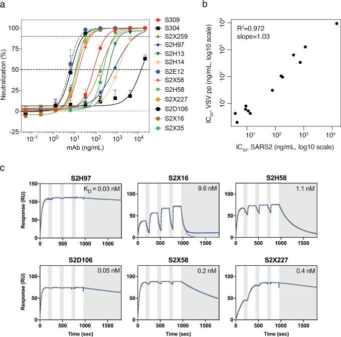
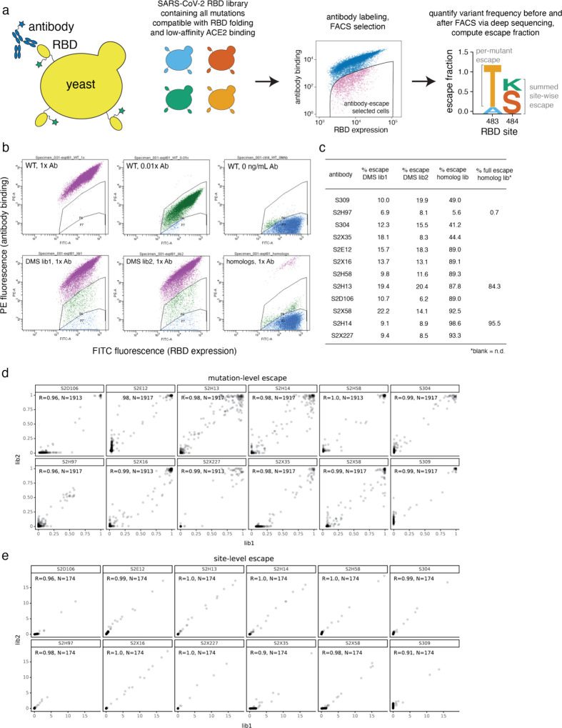
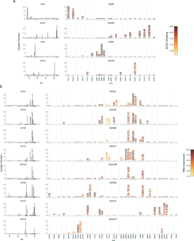
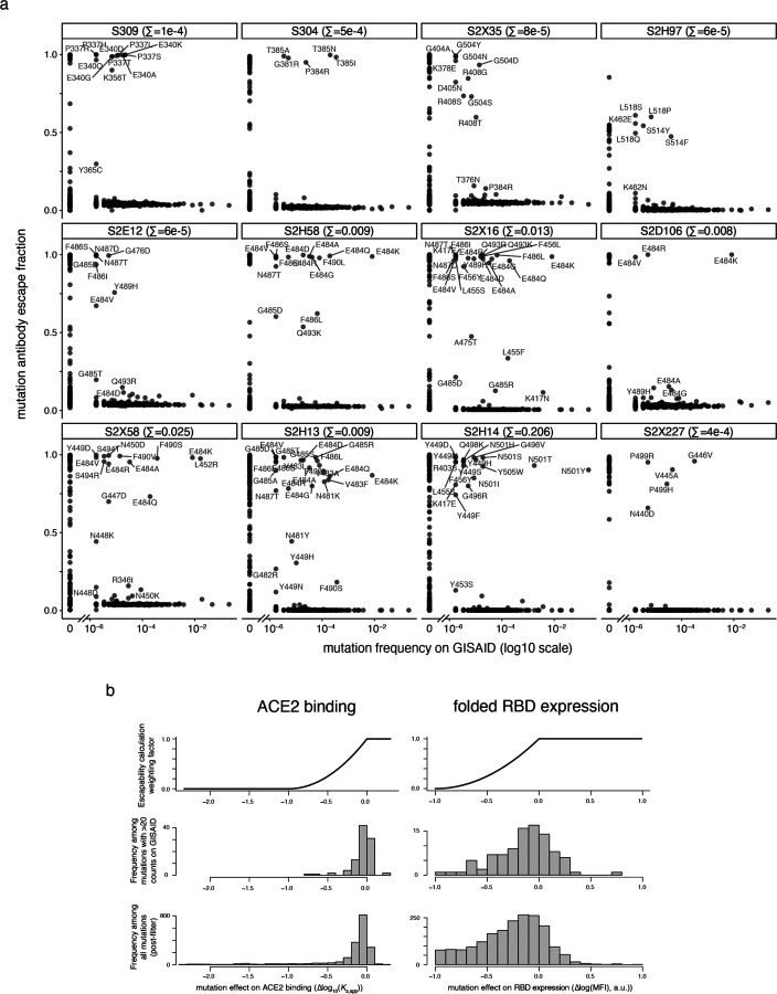
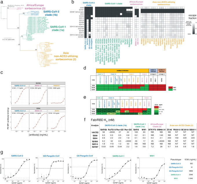
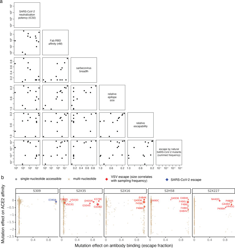
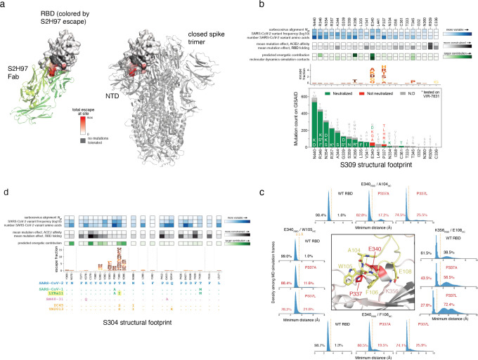
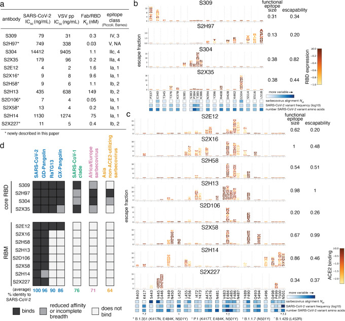
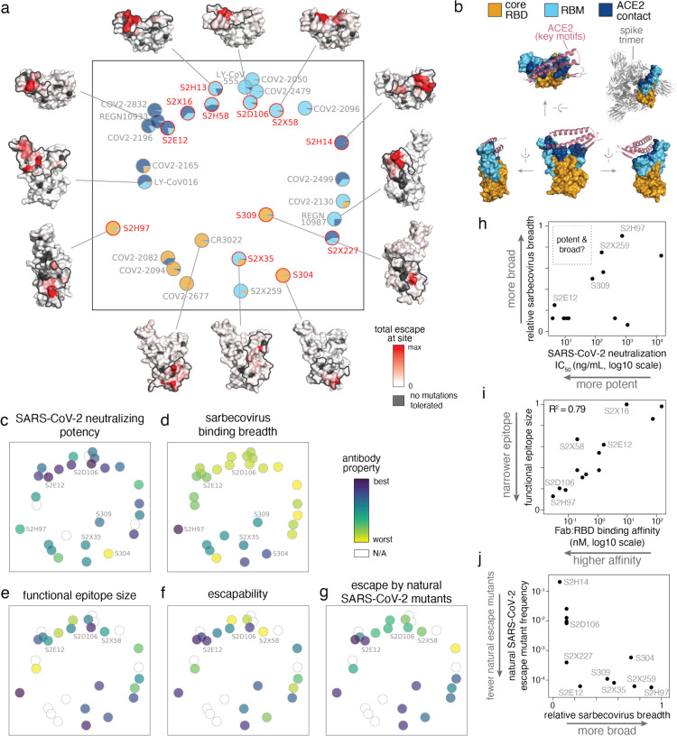
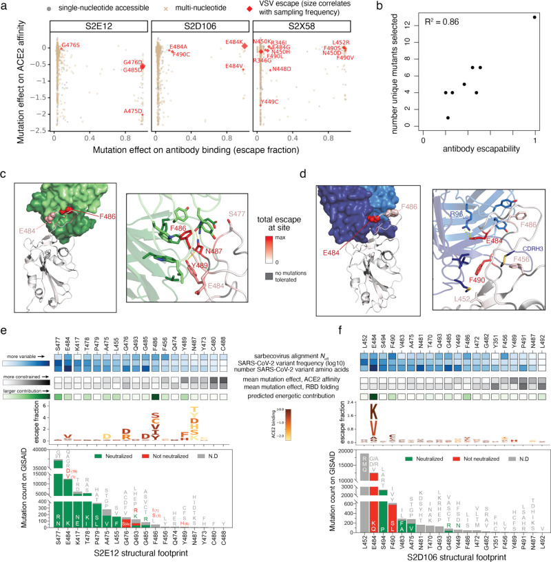
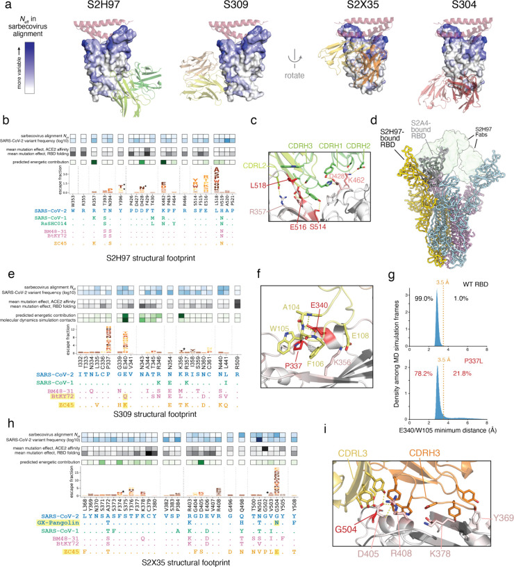
An ideal anti-SARS-CoV-2 antibody would resist viral escape 1-3 , have activity against diverse SARS-related coronaviruses 4-7 , and be highly protective through viral neutralization 8-11 and effector functions 12,13 . Understanding how these properties relate to each other and vary across epitopes would aid development of antibody therapeutics and guide vaccine design. Here, we comprehensively characterize escape, breadth, and potency across a panel of SARS-CoV-2 antibodies targeting the receptor-binding domain (RBD), including S309 4 , the parental antibody of the late-stage clinical antibody VIR-7831. We observe a tradeoff between SARS-CoV-2 in vitro neutralization potency and breadth of binding across SARS-related coronaviruses. Nevertheless, we identify several neutralizing antibodies with exceptional breadth and resistance to escape, including a new antibody (S2H97) that binds with high affinity to all SARS-related coronavirus clades via a unique RBD epitope centered on residue E516. S2H97 and other escape-resistant antibodies have high binding affinity and target functionally constrained RBD residues. We find that antibodies targeting the ACE2 receptor binding motif (RBM) typically have poor breadth and are readily escaped by mutations despite high neutralization potency, but we identify one potent RBM antibody (S2E12) with breadth across sarbecoviruses closely related to SARS-CoV-2 and with a high barrier to viral escape. These data highlight functional diversity among antibodies targeting the RBD and identify epitopes and features to prioritize for antibody and vaccine development against the current and potential future pandemics.


Figures

Extended Data Fig. 1.
Extended Data Fig. 1.. Antibody neutralization and binding data.
a, Neutralization of authentic SARS-CoV-2 (SARS-CoV-2-Nluc) by 13 antibodies. Shown are representative live virus neutralization plots. Symbols are means ± SD of triplicates. Dashed lines indicate IC50 and IC90 values. b, Correlation in antibody neutralization IC50 as determined in spike-pseudotyped VSV particles (n = 3 to 8) versus authentic SARS-CoV-2 (n = 3). c, SPR analysis of Fab fragments of the six newly described antibodies binding to the SARS-CoV-2 RBD, summarized in Fig. 1a. White and gray stripes indicate association and dissociation phases, respectively.
Extended Data Fig. 2.
Extended Data Fig. 2.. Yeast-display deep mutational scanning to comprehensively map mutations that escape antibody binding.
a, Scheme of the deep mutational scanning assay. Conformationally intact RBD is expressed on the surface of yeast, where RBD expression and antibody binding is detectable via fluorescent labeling. We previously constructed mutant libraries containing virtually all of the 3,819 possible amino acid mutations in the SARS-CoV-2 RBD and sorted the library to eliminate mutations that destabilize the RBD or reduce ACE2-binding affinity by more than two orders of magnitude. To identify mutations that escape antibody binding, we incubate the library with a sub-saturating antibody concentration (EC90 as determined by pilot yeast-displayed SARS-CoV-2 RBD binding assays) and use fluorescence-activated cell sorting (FACS) to isolate yeast cells expressing RBD mutants with reduced antibody binding. We use deep sequencing to quantify mutant frequencies before and after FACS selection, enabling calculation of the “escape fraction” of each amino acid mutation, which reflects the fraction of cells carrying that mutation that fall into the antibody-escape bin. Mutation escape fractions are represented in logoplots, where the height of a letter reflects the extent of escape from antibody binding. In this work, we created analogous libraries of yeast expressing all sarbecovirus RBDs, which we select via the same approach of FACS and deep sequencing to calculate the escape fraction of each sarbecovirus RBD. b, Representative selection gates, after gating for single cells expressing RBD as in Greaney et al.. Yeast expressing the SARS-CoV-2 RBD (top panels) are labeled at 1x, 0.01x and no antibody to guide selection gates. Mutant RBDs that reduce binding (green, gate drawn to capture 0.01x WT control) are sorted and sequenced for calculation of mutant escape fractions. This same gate was used to quantify escape within libraries of yeast expressing all sarbecovirus RBD homologs. For several antibodies, we also selected the sarbecovirus RBD library with a more stringent “full escape” gate (blue, gate drawn to capture 0 ng/mL WT control), to enable differentiation between homologs that partially or completely escape antibody binding. c, Fraction of library cells falling into escape bins for each antibody selection. d, Correlation in per-mutation escape fractions for duplicate libraries that were independently generated and assayed. e, Correlation in per-site escape (sum of per-mutation escape fractions) for duplicate libraries.
Extended Data Figure 3.
Extended Data Figure 3.. Expanded antibody escape profiles.
a,b, Complete maps of escape, with mutations colored by their effects on the opposite RBD property illustrated in Fig. 1b,c. Line plots to the left of each logoplot illustrate the total escape at each RBD site. Pink bars illustrate sites shown in logoplots at right. Sites in logoplots are more expansive than Fig. 1b,c, as they reflect a more sensitive threshold of site-wise escape for logoplot visualization due to space constraints in Fig. 1b,c.
Extended Data Fig. 4.
Extended Data Fig. 4.. Antibody escapability in natural SARS-CoV-2 mutants and from deep mutational scanning measurements.
a, For each antibody, scatterplots illustrate the degree to which a mutation escapes antibody binding (escape fraction, y-axis) versus its frequency among 582,276 high-quality human-derived SARS-CoV-2 sequences present on GISAID as of March 4, 2021. Large escape mutations (>5x global median escape fraction) for each antibody with non-zero mutant frequencies are labeled. The sum in each plot label gives the sum of mutant frequencies for all labeled mutations, corresponding to the natural SARS-CoV-2 mutant escape frequency for antibodies shown in Figs. 2g,j. b, To calculate antibody escapability (Fig. 1b,c), mutation escape fractions were weighted by their deleterious consequences for ACE2 binding or RBD expression. Top plots show the weighting factor (y-axis) for mutation effects on ACE2 binding (left) and RBD expression (right). This scaling weight factor was multiplied by the mutation escape fraction in the summation to calculate antibody escapability, as described in the Methods. For contextualizing this weighting penalization, histograms show the distribution of mutation effects on ACE2 binding (left) and RBD expression (right) for all mutations that pass our computational filtering steps (bottom), and mutations that are found with at least 20 sequence counts on GISAID (middle).
Extended Data Fig. 5.
Extended Data Fig. 5.. Breadth of antibody binding across sarbecoviruses.
a, Phylogenetic relationship of sarbecovirus RBDs inferred from aligned RBD nucleotide sequences, with the four clades of sarbecovirus RBD labeled in separate colors used throughout text. Node support values are rapid bootstrap support values, illustrating substantial ambiguity in the exact relationship between the three clades of sarbecovirus in Asia. b, A yeast-display library containing all sarbecovirus RBDs was assayed for antibody escape analogous to SARS-CoV-2 mutant selections, as shown in Extended Data Fig. 2. Heatmaps show escape (white) versus binding (black) within the affinity threshold of the FACS escape bins (Extended Data Fig. 2b). For S2H97, S2H13, and S2H14, we repeated selections with a more stringent “full escape” bin (0 ng/mL WT control, Extended Data Fig. 2b,c), enabling differentiation of RBDs with intermediate binding (e.g., S2H97/RsSHC014) versus complete loss of binding. c-e, Because the high-throughput assay in (b) yields binary measures of binding versus escape at a set threshold determined by FACS gate selection, we performed follow-up quantitative binding assays for select antibody/RBD combinations including flow cytometry detection of antibody binding to isogenic yeast-displayed RBD variants (c), flow cytometry detection of antibody binding to mammalian-surface displayed spikes (d), and ELISA using purified RBD proteins (e). These experiments validate the affinity thresholds of “escape” illustrated in (b) while adding additional context to interactions that are still present but with reduced binding strength. f, Binding of cross-reactive antibodies (Fab) and human ACE2 to select sarbecovirus RBDs was determined via SPR. NB, no binding; NT, not tested. g, S2H97 neutralization of VSV pseudotyped with select sarbecovirus spikes, with entry measured in VeroE6 (SARS-CoV-2, GD-Pangolin-CoV, and SARS-CoV-1) or ACE2-transduced BHK-21 cells (GX-Pangolin-CoV and WIV1). Curves are representative of at least two independent experiments. Error bars represent standard deviation from three technical replicates from one representative experiment.
Extended Data Fig. 6.
Extended Data Fig. 6.. Antibody escapability and its relationship to other properties.
a, Pairwise scatterplots between all antibody properties discussed in the main text. Select scatterplots from this panel are shown in Figs. 2h–j. Details of each property described in Methods. All axes are oriented such that moving to up on the y-axis and right on the x-axis corresponds to moving in the “preferred” direction for an antibody property (lower neutralization IC50, lower KD, higher breadth, narrower epitope size, lower escapability, lower total frequency of SARS-CoV-2 escape mutants among sequences on GISAID). b, Additional viral escape selections, as in Fig. 3a. For S309, we also include the E340A mutation identified in selections of authentic SARS-CoV-2 by Cathcart et al..
Extended Data Fig. 7.
Extended Data Fig. 7.. Data collection and processing of the S/S2D106 and S/S2H97 complex cryoEM datasets.
a,b, Representative electron micrograph and 2D class averages of SARS-CoV-2 S in complex with the S2D106 Fab embedded in vitreous ice. Scale bar: 400 Å. c, Gold-standard Fourier shell correlation curves for the S2D106-bound SARS-CoV-2 S trimer (black line) and locally refined RBD/S2D106 variable domains (gray line). The 0.143 cutoff is indicated by a horizontal dashed line. d, Local resolution map calculated using cryoSPARC for the whole reconstruction and the locally refined RBD/S2D106 variable domain region. e,f, Representative electron micrograph and 2D class averages of SARS-CoV-2 S in complex with the S2H97 Fab embedded in vitreous ice. Scale bar: 400 Å. g, Gold-standard Fourier shell correlation curve for the S2H97-bound SARS-CoV-2 S trimer reconstruction. The 0.143 cutoff is indicated by a horizontal dashed line. h, Local resolution map calculated using cryoSPARC for the whole reconstruction with two orthogonal orientations.
Extended Data Fig. 8.
Extended Data Fig. 8.. Genetic and structural basis for broad sarbecovirus binding.
a, Quaternary context of the S2H97 epitope. Left, S2H97-bound RBD, with RBD sites colored by S2H97 escape (scale bar, bottom). Right, RBD in the same angle as left, in the closed spike trimer. The S2H97 epitope is buried with the NTD of the neighboring protomer, requiring extensive RBD opening to enable access to the S2H97 epitope (Fig. 4d). b, Integrative genetic and functional features of the structural epitope of S309. Details as in Fig. 3e,f. c, Occupancy of key S309/RBD contacts during molecular dynamics simulation of S309 bound to the wildtype (42-μs simulation), P337A (56-μs) and P337L (91-μs) mutated RBD. Details as in Fig. 4g. d, Integrative genetic and functional features of the structural epitope of S304. Details as in Fig. 4b.
Fig. 1.
Fig. 1.. Potency, escapability, and breadth in a diverse panel of RBD antibodies.
a, For a panel of 12 SARS-CoV-2 antibodies, summary of neutralization potency (authentic SARS-CoV-2-NLuc [n=3] and SARS-CoV-2 spike-pseudotyped VSV particles [n = 3 to 8] on Vero E6 cells, Extended Data Fig. 1a,b), 1:1 Fab:RBD binding affinities (SPR, Extended Data Fig. 1c), and epitope class according to the schemes of Piccoli et al. and Barnes et al. See Extended Data Table 1 for additional antibody details. Binding affinities for previously described antibodies measured in Piccoli et al. (S304, S2X35, S2H13, S2H14), Tortorici et al. (S2E12) and Cathcart et al. (S309) b,c, Complete maps of mutations that escape binding by antibodies targeting the core RBD (b) or receptor-binding motif [RBM] (c), as determined by a yeast-display deep mutational scanning method (Extended Data Fig. 2). In each map, the height of a letter indicates that mutation’s strength of escape from antibody binding. Letters are colored by their effects on folded RBD expression (b) or ACE2 binding affinity (c) [scale bars, right], as determined in our prior deep mutational scans. See Extended Data Fig. 3 for escape maps colored by the opposite functional property. For each antibody, the relative “functional epitope size” and “escapability” are tabulated at right (see Methods for details). Heatmaps at bottom illustrate variability of each position within the sarbecovirus alignment or among globally sampled SARS-CoV-2 mutants. See Extended Data Fig. 4a for mutation-level variability and escape among observed SARS-CoV-2 mutants. Interactive escape maps and structural visualizations can be found at: https://jbloomlab.github.io/SARS-CoV-2-RBD_MAP_Vir_mAbs. d, Breadth of sarbecovirus binding by each antibody, summarizing comprehensive pan-sarbecovirus RBD yeast-display, ELISA, mammalian-display, and SPR binding assays. See Extended Data Fig. 5 for all data and phylogenetic definition of RBD clades. Black indicates an antibody binds an RBD or all RBDs within a clade with binding strength similar to SARS-CoV-2, gray indicates binding is reduced in affinity, or not all homologs within a clade are strongly bound, and white indicates no binding detected to a homolog or within a clade. The percent identity between RBD amino acid sequences with SARS-CoV-2 (or average % identity for clades) is shown below each column.
Fig. 2.
Fig. 2.. Relationship between antibody epitope, potency, breadth, and escapability.
a, Multidimensional scaling projection of antibody epitopes based on similarities in sites of binding-escape as mapped in this (red) or prior (gray) studies. Pie charts illustrate the RBD sub-domains where mutations confer escape for each antibody [see key, (b)]. Structural projections of escape from representative antibodies are arrayed around the perimeter (scale bar, bottom right), with gray outlines tracing the structural footprint for antibodies with solved structures. b, Structural key for (a), illustrating the relative angles of structural views, the classification of sub-domains, and the context of ACE2 binding (only the interacting structural elements are shown) and spike quaternary structure. c-g, Projected epitope space from (a) annotated by antibody properties. For each property, antibodies are colored such that purple (scale bar, upper-right) reflects the most desirable antibody: most potent neutralization (IC50, log10 scale), highest breadth, narrowest functional epitope size, lowest escapability, and lowest natural frequency of escape mutants (log10 scale). See Methods for definition of each property, and Extended Data Fig. 6a for quantitative scales of each property and all of their pairwise correlations. h, Relationship between sarbecovirus breadth and SARS-CoV-2 neutralization potency for antibodies characterized in this study and S2X259 (see accompanying paper, Tortorici et al.). i, Relationship between functional epitope size and RBD binding affinity for antibodies characterized in this study and S2X259. Note that binding-escape selections were calibrated independently for each antibody (Extended Data Fig. 2, Methods), so this correlation is not a simple consequence of high-affinity antibodies having fewer mutations that reduce affinity below some global threshold of escape applied universally to all antibodies. j, Relationship between natural SARS-CoV-2 mutation escape (summed frequency of all binding-escape mutations on GISAID, Extended Data Fig. 4a) and sarbecovirus breadth for antibodies characterized in this study and S2X259.
Fig. 3.
Fig. 3.. Structural basis for variation in antibody escapability.
a, Escape mutations identified in spike-expressing VSV passaged in the presence of monoclonal antibody, illustrated with respect to their effects on antibody (x-axis) and ACE2 (y-axis) binding. Amino acid mutations are coded by whether they are accessible via single-nucleotide mutation from the wildtype spike gene sequence used in the VSV selections (Wuhan-Hu-1+D614G). See additional selections, Extended Data Fig. 6b. b, Correlation between the number of unique mutations selected across viral escape selection experiments and antibody escapability as tabulated in Fig. 1b,c, plus S2X259 (see accompanying paper, Tortorici et al.). c,d, Structures of S2E12 (c) and S2D106 (d) bound to RBD. RBD sites are colored by escape (scale bar, center). Antibody heavy chains colored dark green (S2E12) or blue (S2D106), with light chains in lighter shades. Righthand images show zoomed-in context of key structural features impacting antibody escapability. e,f, Integrative genetic and functional features of the structural epitopes of S2E12 (e) and S2D106 (f). Sites within the structural footprint of each antibody (5 Å cutoff) are ordered by the frequency of observed mutants among SARS-CoV-2 sequences present on GISAID. Heatmaps illustrate evolutionary variability (blue), functional constraint from prior deep mutational scanning measurements (gray) and predicted energetic contribution of a residue derived from the structures (green). Logoplots illustrate escape from antibody binding as in Fig. 1c, but only showing amino acid mutations accessible via single-nucleotide mutation from Wuhan-Hu-1 for comparison with (a). Barplots illustrate the frequency of SARS-CoV-2 mutants at each position and their validated effects on antibody neutralization measured in spike-pseudotyped VSV particles with Vero E6 cells (red, >3-fold increase in IC50 due to mutation).
Fig. 4.
Fig. 4.. Structural basis for broad sarbecovirus binding.
a, Overview of the surfaces targeted by broadly binding RBD antibodies. RBD surface is colored by site variability in the sarbecovirus alignment (effective number of amino acids, scale bar at left). ACE2 (key motifs) shown in transparent cartoon. Antibody variable domains shown as cartoon, with darker shade indicating the heavy chain. b, Integrative genetic and structural features of the S2H97 structural epitope (5 Å cutoff). Heatmap details and scale bars as in Fig. 3e,f. Logoplots are colored by mutation effects on folded RBD expression (see scale bar, Fig. 1b). Asterisks in logoplot indicate escape mutations that introduce N-linked glycosylation motifs (NxS/T). Below the logoplot is a selection of aligned sarbecovirus RBDs (sequenced colored by clade as in Fig. 1d, Extended Data Fig. 5a). c, Zoomed in view of the S2H97/RBD interface, with important contact and escape residues labeled. d, CryoEM structure of S2H97-bound SARS-CoV-2 S. Spike protomers are shown in yellow, blue, and pink, and S2H97 Fabs in transparent green surface. S2A4-bound spike protomer from PDB 7JVC is shown in gray and aligned to the yellow subunit, indicating the additional extent of RBD opening necessary to access the S2H97 epitope compared to a class II antibody. e, Integrative features of the S309 structural epitope, details as in (b). An additional row in the heatmap overlay reflects the proportion of all close S309/RBD contacts (<3.5 Å) made by each residue during molecular dynamics simulation. Highlighted sarbecoviruses identify those that escape S309 binding, and highlighted mutation in the alignment is the likely contributor according to our escape map. f, Zoomed in view of the S309/RBD interface, with important contact and escape residues labeled. g, Molecular dynamics simulations of the S309/RBD structure. Histograms show the distribution of minimum distance between E340RBD and W105HC heavy atoms across 1-ns frames during the simulation of the unmutated (top, 42-μs simulation) and P337L mutated (bottom, 91-μs simulation) RBD bound to S309. Orange line reflects the 3.5 Å distance cutoff used to define close contact. Percentage of frames in which E340 and W105 are or are not in close contact is labeled. See Extended Data Fig. 8c for the occupancy of other S309:RBD contacts across the simulations. h, Integrative features of the S2X35 structural epitope, details as in (b). i, Zoomed in view of the S2X35/RBD interface, with important contact and escape residues labeled.

References

    1. Liu Z. et al. Identification of SARS-CoV-2 spike mutations that attenuate monoclonal and serum antibody neutralization. Cell Host Microbe 29, 477–488.e4 (2021). - PMC - PubMed
    1. Weisblum Y. et al. Escape from neutralizing antibodies by SARS-CoV-2 spike protein variants. Elife 9, e61312 (2020). - PMC - PubMed
    1. Greaney A. J. et al. Complete Mapping of Mutations to the SARS-CoV-2 Spike Receptor-Binding Domain that Escape Antibody Recognition. Cell Host Microbe 29, 44–57.e9 (2021). - PMC - PubMed
    1. Pinto D. et al. Cross-neutralization of SARS-CoV-2 by a human monoclonal SARS-CoV antibody. Nature 583, 290–295 (2020). - PubMed
    1. Rappazzo C. G. et al. Broad and potent activity against SARS-like viruses by an engineered human monoclonal antibody. Science 371, 823–829 (2021). - PMC - PubMed

Publication types