[Preprint]. 2021 Apr 10:2021.04.08.438911.
doi: 10.1101/2021.04.08.438911.

Nanobody Repertoires for Exposing Vulnerabilities of SARS-CoV-2

Affiliations

Nanobody Repertoires for Exposing Vulnerabilities of SARS-CoV-2

Fred D Mast et al. bioRxiv. .

Update in

  • Highly synergistic combinations of nanobodies that target SARS-CoV-2 and are resistant to escape.
    Mast FD, Fridy PC, Ketaren NE, Wang J, Jacobs EY, Olivier JP, Sanyal T, Molloy KR, Schmidt F, Rutkowska M, Weisblum Y, Rich LM, Vanderwall ER, Dambrauskas N, Vigdorovich V, Keegan S, Jiler JB, Stein ME, Olinares PDB, Herlands L, Hatziioannou T, Sather DN, Debley JS, Fenyö D, Sali A, Bieniasz PD, Aitchison JD, Chait BT, Rout MP. Mast FD, et al. Elife. 2021 Dec 7;10:e73027. doi: 10.7554/eLife.73027. Elife. 2021. PMID: 34874007 Free PMC article.

Abstract

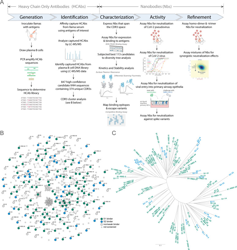
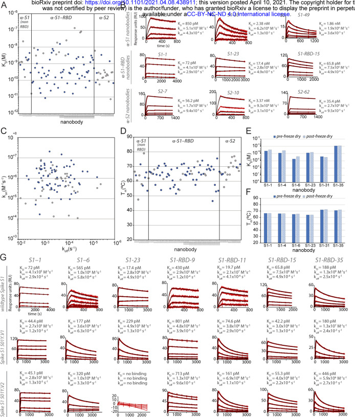
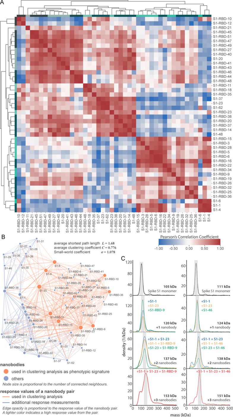
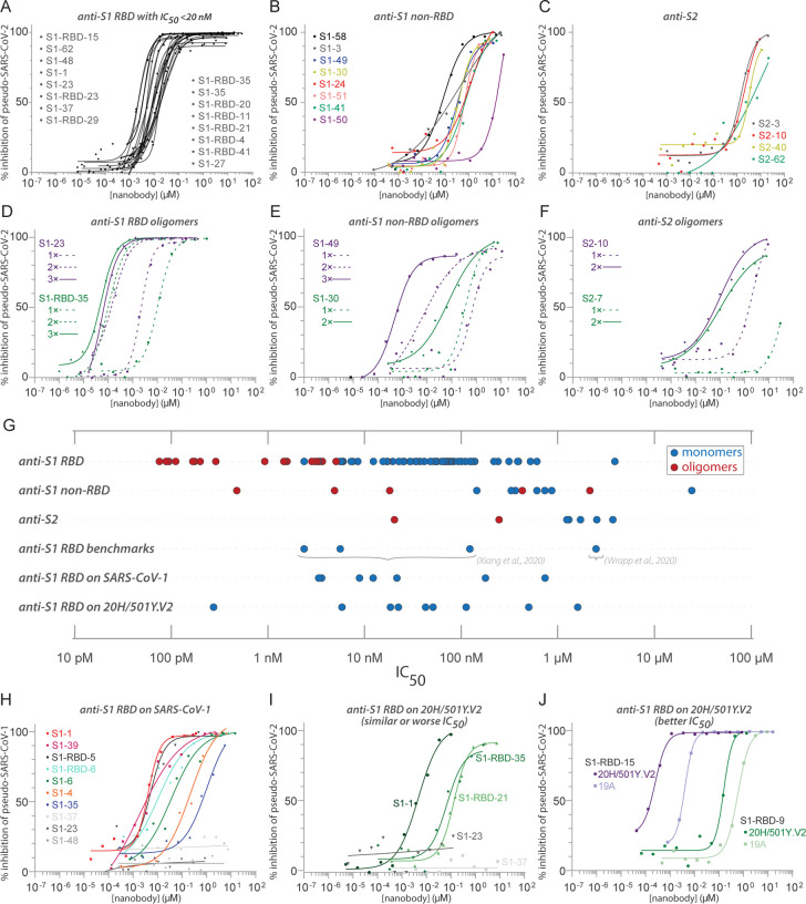
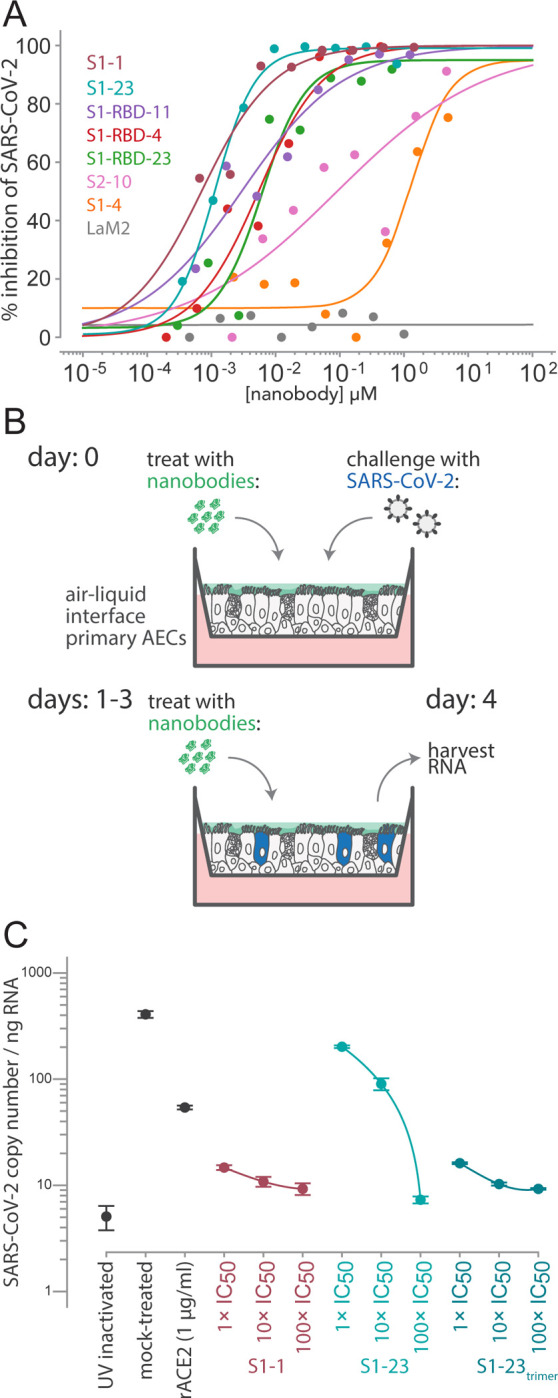
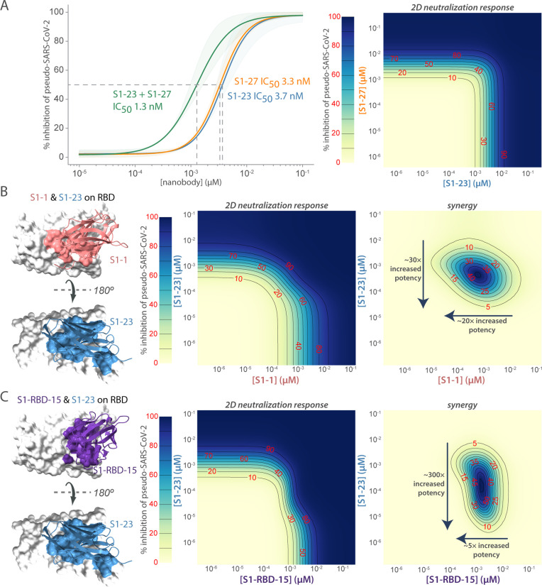
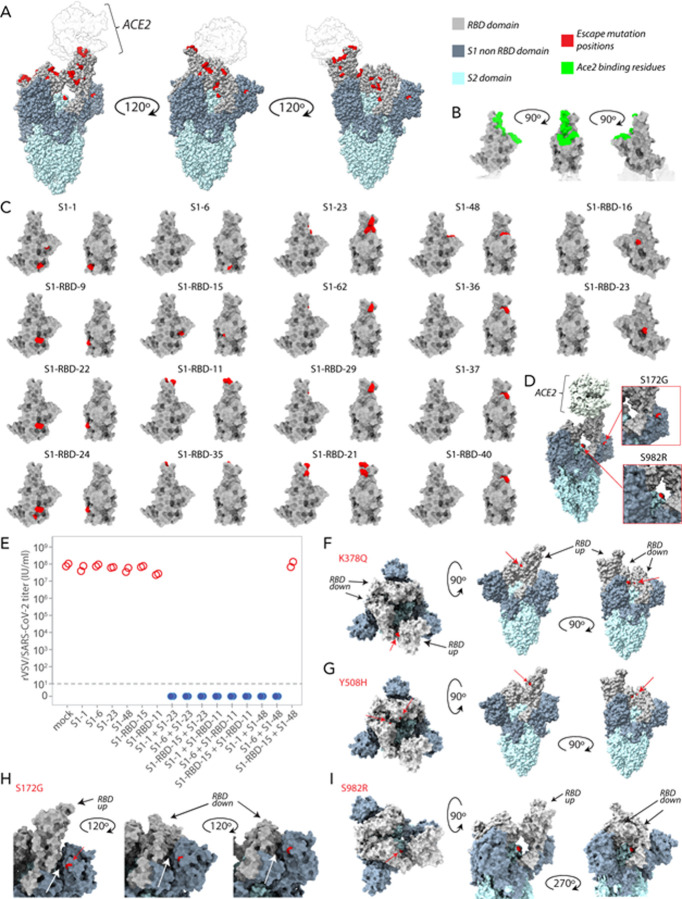
Despite the great promise of vaccines, the COVID-19 pandemic is ongoing and future serious outbreaks are highly likely, so that multi-pronged containment strategies will be required for many years. Nanobodies are the smallest naturally occurring single domain antigen binding proteins identified to date, possessing numerous properties advantageous to their production and use. We present a large repertoire of high affinity nanobodies against SARS-CoV-2 Spike protein with excellent kinetic and viral neutralization properties, which can be strongly enhanced with oligomerization. This repertoire samples the epitope landscape of the Spike ectodomain inside and outside the receptor binding domain, recognizing a multitude of distinct epitopes and revealing multiple neutralization targets of pseudoviruses and authentic SARS-CoV-2, including in primary human airway epithelial cells. Combinatorial nanobody mixtures show highly synergistic activities, and are resistant to mutational escape and emerging viral variants of concern. These nanobodies establish an exceptional resource for superior COVID-19 prophylactics and therapeutics.


Figures

Figure 1.
Figure 1.. Approach.
(A) Schematic of our strategy for generating, identifying and characterizing large, diverse repertoires of nanobodies that bind the Spike protein of CoV-2. The highest quality nanobodies were assayed for their ability to neutralize CoV-2 pseudo-virus, CoV-2 virus and viral entry into primary human airway epithelial cells. We also measured the activities of homodimers/homotrimers and mixtures. (B) A network visualization of 374 high confidence CDR3 sequences identified from Mass Spectrometry workflow. Nodes (CDR3 sequences) were connected by edges defined by a Damerau-Levenshtein distance of no more than 3, forming 183 isolated components. A thicker edge indicates a smaller distance value, i.e. a closer relation. (C) Dendrogram showing sequence relationships between the 113 selected nanobodies, demonstrating that the repertoire generally retains significant diversity in both anti-S1 (green) and anti-S2 (blue) nanobodies, albeit with a few closely related members. Scale, 0.2 substitutions per residue.
Figure 2.
Figure 2.. Biophysical characterization of anti-SARS-CoV-2 Spike nanobodies.
(A) Each nanobody plotted against their affinity (KD) for their antigen separated into three groups based on their binding region on SARS-CoV-2 Spike protein. The data points highlighted in blue correspond to nanobodies that neutralize. The majority of nanobodies have high affinity for their antigen with KDs below 1 nm. 10 nanobodies are not included in this plot as they were unable to be analyzed successfully using SPR. (B) SPR sensorgrams for each of the three targets on SARS-CoV-2 Spike protein of our nanobody repertoire, showing three representatives for each binding region. (C) The association rate of each nanobody (kon) versus the corresponding dissociation rate (koff). The majority of our nanobodies have fast association rates (≥10+5 M−1s−1), with many surpassing the kon of high performing monoclonal antibodies and nanobodies with a kon > M−1s−1. (D) Each nanobody plotted against their Tm as measured by DSF, revealing all but two nanobodies fall within the optimal Tm range (between 50°C and 80°C), where the bulk of our nanobodies have a Tm ≥60°C. No data could be collected for two nanobodies and 10 nanobodies exhibited two dominant peaks in the thermal shift assay and were not included in this plot (a full summary of this data can be seen in Suppl. Tables 1 and 2). The KD (E) and TM (F) of six nanobodies was assessed pre- and post- freeze-drying, revealing no significant change in affinity or Tm after freeze-drying. (G) SPR sensorgrams comparing the kinetic and affinity analysis of seven nanobodies against wildtype Spike S1 (Wuhan str.), Spike 20I/S1 501Y.V1 (United Kingdom variant) and 20H/Spike S1 501Y.V2 (Sth. African variant).
Figure 3.
Figure 3.. Epitope characterization of nanobodies against the S1-RBD of SARS-CoV-2 Spike.
(A) Major epitope bins are revealed by a clustered heat map of Pearson’s Correlation Coefficients computed from the response values of nanobodies binding to the Spike RBD in pairwise cross-competition assays on a biolayer interferometer. Correlated values (red) indicate that the two nanobodies respond similarly when measured against a panel of nine RBD nanobodies that bind to distinct regions of the RBD. A strong correlation score indicates binding to a similar/overlapping region on the RBD. Anti-correlated values (blue) indicate that a nanobody pair responds divergently when measured against nanobodies in the representative panel, and indicate binding to distinct or non-overlapping regions on the RBD. Hierarchical clustering analysis reveals 15 separate and partially overlapping epitope bins as visualized by alternating dark and light teal bars linked together by a dendrogram. (B) A network visualization of anti-S1-RBD nanobodies. Each node is a nanobody and each edge is a response value measured by biolayer interferometry from pairwise cross-competition assays. Orange nodes represent nine nanobodies used as a representative panel for clustering analysis in A. Blue nodes represent the other anti-S1-RBD nanobodies present in the dataset. The average shortest distance between any nanobody pair in the dataset of 1.68. An average clustering coefficient of 0.776 suggests that the measurements are well-distributed across the dataset. The small world coefficient of 1.078 indicates that the network is more connected than to be expected from random, but the average path length is what you would expect from a random network, together indicating that the relationship between nanobody pairs not actually measured can be inferred from the similar/neighboring nanobodies. (C) Mass photometry (MP) analysis of Spike S1 monomer incubated with different anti-Spike S1 nanobodies. Two examples of an increase in mass as Spike S1 monomer (black line) is incubated with one to three nanobodies. The accumulation in mass upon addition of each different nanobody on Spike S1 monomer is due to each nanobody binding to non-overlapping space on Spike S1, an observation consistent with Octet binning data. As a control, using MP, each individual nanobody was shown to bind Spike S1 monomer on its own (data not shown).
Figure 4.
Figure 4.. Diverse and potent nanobody-based neutralization of SARS-CoV-2.
Nanobodies targeting the S1-RBD, S1 non-RBD, and S2 portions of Spike effectively neutralize lenti-virus pseudotyped with various SARS-CoV Spikes and their variants from infecting ACE2 expressing HEK293T cells. (A) Of the 113 nanobodies, monomers that neutralize SARS-CoV-2 pseudovirus with IC50 values 20nM and lower are displayed. (B) Representative nanobodies targeting the non-RBD portions of S1 and (C) the S2 domain of SARS-CoV-2 neutralize SARSCoV-2 pseudovirus. (D-F) Oligomerization of RBD, S1 non-RBD and S2 nanobodies significantly increases neutralization potency. (G) Summary scatter plot of all nanobody IC50s across the major domains of SARS-CoV-2 Spike and where tested, across SARS-CoV-2 variant 20H/501Y.V2 and SARS-CoV-1. Representative published nanobodies were also tested in our neutralization assays and show similar potency towards SARS-CoV-2 pseudovirus. (H) Representative SARS-CoV-2 RBD targeting nanobodies cross-neutralize SARS-CoV-1 pseudotyped lentivirus and (I-J) the 20H/501Y.V2 SARS-CoV-2 variant (B.1.351) with L18F, D80A, K417N, E484K, and N501Y amino acid substitutions in Spike. 19A (I-J) is the initial SARS-CoV-2 clade that includes the prototypical Wuhan-Hu-1 Spike used as “wild-type” in these pseudovirus assays. In all cases, n>/=2 biological replicates of each nanobody monomer/oligomer with a representative biological replicate with n=4 technical replicates per dilution displayed.
Figure 5.
Figure 5.. Authentic SARS-CoV-2 neutralization by anti-Spike nanobodies.
(A) Neutralization curves are plotted from the results of a focus-forming reduction neutralization assay with the indicated nanobodies. Serial dilutions of each nanobody were incubated with SARS-CoV-2 (MOI 0.5) for 60 min and then overlaid on a monolayer of Vero E6 cells and incubated for 24 h. LaM2, an anti-mCherry nanobody (Fridy et al., 2014) was used as a non-neutralizing control. After 24 h, cells were collected and stained with anti-Spike antibodies and the ratio of infected to uninfected cells was quantified by flow cytometry. (B) A schematic of an air-liquid interface (ALI) culture of primary human airway epithelial cells (AECs) as a model for SARS-CoV-2 infection. Cells were incubated with nanobodies and then challenged with SARSCoV-2 (MOI 0.5). After daily treatment with nanobodies for three more days, the cultures are harvested to isolate RNA and quantify the extent of infection. (C) AECs were infected with the indicated concentrations of anti-SARS-CoV Spike nanobodies. The infected cultures were maintained for five days with a daily 1 h incubation of nanobodies before being harvested for RNA isolation and determination of the SARS-CoV-2 copy number by qPCR. SARS-CoV-2 copy number was normalized to total RNA measured by spectrophotometry. Mock-treated samples exposed to infectious and UV-inactivated SARS-CoV-2 virions served as positive and negative controls. Recombinant soluble angiotensin converting enzyme 2 (rACE2) was used as a positive treatment control. The indicated nanobodies were used at 1, 10, and 100× their IC50 values determined in pseudovirus neutralization assays.
Figure 6.
Figure 6.. Synergistic neutralization of Spike with nanobody cocktails.
(A) An example of additive effects between two anti-SARS-CoV2 Spike nanobodies. S1–23 and S1–27 were prepared in a two-dimensional serial dilution matrix and then incubated with SARS-CoV-2 pseudovirus for 1 h before adding the mixture to cells. After 56 h, the expression of luciferase in each well was measured by addition of Steady-Glo reagent and read out on a spectrophotometer. Neutralization curves and the calculated IC50 of each nanobody alone, or in a 1:1 combination was determined. The right panel shows a heat map of pseudovirus neutralization by a two-dimensional serial dilution of combinations of S1–23 and S1–27. Lines and red numbers demarcate the % inhibition, that is, inhibitory concentration where X% of the virus is neutralized, e.g. IC50. Dark blue regions are concentrations that potently neutralize the pseudovirus, as per the heat map legend. (B) The left panel shows a model of S1–1 and S1–23 neutralizing nanobodies binding to distinct epitopes of the RBD. The middle panel shows the heatmap of pseudovirus neutralization observed by a two-dimensional serial dilution of combinations of S1–1 and S1–23. The right panel shows a heat map with the difference between the observed neutralization and that expected in a null model of only additive effects. The lines and red numbers demarcate regions in the heat map where the observed neutralization is greater than additive by the indicated percentages (red numbers). Overall, S1–1 enhances the effect of S1–23 ~30-fold, whereas S1–23 enhances the effect of S1–1 ~20 fold. (C) As in B, but comparing S1-RBD-15 with S1–23. The left panel shows a model of S1-RBD-15 and S1–23 neutralizing nanobodies binding to distinct epitopes of the RBD. The middle panel shows a heatmap of pseudovirus neutralization observed for 2D serial dilution of S1-RBD-15 and S1–23. The right panel shows the synergy observed between S1-RBD-15 and S1–23. Overall, S1-RBD-15 enhances the effect of S1–1 ~300 fold, whereas S1–23 enhances the effect of S1-RBD-15 ~5 fold.
Figure 7.
Figure 7.. Mapping of Spike substitutions in rVSV/SARS-CoV-2/GFP escape mutants obtained in the presence of the corresponding nanobody.
(A) Mapped on to the structure of SARS-CoV2 Spike trimer in complex with one ACE2 molecule (PDB ID 7KNB, used for all SARS-CoV2 Spike trimer representations) is the position of neutralization-resistant amino acid substitutions (in red), also known as ‘escape mutants’ that were generated in response to cultivation of rVSV/SARS-CoV-2/GFP in the presence of each nanobody, and were subsequently shown to confer resistance to the same nanobody. (B) Structure of the SARS-CoV-2 RBD (PDB ID 6M0J) showing the positions of amino acid residues (in green) that form the ACE2 binding site, for reference. (C) Structure of the SARS-CoV-2 RBD (PDB ID: 6M0J) showing the positions (in red) of the location of substitutions that confer resistance for each nanobody tested in two orientations 90° apart. For structure pairs of S1-RBD-16 and S1-RBD-23 escape mutants, the rotation is 90° from the structure to the left in the pair. (D) The location of two key non-RBD escape mutants S172G and S982K resulting from assays performed with an anti-S1 non RBD (S1–49) and anti-S2 (S2–10) nanobody respectively. (E) Infectious rVSV/SARS-CoV-2/GFP yield (IU/ml) following two passages in the presence of the indicated individual nanobodies or nanobody combinations, at 100× IC50 of the individual nanobodies, or 50× IC50 of each of the nanobodies in the combinations. Each data point represents an independent titer measurement. Red open circles represent virus escapes while blue circles represent nanobody combinations for which no escapes (titer = 0) were detected. The location of two escape mutants K378Q (F) and Y508H (G) mapped onto SARS-CoV2 Spike trimer for the two corresponding nanobodies S1-RBD-9 and S1-RBD-15 respectively, revealing an exposed putative nanobody binding site on RBD when in the “up” position that is hidden when RBD is in the “down” position. (H) A close up of escape mutant S172G on each monomer of SARS-CoV2 Spike trimer revealing a larger crevice between the NTD of Spike S1 and RBD when the RBD is in the “down” position compared to the “up” position. (I) Three orientations of SARS-CoV2 Spike trimer revealing the position in all three orientations of escape mutant S982R revealing the putative binding site for nanobody S2–10 is accessible regardless of whether the RBD is the “up” or “down” position.

References

    1. Andreano E., Nicastri E., Paciello I., Pileri P., Manganaro N., Piccini G., Manenti A., Pantano E., Kabanova A., Troisi M., et al. (2021). Extremely potent human monoclonal antibodies from COVID-19 convalescent patients. Cell 184, 1821–1835 e1816. - PMC - PubMed
    1. Bannas P., Hambach J., and Koch-Nolte F. (2017). Nanobodies and nanobody-based human heavy chain antibodies as antitumor therapeutics. Frontiers in immunology 8, 1603. - PMC - PubMed
    1. Bard G.V. (2007). Spelling-error tolerant, order-independent pass-phrases via the Damerau–Levenshtein string-edit distance metric. In Proceedings of the Fifth Australasian Symposium on ACSW Frontiers: 2007, Ballarat, Australia, January 30 - February 2, 2007 Conferences in Research and Practice in Information Technology (Darlinghurst, Australia: Australian Computer Society, Inc.), pp. 117–124.
    1. Barnes C.O., Jette C.A., Abernathy M.E., Dam K.A., Esswein S.R., Gristick H.B., Malyutin A.G., Sharaf N.G., Huey-Tubman K.E., Lee Y.E., et al. (2020). SARS-CoV-2 neutralizing antibody structures inform therapeutic strategies. Nature 588, 682–687. - PMC - PubMed
    1. Barrow K.A., Rich L.M., Vanderwall E.R., Reeves S.R., Rathe J.A., White M.P., and Debley J.S. (2021). Inactivation of Material from SARS-CoV-2-Infected Primary Airway Epithelial Cell Cultures. Methods Protoc 4. - PMC - PubMed

Publication types

LinkOut - more resources