

Neurovascular-glymphatic dysfunction and white matter lesions
- PMID: 33851307
- PMCID: PMC8492845
- DOI: 10.1007/s11357-021-00361-x
Neurovascular-glymphatic dysfunction and white matter lesions
Abstract
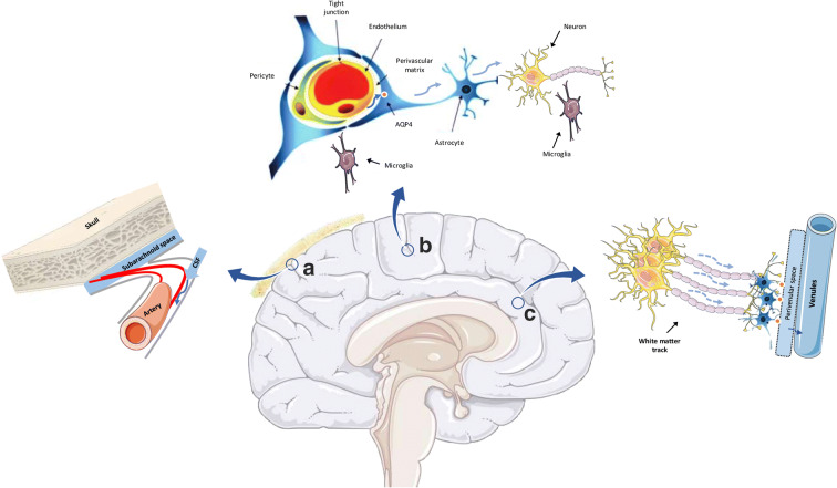
Cerebral white matter lesions (WML) represent a spectrum of age-related structural changes that are identified as areas of white matter high signal intensity on brain magnetic resonance imaging (MRI). Preservation of white matter requires proper functioning of both the cerebrovascular and glymphatic systems. The cerebrovascular safeguards adequate cerebral blood flow to supply oxygen, energy, and nutrients through a dynamic process of cerebral autoregulation and neurovascular coupling to keep up with global and regional demands of the brain. The glymphatic system maintains white matter integrity by preserving flow of interstitial fluid, exchanging metabolic waste and eventually its clearance into the venous circulation. Here, we argue that these two systems should not be considered separate entities but as one single physiologically integrated unit to preserve brain health. Due to the process of aging, damage to the neurovascular-glymphatic system accumulates over the life course. It is an insidious process that ultimately leads to the disruption of cerebral autoregulation, to the neurovascular uncoupling, and to the accumulation of metabolic waste products. As cerebral white matter is particularly vulnerable to hypoxic, inflammatory, and metabolic insults, WML are the first recognized pathologies of neurovascular-glymphatic dysfunction. A better understanding of the underlying pathophysiology will provide starting points for developing effective strategies to prevent a wide range of clinical disorders among which there are gait disturbances, functional dependence, cognitive impairment, and dementia.
Keywords: Aging; Glymphatic system; Neurovascular unit; Small vessel disease; White matter.
© 2021. American Aging Association.
Conflict of interest statement
Behnam Sabayan declares no conflict of interest. Rudi Westendorp is supported by grants from Nordea Fonden [02–2017-1749] and Novo Nordisk Fonden Challenge Programme: Harnessing the Power of Big Data to Address the Societal Challenge of Aging [NNF17OC0027812]. These funding bodies had no influence on writing this manuscript.
Figures
References
-
- de Leeuw FE, de Groot JC, Achten E, Oudkerk M, Ramos LM, Heijboer R, et al. Prevalence of cerebral white matter lesions in elderly people: a population based magnetic resonance imaging study. The Rotterdam Scan Study. J Neurol Neurosurg Psychiatry. 2001;70:9–14. doi: 10.1136/jnnp.70.1.9. - DOI - PMC - PubMed
Publication types
MeSH terms
LinkOut - more resources
Full Text Sources
Other Literature Sources
Research Materials
