. 2021 Jun 1;320(6):L1118-L1125.
doi: 10.1152/ajplung.00598.2020. Epub 2021 Apr 14.

PINK1 mediates the protective effects of thyroid hormone T3 in hyperoxia-induced lung injury

Affiliations

PINK1 mediates the protective effects of thyroid hormone T3 in hyperoxia-induced lung injury

Yi Zhang et al. Am J Physiol Lung Cell Mol Physiol. .

Abstract

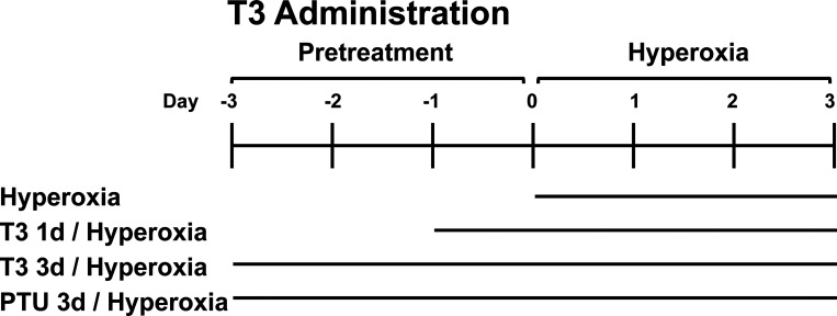
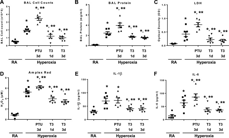
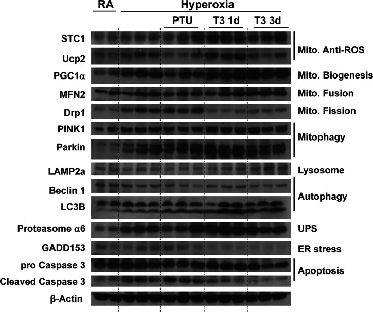
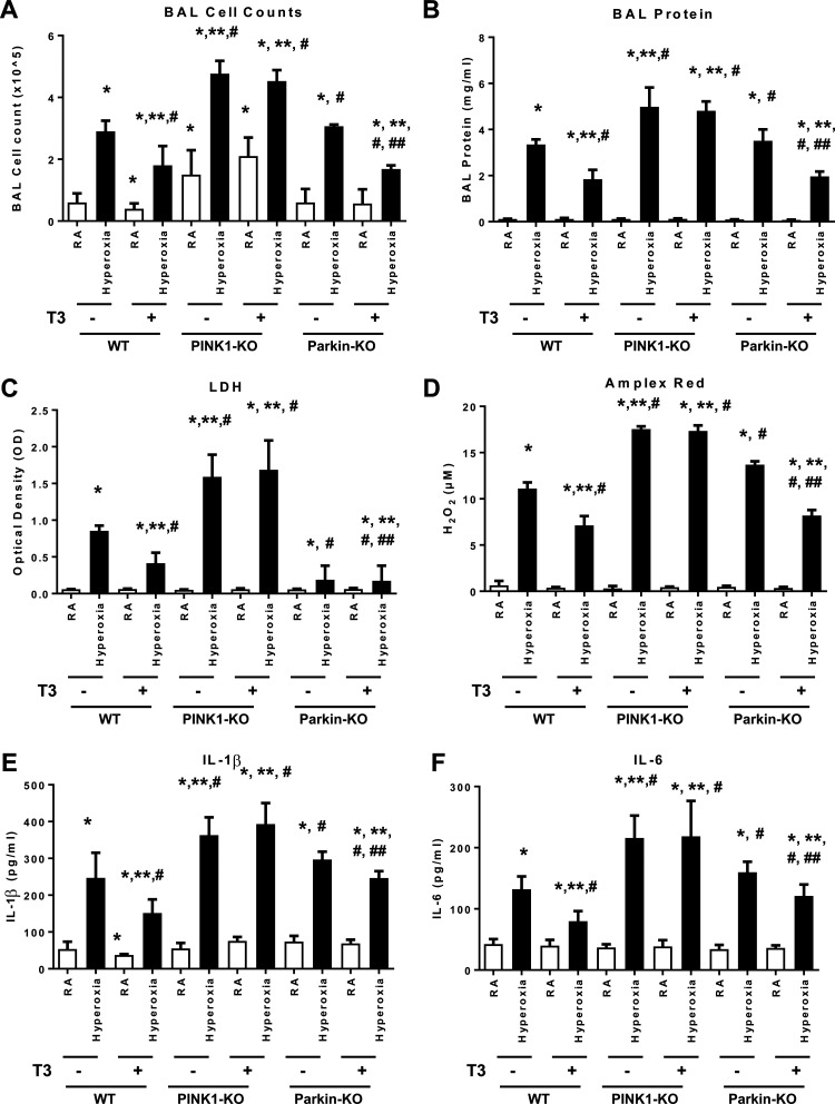
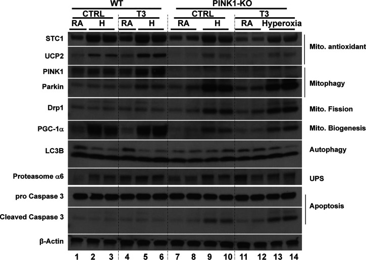
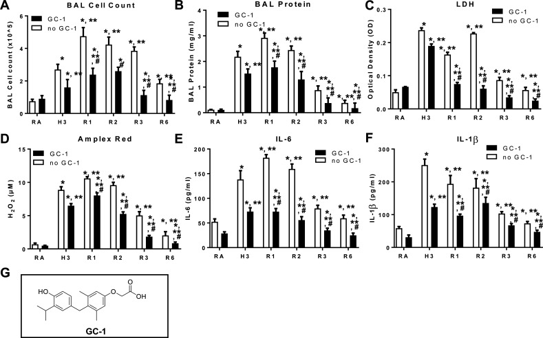
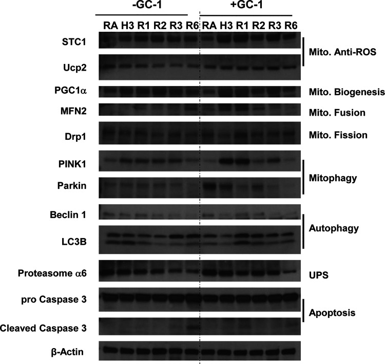
Hyperoxia can lead to respiratory failure and death. Our previous work demonstrates that oxidant and mitochondrial injury play a critical role in hyperoxia-induced acute lung injury (HALI). Recently, thyroid hormone has been demonstrated to promote mitochondrial survival in other models of lung injury, but its role in hyperoxia is unknown. Adult wild-type (WT) mice were pretreated with either nebulized triiodothyronine (T3, 40 μg/kg) for 1 or 3 days, or with propylthiouracil (PTU, 100 μg/kg), for 3 days. Following pretreatment, WT mice underwent 72 h of hyperoxia exposure. WT and PINK1-/- mice were pretreated with either nebulized T3 (40 μg/kg) for 3 days or no pretreatment before 72 h continuous hyperoxia exposure. Bronchoalveolar lavage (BAL), histological changes in cellular composition, and type I cytokine induction were assessed. Lung lysates for mitochondrial cellular bioenergetics markers were analyzed by Western blot. Hyperoxia caused a significant increase in BAL total cell counts and lung cellular infiltrates. Administration of PTU enhanced HALI, whereas T3 attenuated HALI, inflammation, and oxidants in WT mice. In addition, T3 pretreatment increased mitochondrial biogenesis/fusion/mitophagy and decreased ER stress and apoptosis. PINK1-/- mice were more susceptible to hyperoxia than WT mice. Notably, pretreatment with T3 did not attenuate HALI in PINK1-/- mice. In addition, T3 pretreatment increased mitochondrial anti-ROS potential, improved mitochondrial bioenergetics and mitophagy, and attenuated mitochondria-regulated apoptosis, all in a PINK1-dependent manner. Our results highlight a novel protective role for PINK1 in mediating the cytoprotective effects of thyroid hormone in HALI. Therefore, thyroid hormone may represent a potential therapy for ALI.

Keywords: HALI; PINK1; hyperoxia; lung injury; thyroid hormone.


Conflict of interest statement

Y.Z., G.Y., N.K., and P.J.L. have IP on the use of thyroid hormone mimetics in IPF and ARDS licensed to biotechnology. There are no conflicts of interest, financial or otherwise, to disclose.

Figures

Figure 1.
Figure 1.
Schema for triiodothyronine (T3) delivery. Wild-type (WT) mice were nebulized with triiodothyronine (T3, 40 µg/kg) for 1 day or 3 days, or propylthiouracil (PTU, 100 µg/kg) for 3 days before 72 h continuous hyperoxia exposure. Control mice were exposed to room air (RA).
Figure 2.
Figure 2.
Triiodothyronine (T3) pretreatment attenuates hyperoxia-induced lung injury, inflammation, and oxidants in wild-type (WT) mice compared with mice treated with propylthiouracil (PTU). WT mice were nebulized with triiodothyronine (T3, 40 µg/kg) for 1 day or 3 days or propylthiouracil (PTU, 100 µg/kg) for 3 days before 72 h continuous hyperoxia exposure. Control mice were exposed to room air (RA). A: total cells recovered from bronchoalveolar lavage (BAL) were counted. B: lung permeability was assessed by BAL protein content. C) lactate dehydrogenase (LDH) activity assays were performed on BAL fluid. D: oxidant generation was detected by Amplex Red from BAL fluid. IL-1β (E) and IL-6 (F) were detected by ELISA in BAL fluid. The values are expressed as means ±SD and analyzed by Mann–Whitney test (n = 6 for each group). *P < 0.05, vs. RA; **P < 0.05, vs. hyperoxia without pretreatment.
Figure 3.
Figure 3.
Triiodothyronine (T3) pretreatment increases mitochondrial biogenesis/fusion/mitophagy and decreases ER stress and apoptosis compared to mice treated with propylthiouracil (PTU). WT, PINK1−/−, and Parkin−/− mice were nebulized with triiodothyronine (T3, 40 µg/kg) for 3 days or no pretreatment before 72 h continuous hyperoxia exposure. Lysates were isolated and immunoblotted against antibodies as listed. β-Actin was used as protein loading control. Quantification based on densitometry of the Western blot is shown in Supplemental Fig. S1. The uncut Western blots listed in the Supplemental Fig. S3. PINK1, PTEN-induced kinase I; RA, room air; WT, wild-type.
Figure 4.
Figure 4.
PINK1, not Parkin, mediates triiodothyronine (T3) effects against hyperoxia-induced lung injury. WT, PINK1−/−, and Parkin−/− mice were nebulized with triiodothyronine (T3, 40 µg/kg) for 3 days or no pretreatment before 72 h continuous hyperoxia exposure. A: total cells recovered from bronchoalveolar lavage (BAL) were counted. B: lung permeability was assessed by BAL protein content. C: lactate dehydrogenase (LDH) activity assays were performed on BAL fluid. D: oxidant generation was detected by Amplex Red from BAL fluid. IL-1β (E) and IL-6 (F) were detected by ELISA in BALF. The values are expressed as means ± SD and analyzed by Mann–Whitney test (n = 6 for each group). *P < 0.05, vs. WT no T3 RA; **P < 0.05, vs. WT no T3 hyperoxia; #P < 0.05, vs. corresponding RA; ##P < 0.05, vs. corresponding no T3 hyperoxia. PINK1, PTEN-induced kinase I; RA, room air; WT, wild-type.
Figure 5.
Figure 5.
Triiodothyronine (T3) pretreatment increases in mitochondrial anti-ROS potential, biogenesis and mitophagy under hyperoxia are dependent upon PINK1. WT and PINK1−/− mice were nebulized with triiodothyronine (T3, 40 µg/kg) for 3 days before 72 h continuous hyperoxia exposure. Lysates were isolated and immunoblotted against antibodies as listed. β-Actin was used as protein loading control. Quantification based on densitometry of the western blots shown in Supplemental Fig. S2. The uncut Western blots are shown in Supplemental Fig. S4. PINK1, PTEN-induced kinase I; ROS, reactive oxygen species; WT, wild-type.
Figure 6.
Figure 6.
Triiodothyronine (T3) agonist GC-1 pretreatment prevents hyperoxia induced lung injury and aids recovery. WT mice were orally gavaged with GC-1 (5 mg/kg) and then exposed to RA or to hyperoxia for 72 h (H3), followed by a recovery phase (R1–3, 1, 2, 3, and R6, 6 days posthyperoxia). Cells recovered from bronchoalveolar lavage (BAL) were counted as BAL total cell counts (A). B: lung permeability was assessed by BAL protein content. C: lactate dehydrogenase (LDH) activity assays were performed on BAL fluid. D: oxidant generation was detected by Amplex Red from BAL fluid. IL-6 (E) and IL-1β (F) was detected by ELISA in BALF. G: structural formula of GC-1. The values are expressed as means ± SD and analyzed by Mann–Whitney test (n =4 for each group). *P < 0.05 vs. no GC-1 RA; **P < 0.05 vs. no GC-1 H3; #P < 0.05 vs. corresponding no GC-1. RA, room air.
Figure 7.
Figure 7.
GC-1 pretreatment prevents hyperoxia induced lung injury and aids recovery. Wild-type (WT) mice were orally gavaged with GC-1 (5 mg/kg) and then exposed to RA or to hyperoxia for 72 h (H3), followed by a recovery phase (R1-3, 1, 2, 3, and R6, 6 days posthyperoxia). Lysates from mouse lungs were immunoblotted against the listed Abs. The uncut Western blots are shown in Supplemental Fig. S5. All of the fold changes are compared with the room air control set as 1. For LC3B, comparison was made between the two bands of LC3B, but not to the loading control; the ratio of the two bands indicates the change in autophagy.

Similar articles

Cited by

References

    1. Dias-Freitas F, Metelo-Coimbra C, Roncon-Albuquerque R. Molecular mechanisms underlying hyperoxia acute lung injury. Respir Med 119: 23–28, 2016. doi:10.1016/j.rmed.2016.08.010. - DOI - PubMed
    1. Ornatowski W, Lu Q, Yegambaram M, Garcia AE, Zemskov EA, Maltepe E, Fineman JR, Wang T, Black SM. Complex interplay between autophagy and oxidative stress in the development of pulmonary disease. Redox Biol 36: 101679, 2020. doi:10.1016/j.redox.2020.101679. - DOI - PMC - PubMed
    1. Sinha RA, Singh BK, Zhou J, Wu Y, Farah BL, Ohba K, Lesmana R, Gooding J, Bay B-H, Yen PM. Thyroid hormone induction of mitochondrial activity is coupled to mitophagy via ROS-AMPK-ULK1 signaling. Autophagy 11: 1341–1357, 2015. doi:10.1080/15548627.2015.1061849. - DOI - PMC - PubMed
    1. Yau WW, Singh BK, Lesmana R, Zhou J, Sinha RA, Wong KA, Wu Y, Bay B-H, Sugii S, Sun L, Yen PM. Thyroid hormone (T3) stimulates brown adipose tissue activation via mitochondrial biogenesis and MTOR-mediated mitophagy. Autophagy 15: 131–150, 2019. doi:10.1080/15548627.2018.1511263. - DOI - PMC - PubMed
    1. Zhang Y, Sauler M, Shinn AS, Gong H, Haslip M, Shan P, Mannam P, Lee PJ. Endothelial PINK1 mediates the protective effects of NLRP3 deficiency during lethal oxidant injury. J Immunol 192: 5296–5304, 2014. doi:10.4049/jimmunol.1400653. - DOI - PMC - PubMed

Publication types

LinkOut - more resources