

Primer malignant giant cell tumour of kidney: a case report
- PMID: 33851880
- PMCID: PMC10335276
- DOI: 10.1308/rcsann.2020.7113
Primer malignant giant cell tumour of kidney: a case report
Abstract
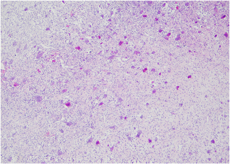
Osteoclast-like giant cell tumours of the kidney are extremely rare and usually accompanied by a conventional urothelial neoplasm such as papillary, transitional renal cell, or sarcomatoid carcinoma. Although they have morphological features similar to those of the giant cell tumours in the skeletal system, their counterparts in the urinary system show highly malignant features. Our case is the third primer malignant giant cell tumour of the kidney in the literature. The patient was a 50-year-old male and underwent nephroureterectomy for a mass of 18×14×13cm in his left kidney. However, the patient died in the second month postoperatively as a result of local recurrences and multiple distant metastases. The general condition of the patient deteriorated progressively; hence, he could not have any adjuvant therapy. Having more information about the pathological and clinical findings of these exceedingly rare tumours can help inform treatment steps.
Keywords: Giant cell tumour; Kidney; Malign; Osteoclast-like tumour.
Conflict of interest statement
None.
Figures


Similar articles
-
Primary de novo malignant giant cell tumor of kidney: a case report.BMC Urol. 2004 Jun 18;4:7. doi: 10.1186/1471-2490-4-7. BMC Urol. 2004. PMID: 15207006 Free PMC article. Review.
-
Osteoclast-rich undifferentiated carcinomas of the urinary tract.Mod Pathol. 2006 Feb;19(2):161-71. doi: 10.1038/modpathol.3800521. Mod Pathol. 2006. PMID: 16322750
-
Solitary osteoclast-like giant cell tumor of the kidney: a case report.Urology. 2012 Nov;80(5):e67-8. doi: 10.1016/j.urology.2012.06.047. Urology. 2012. PMID: 23107416
-
Malignant osteoclastoma-like giant cell tumour of the renal pelvis.Histopathology. 1987 Apr;11(4):415-25. doi: 10.1111/j.1365-2559.1987.tb02646.x. Histopathology. 1987. PMID: 3297968
-
Malignant osteoclast-like giant cell tumor of the kidney.Urology. 1998 Mar;51(3):495-8. doi: 10.1016/s0090-4295(97)00649-3. Urology. 1998. PMID: 9510362 Review.
Cited by
-
Undifferentiated carcinoma of the liver with osteoclast-like giant cells: a case report and literature review.Infect Agent Cancer. 2024 Apr 20;19(1):14. doi: 10.1186/s13027-024-00582-7. Infect Agent Cancer. 2024. PMID: 38643211 Free PMC article. Review.
-
A Primary Kidney Giant Cell Tumor of Soft Tissue Caused Peritoneal Dissemination, Considered to Be Malignant Transformation: A Case Report.Diagnostics (Basel). 2023 Feb 16;13(4):752. doi: 10.3390/diagnostics13040752. Diagnostics (Basel). 2023. PMID: 36832239 Free PMC article.
References
-
- Meis JM, Dorfman HD, Nathanson SDet al. . Primary malignant giant cell tumor of bone: ‘dedifferentiated’ giant cell tumor. Mod Pathol 1989; 2: 541–546. - PubMed
-
- Jang HW, Park WK, Chang JCet al. . Undifferentiated carcinoma with osteoclast-like giant cells of the pancreas. Korean J Gastroenterol 2006; 48: 355–359. - PubMed
Publication types
MeSH terms
LinkOut - more resources
Full Text Sources
Other Literature Sources
Medical