. 2021 Sep 8;45(5):fuab021.
doi: 10.1093/femsre/fuab021.

Cofactor F420: an expanded view of its distribution, biosynthesis and roles in bacteria and archaea

Affiliations

Cofactor F420: an expanded view of its distribution, biosynthesis and roles in bacteria and archaea

Rhys Grinter et al. FEMS Microbiol Rev. .

Abstract

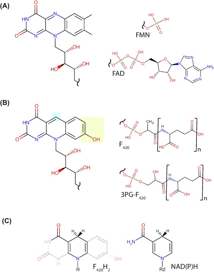
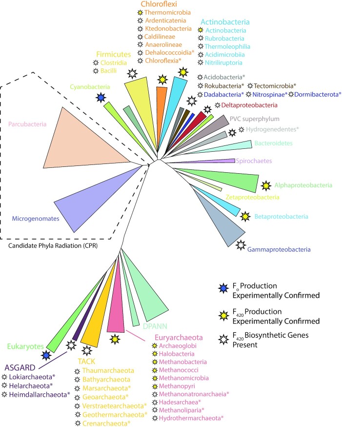
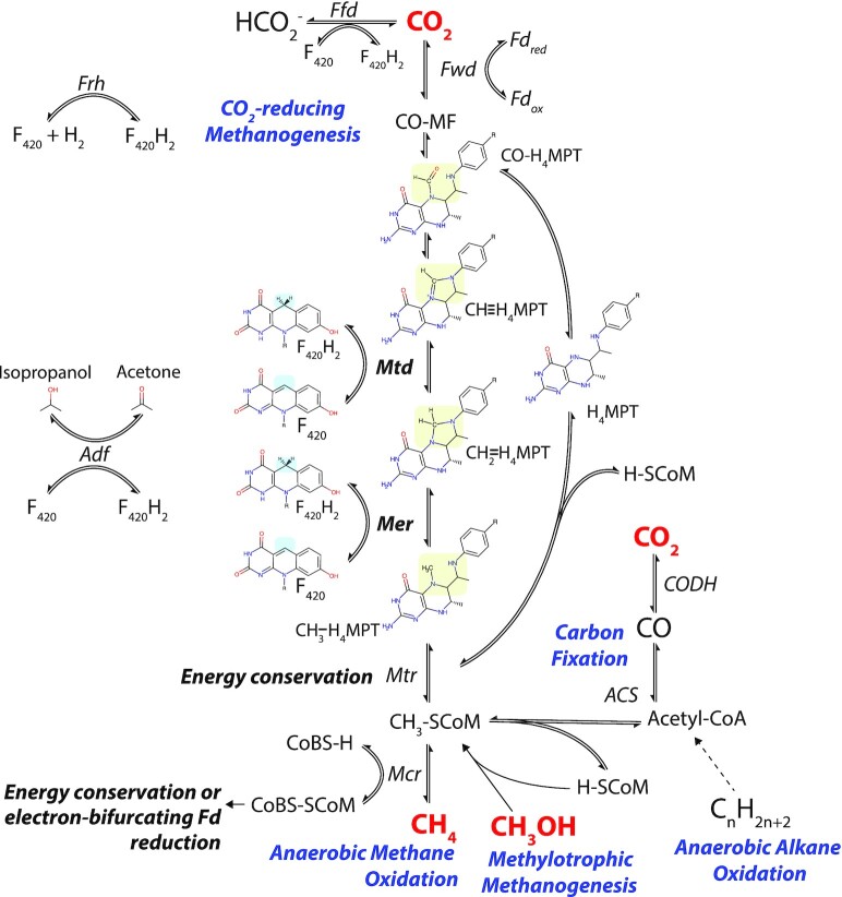
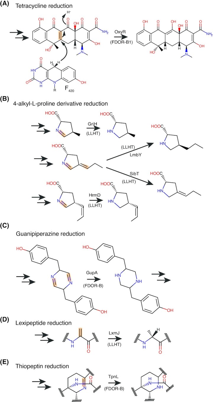
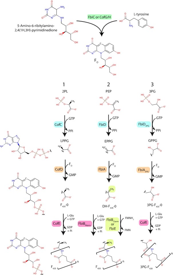
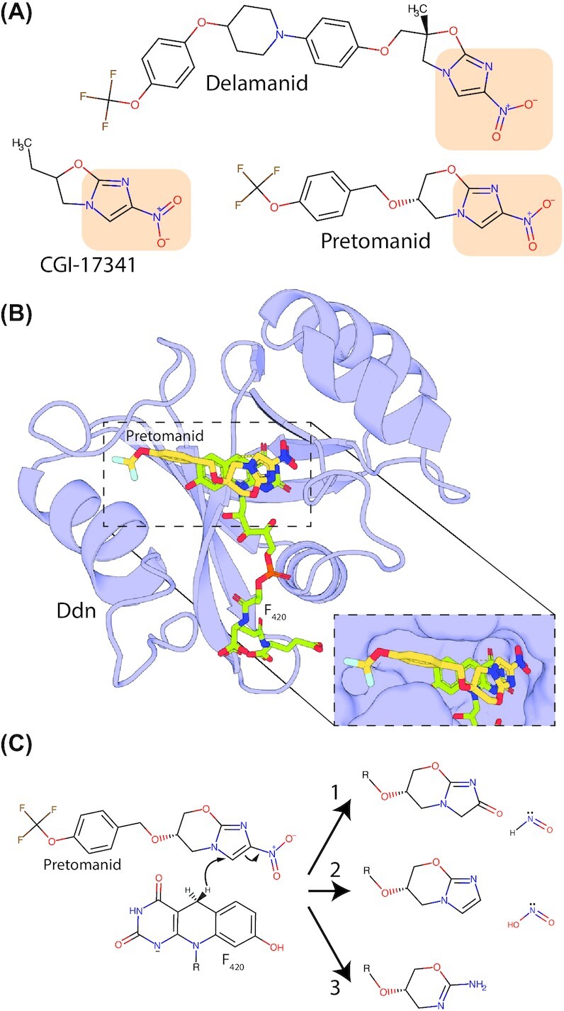
Many bacteria and archaea produce the redox cofactor F420. F420 is structurally similar to the cofactors FAD and FMN but is catalytically more similar to NAD and NADP. These properties allow F420 to catalyze challenging redox reactions, including key steps in methanogenesis, antibiotic biosynthesis and xenobiotic biodegradation. In the last 5 years, there has been much progress in understanding its distribution, biosynthesis, role and applications. Whereas F420 was previously thought to be confined to Actinobacteria and Euryarchaeota, new evidence indicates it is synthesized across the bacterial and archaeal domains, as a result of extensive horizontal and vertical biosynthetic gene transfer. F420 was thought to be synthesized through one biosynthetic pathway; however, recent advances have revealed variants of this pathway and have resolved their key biosynthetic steps. In parallel, new F420-dependent biosynthetic and metabolic processes have been discovered. These advances have enabled the heterologous production of F420 and identified enantioselective F420H2-dependent reductases for biocatalysis. New research has also helped resolve how microorganisms use F420 to influence human and environmental health, providing opportunities for tuberculosis treatment and methane mitigation. A total of 50 years since its discovery, multiple paradigms associated with F420 have shifted, and new F420-dependent organisms and processes continue to be discovered.

Keywords: cofactor 420; cofactor biosynthesis; cofactor distribution; enzymology; redox chemistry; redox cofactor.


Figures

Figure 1.
Figure 1.
Structural comparison of F420 with flavins and nicotinamides. (A) Structures of the riboflavin head group and tail groups of the flavins FMN and FAD. (B) Structures of the 5-deazaflavin head group and tail groups of F420 and 3PG-F420. Locations of chemical substitutions between riboflavin and 5-deazaflavin are highlighted. N = 2–9 depending on the microbial species. (C) Structural similarity between the nicotinamides NAD(P)H and the central redox-active portion of F420H2. For F420H2, R represents the phospholactyl and oligoglutamate tail shown in panel B. For NAD(P)H, R2 represents the ribose-5-phosphate of the nicotinamide nucleotide and the adenosine nucleobase as shown in (Bogan and Brenner 2013).
Figure 2.
Figure 2.
F420 protonaiton states, redox transitions and associated spectral shifts. (A) Changes in the protonation state of F420 and F420H2 as a result of the change in external pH. R = F420 tail group as depicted in Fig. 1B. (B) Spectral changes of F420 between pH 5.5 and 9.0 resulting from a change in protonation state depicted in panel A. Inset graph shows a change in absorbance at 420 nm. (C) Spectral change in F420H2 as in panel B. Inset graph shows changes in absorbance at 280 nm. Panels B and C are adapted from Mohamed et al. (2016a).
Figure 3.
Figure 3.
F 420-dependent enzyme families that reduce or oxidize substrates via direct hydride transfer. Representative structures are shown of families of F420-dependent oxidoreductases in complex with F420 and substrate, inhibitor, or substrate analog. Inhibitors or substrate analogs are indicated with *. The secondary structural elements are highlighted (blue = α-helix, yellow = β-sheet or coil). (A) FDOR-A family F420H2-dependent menaquinone reductase (Ddn) from M. tuberculosis docked with menadione (PDB ID = 3R5R; Cellitti et al. 2012). (B) FDOR-B family enzyme of unknown function MSMEG_6526 from M. smegmatis in complex with 2-methyl-2,4-pentanediol (MPD; PDB ID = 4ZKY; Ahmed et al. 2015). (C) Rossmann-A fold enzyme NADPH:F420 oxidoreductase (Fno) from A. fulgidus in complex with NADPH (PDB ID = 1JAY; Warkentin et al. 2001). (D) LLHT family F420-reducing glucose-6-phosphate dehydrogenase (Fgd) from M. tuberculosis in complex with citrate (PDB ID = 3B4Y; Bashiri et al. 2008). The region of protein capping the active site is rendered transparent for clarity. (E) Rossmann-B fold enzyme F420-dependent CH2=H4MPT dehydrogenase (Mtd) from Methanopyrus kandleri in complex with CH2=H4MPT (PDB ID = 3IQE; Ceh et al. 2009).
Figure 4.
Figure 4.
F420-dependent enzymes that mediate oxidation or reduction indirectly via flavin. Representative structures or models of families F420-dependent oxidoreductases that mediate hydride transfer via a bound flavin cofactor. Structures generated via homology modeling using Phyre2 (Kelley et al. 2015) are indicated with *. The secondary structural elements are highlighted (blue = α-helix, yellow = β-sheet or coil), FMN or FAD colored red and FeS clusters and metal ions are shown as spheres. (A) FDFO family F420H2-dependent flavodiiron oxidase (FprA) from Methanothermobacter thermautotrophicus responsible for the reduction of O2 to H2O (PDB ID = 2OHJ; Seedorf et al. 2007). (B) FDRC domain-containing F420-reducing NiFe hydrogenase (Frh) from Methanothermobacter marburgensis (PDB ID = 4CI0; Allegretti et al. 2014). (C) F420H2-dependent thioredoxin reductase (DFTR) from M. jannaschii (homology model; Susanti, Loganathan and Mukhopadhyay 2016).
Figure 5.
Figure 5.
Phylogenetic distribution of FO and F420 producing organisms. A simplified two-domain tree of life depicted the organisms shown or predicted to produce the 5-deazaflavins FO or F420. This is based on currently available data from published work (Greening et al. ; Ney et al. 2017a), and genomic and metagenomic data in the NCBI database (as of October 2020). Tree topography is based on Hug et. al. (Hug et al. 2016) and Castelle and Banfield (2018), with additional reference to Zhou et al. (2020), Wang et al. (2019) and Momper, Aronson and Amend (2018). * = F420 biosynthesis genes detected only in multiple metagenome-assembled genomes (MAGs) or single-amplified genomes (SAGs) from these archaea and bacteria, rather than genomes derived from pure culture.
Figure 6.
Figure 6.
F420-dependent reactions of one-carbon metabolism in archaea. F420 is a cofactor involved in key steps in hydrogenotrophic methanogenesis, methylotrophic methanogenesis, anaerobic methanotrophy and anaerobic alkane oxidation in archaea. Hydride transfer reactions involving F420-dependent enzymes are indicated as is the enzyme responsible. F420H2 reduced through the oxidation of formate (Ffd), H2 (Frh), or secondary alcohols (Adf) can be utilized for reactions mediated by Mtd, Mer, or for other cellular processes. Only reactions mediated by F420-dependent enzymes are shown in detail. For a full outline of methanogenesis pathways, refer to the following reviews on the subject (Deppenmeier ; Thauer et al. ; Timmers et al. ; Evans et al. 2019).
Figure 7.
Figure 7.
Physiological reactions proposed to be mediated by F420-dependent enzymes in mycobacteria. The bond oxidized or reduced is highlighted in orange for each substrate, with the enzyme responsible for the reaction indicated. For the reactions shown in A, B and D, F420H2 is generated by Fgd through oxidation of G6P. For the reaction shown in C, F420 oxidizes hydroxymycolic acid to ketomycolic acid at the extracellular face of the cytoplasmic membrane, yielding F420H2.
Figure 8.
Figure 8.
Reactions proposed to be mediated by F420-dependent reductases in streptomycetes. The bond reduced is highlighted in orange for each substrate, with the enzyme responsible for the reaction indicated. F420H2 for the reactions shown is generated by the enzyme Fno via the oxidation of NADPH.
Figure 9.
Figure 9.
Diverse routes to F420 biosynthesis employed by bacteria and archaea. 1 = classical archaeal pathway (Euryarchaeota), 2 = bacterial pathway a (Actinobacteria, Chloroflexi), 3 = bacterial pathway b (Betaproteobacteria). The substrates and mechanisms for FO biosynthesis are shared between all identified pathways. Abbreviated compounds are as follows: PEP, phosphoenolpyruvate; 2PL, 2-phospho-L-lactate; 3PG, 3-phosphoglycerate; EPPG, enolpyruvyl-diphospho-5’-guanosine; LPPG, lactyl-diphospho-5’-guanosine; GPPG, 3-guanosine-5’-disphospho-D-glycerate. The enzymes involved in each biosynthesis step are indicated.
Figure 10.
Figure 10.
Genetic organization of F420 biosynthetic genes in bacteria and archaea. A schematic of the generalized the genetic context of F420 biosynthetic genes in experimentally confirmed (panel A) and predicted (panel B) F420-producing bacteria (left) and archaea (right). F420 biosynthetic genes are labeled and color-coded. Additional F420 related genes are colored grey and labeled as follows: Fgd, F420-reducing glucose 6-phosphate dehydrogenase; Fno, F420-reducing NADPH dehydrogenase; FDOR, predicted F420H2-dependent reductase; LLHT, predicted F420H2-dependent luciferase-like hydride transferase; Mer, F420H2-dependent CH2=H4MPT reductase. Hypothetical genes or those with no known F420-related function are shown and colored white. Black tilde symbols designate undefined intergenomic space. Gene context is adapted from Ney et al. (2017a) or determined directly from available genome sequences.
Figure 11.
Figure 11.
Production of the deazaflavin FO is catalyzed by dual SAM-radical domains. (A) Structural model of the SAM radical domains that mediate Fo synthesis, consisting of the two separate proteins CofG and CofH (in archaea and some bacteria) or a single fusion protein FbiC (in bacteria and eukaryotes). Structural models constructed based on homology modeling using Phyre2 based on the structure of MqnE from Pedobacter heparinus (PDB ID = 6XI9; Kelley et al. 2015). (B) A summary of the proposed reaction performed by CofH. (C) A summary of the proposed reaction performed by CofG. For the full reaction scheme summarized in panels B and C refer to Philmus et al. (2015). R = The FO ribose tail as shown in Fig. 9.
Figure 12.
Figure 12.
Crystal structure of FbiD from M. tuberculosis in complex with PEP substrate. (A) Cartoon view of the crystal structure of FbiD in complex with PEP (PDB ID = 6BWH). (B) A surface model of the FbiB active site showing the region predicted to bind GTP adopts an occluded conformation. (C) Key residues of FbiD involved in the coordination of the PEP substrate and catalytic Mg2+ ions. Atomic distances less than 3.2 Å are shown as dashed yellow lines.
Figure 13.
Figure 13.
Crystal structure of the FbiA-substrate complex from M. smegmatis. (A) Crystal structure of the FbiA dimer [PDB ID = 6UW5] in complex with GDP in Mol. A and Fo and EPPG (modeled in place of co-crystallized GDP) in Mol. B. FbiA is shown as a cartoon model, substrate molecules are shown as sticks and a Ca2+ ion (likely Mg2+ in the active enzyme) is shown as a sphere. (B) A cross-eye stereo view of the active site of FbiA Mol. B from panel A, showing key residues for coordinating the FbiA substrate complex as sticks and coordination distances as dotted lines. (C) A proposed reaction mechanism for synthesis DH-F420-0 by FbiA, in which the carboxyl group of EPPG donates an electron to FO, activating it to perform nucleophilic attack on the EPPG β-phosphate.
Figure 14.
Figure 14.
Crystal structure of CofE from A. fulgidus in complex with GDP and Mn2+. (A) Cartoon view of the crystal structure of the functional dimer of CofE [PDB ID = 2PHN]. CofE subunits are shown in pink (Mol. A) and red (Mol. B). Bound GDP and catalytic Mn2+ ions are shown for Mol. B only, as stick and sphere representation respectively. (B) Electrostatic surface view of the CofE active site showing bound GDP, catalytic Mn2+ ions and predicted F420-0 binding pocket. (C) Proposed catalytic mechanism for the first γ-linked glutamate addition mediated by CofE, R = FO minus terminal hydroxyl.
Figure 15.
Figure 15.
Crystal structure of FbiBC-term from M. tuberculosis. (A) Cartoon view of the crystal structure of the functional dimer of the FbiB C-terminal domain responsible for the FMNH2 mediated reduction of DH-F420-0 [PDB IDs = 4XOO, 4XOQ]. FbiBC-term is shown in green and FMN and DH-F420-0 (modeled based on the co-crystal structure of FO) are shown as sticks. (B) A zoomed view of the FbiBC-term active site in complex with FMN and DH-F420-0 as in panel A, with a cartoon and transparent atomic surface of the FbiBC-term shown.
Figure 16.
Figure 16.
Schematic showing possible events in the evolution of F420 and its acquisition by different bacterial and archaeal lineages. Arrows with solid lines indicate potential horizontal transfer of ancestral F420 biosynthetic genes. Dashed lines with bidirectional arrows indicate a likely gene transfer event of unknown directionality. Phyla labels are simplified for clarity. Note the figure is a speculative model drawn based on data from sources discussed in the main text and other models are also consistent with these data.
Figure 17.
Figure 17.
Compounds reduced by purified F420-dependent enzymes. (A) Classes of compounds shown to be reduced by purified F420-dependent enzymes. The reduced bond is highlighted in orange for each compound. The enzymes tested, as well as their substrate range and relative activity levels, are provided in Table 5. (B) Proposed reaction schemes for the reduction of menaquinone and malachite green by F420H2-dependent reductases of the FDOR-A superfamily. In both cases, initial hydride transfer to one carbon atom of an activated alkene is followed by tautomerization yielding the final reaction product.
Figure 18.
Figure 18.
Nitroimidazole prodrugs effective against M. tuberculosis and their activation by Ddn. (A) Structure of nitroimidazole-containing prodrugs developed for tuberculosis treatment. Delamanid and pretomanid were recently approved for the treatment of M. tuberculosis infection, while CGI-17341 was abandoned due to toxicity concerns. The nitroimidazole functional group is highlighted in orange. (B) The complex between pretomanid and its activating enzyme Ddn from M. tuberculosiswas generated by molecular docking using AutoDock Vina (Trott and Olson 2010). The proximity between the nitroimidazole group of pretomanid and the hydride transferring C5 carbon of F420 is shown in the inset panel. (C) Proposed products for the breakdown of pretomanid following reduction by Ddn, full reaction schemes leading to product generation refer to Singh et al. (2008).

Similar articles

Cited by

References

    1. Abken H-J, Deppenmeier U. Purification and properties of an F420H2 dehydrogenase from Methanosarcina mazei Gö1. FEMS Microbiol Lett. 1997;154:231–7.
    1. Adam PS, Borrel G, Gribaldo S. An archaeal origin of the Wood–Ljungdahl H4MPT branch and the emergence of bacterial methylotrophy. Nat Microbiol. 2019;4:2155–63. - PubMed
    1. Afting C, Hochheimer A, Thauer R. Function of H2-forming methylenetetrahydromethanopterin dehydrogenase from Methanobacterium thermoautotrophicum in coenzyme F420 reduction with H2. Arch Microbiol. 1998;169:206–10. - PubMed
    1. Ahmed FH, Carr PD, Lee BMet al. . Sequence–structure–function classification of a catalytically diverse oxidoreductase superfamily in Mycobacteria. J Mol Biol. 2015;427:3554–71. - PubMed
    1. Ahmed FH, Mohamed AE, Carr PDet al. . Rv2074 is a novel F420H2-dependent biliverdin reductase in Mycobacterium tuberculosis. Protein Sci. 2016;25:1692–709. - PMC - PubMed

Publication types

Grants and funding

LinkOut - more resources