. 2021 Apr 14;16(4):e0243333.
doi: 10.1371/journal.pone.0243333. eCollection 2021.

Versatile and flexible microfluidic qPCR test for high-throughput SARS-CoV-2 and cellular response detection in nasopharyngeal swab samples

Affiliations

Versatile and flexible microfluidic qPCR test for high-throughput SARS-CoV-2 and cellular response detection in nasopharyngeal swab samples

Julien Fassy et al. PLoS One. .

Abstract

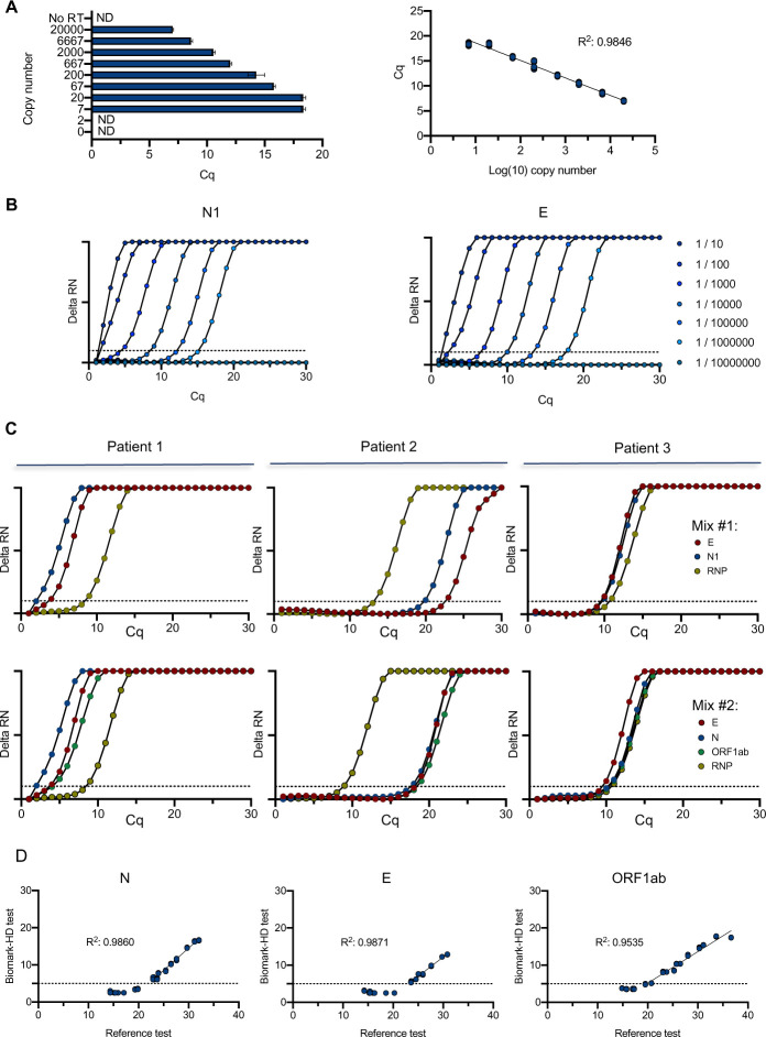
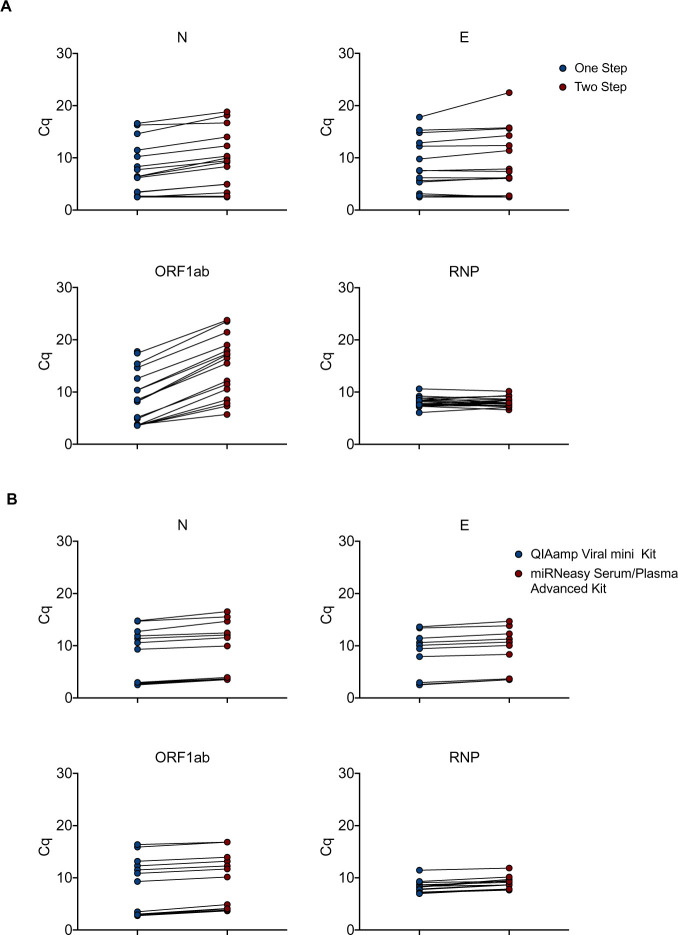
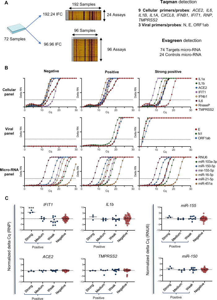
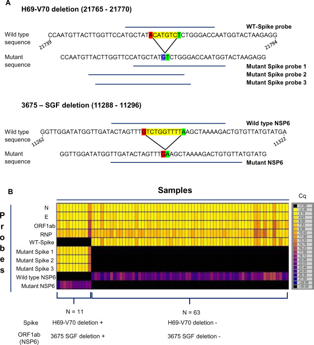
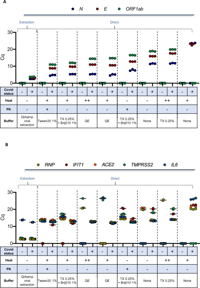
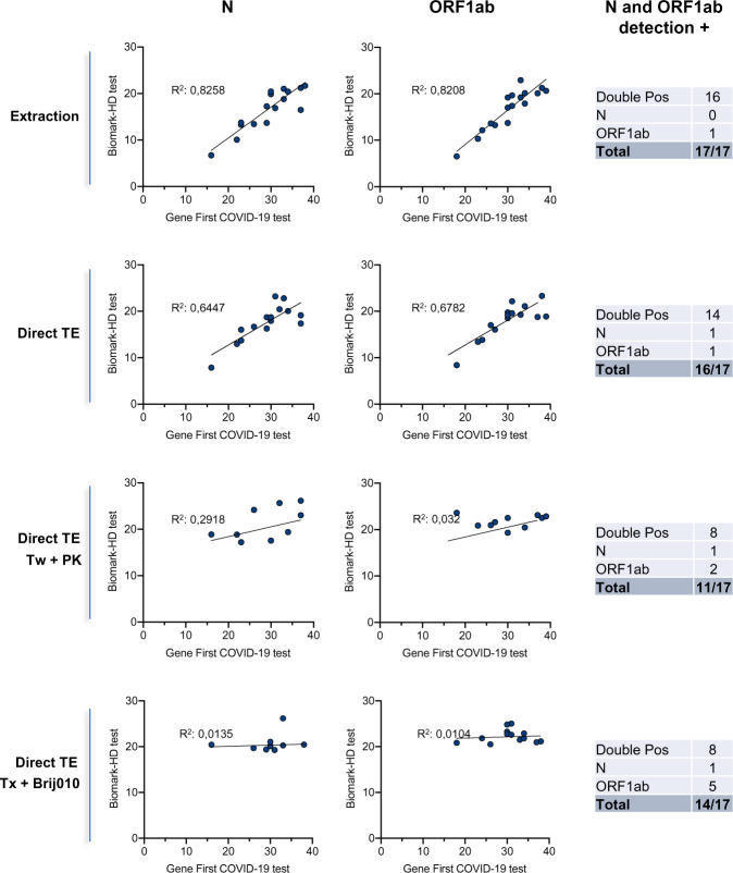
The emergence and quick spread of SARS-CoV-2 has pointed at a low capacity response for testing large populations in many countries, in line of material, technical and staff limitations. The traditional RT-qPCR diagnostic test remains the reference method and is by far the most widely used test. These assays are limited to a few probe sets, require large sample PCR reaction volumes, along with an expensive and time-consuming RNA extraction step. Here we describe a quantitative nanofluidic assay that overcomes some of these shortcomings, based on the BiomarkTM instrument from Fluidigm. This system offers the possibility of performing 4608 qPCR end-points in a single run, equivalent to 192 clinical samples combined with 12 pairs of primers/probe sets in duplicate, thus allowing the monitoring of SARS-CoV-2 including the detection of specific SARS-CoV-2 variants, as well as the detection other pathogens and/or host cellular responses (virus receptors, response markers, microRNAs). The 10 nL-range volume of BiomarkTM reactions is compatible with sensitive and reproducible reactions that can be easily and cost-effectively adapted to various RT-qPCR configurations and sets of primers/probe. Finally, we also evaluated the use of inactivating lysis buffers composed of various detergents in the presence or absence of proteinase K to assess the compatibility of these buffers with a direct reverse transcription enzymatic step and we propose several protocols, bypassing the need for RNA purification. We advocate that the combined utilization of an optimized processing buffer and a high-throughput real-time PCR device would contribute to improve the turn-around-time to deliver the test results to patients and increase the SARS-CoV-2 testing capacities.


Conflict of interest statement

The authors have read the journal’s policy and have the following competing interests: DR is a paid employee of Bayer SAS and VL is employed by LBM Bioesterel (LBM Bioesterel). There are no patents, products in development or marketed products associated with this research to declare. This does not alter our adherence to PLOS ONE policies on sharing data and materials.

Figures

Fig 1
Fig 1. Performance and validation of the Biomark-HD SARS-CoV-2 qPCR assay.
(A) Titration of the diluted (N) nucleocapside spike-in transcript. A serial dilution of the synthetic N transcript from 2.104 to 2 copies was performed and processed through the Biomark-HD protocol. Correlation and amplification curves of detected Cq values according to synthetic N transcript copy number obtained with N1 primers/probe are shown. (B) Amplification curves showing the range of detection of a SARS-CoV-2 positive clinical sample serial dilution with N1 and E primers/probe. (C) Typical amplification curves showing the combination of two primers/probe sets on 3 SARS-CoV-2 positive clinical samples. Mix #1: E, N1, human RNP. Mix #2: E, N, ORF1ab and human RNP. (D) Validation of the Biomark-HD protocol on a cohort of 18 biopsies from positive patients. The correlation of the Cq values obtained for the N, E and the ORF1ab genes are presented. The data presented are representative of at least two independent experiments performed in quadruplicate.
Fig 2
Fig 2. Optimization of the assay.
(A) Total RNA from 18 clinical samples with a wide range of SARS-CoV-2 infection were subjected to either a Two-step reaction (red circles; consecutive Reverse Transcription and Pre-amplification) or One-step reaction (blue circles; combined Reverse Transcription and Pre-amplification). Quantitative PCR reactions were performed on the Biomark-HD using cellular (RNP) and viral (N, E, ORF1ab) primers/probe sets. (B) Total RNA from 18 clinical samples with a wide range of SARS-CoV-2 infection were extracted with either the miRNeasy Advanced Serum Plasma Kit (red circles) or the Virus QIAamp Viral RNA kit (blue circles). RNAs were processed using the One step reaction. Quantitative PCR reactions were performed on the Biomark-HD using cellular (RNP) and viral (N, E, ORF1ab) primers/probe sets. The Cq presented are representative of two independent experiments performed in quadruplicate.
Fig 3
Fig 3. Use of the Biomark-based protocol to analyze the host response to SARS-CoV-2 infection at the mRNA and microRNA levels.
(A) Overview of the analysis strategy on 72 patient samples. They were divided into 4 groups, according to their viral load, from negative, weak (Cq for viral probes >20), medium (20 > Cq for viral probes >10) and strong (Cq for viral probes < 10) SARS-CoV-2 positive. (B) Typical amplification curves of the different genes (cellular, viral and micro-RNA) on three different SARS-CoV-2 patient status. (C) Modulation of cellular markers in the different groups of patients according to their SARS-CoV-2 viral load. IFIT1 expression was statistically elevated (p < 0.001) in the strong COVID-19-positive samples compared to the three other groups considered separately. The Cq presented are representative of two independent experiments performed in quadruplicate.
Fig 4
Fig 4. Use of the Biomark-based protocol to detect mutations associated with SARS-CoV-2 variants.
(A) Overview of the probes design used for the detection of two deletion mutations found in the Spike gene (H69-V70, top panel) and in the NSP6 gene (3675-SGF, bottom panel). Deletions are highlighted in yellow with both nucleotide at the 5’ and 3’ side shown in red and green respectively. Of note: for the H69-V70 mutation an A-to-G punctual deletion is shown in blue. Three mutant probes and one wild type probe were generated for the H69—V70 deletion (21765–21770) and one mutant and one wild type probes were generated for the 3675 –GSF deletion (11288–11296). The nucleotide numbering is based on the reference SARS-CoV-2 complete sequence (NCBI Reference Sequence: NC_045512.2). (B) Heatmap of Cq value from RT-qPCR using various combinations of primer/probes performed on 74 SARS-CoV-2 positive clinical samples. Cq scale range is shown on the right. Using the Biomark-based protocol, 11 positive samples for the mutation associated with the SARS-CoV-2 variants have been detected (on 74 positive clinical samples).
Fig 5
Fig 5. Detection of viral and cellular genes using inactivating lysis buffers-based protocols.
Two clinical samples of known COVID-19 status (one positive and one negative) sampled in a commercial VTM medium, were aliquoted and then treated in parallel using the indicated detergent conditions (selected from S5 Fig) in the presence or absence of PK. RNA was extracted as a control. Biomark-HD RT-qPCR was performed using (A) viral (N, E, ORF1ab) and (B) cellular (ACE2, IFIT1, IL6, TMPRSS and RNP) primers/probe sets. Legend: +: 10 min at 65°C; ++: 10 min at 65°and 5 min at 95°C; TX: Triton X-100; QE: Quick ExtractTM DNA Extraction Solution. The Cq presented are representative of two independent experiments performed in quadruplicate.
Fig 6
Fig 6. Correlation of the detection of two viral genes (N, ORF1ab) obtained using a commercial COVID-19 detection kit and extraction-based or direct Biomark-HD assays.
We used a set of 17 clinical samples from SARS-CoV-2-diagnosed patients collected in saline solution with a wide range of Cq values. To control pH conditions and limit RNA degradation, all samples were diluted 2X in TE buffer. RNA was extracted (as a control) or samples were treated using Tween 20 and PK (Tw + PK) or Triton X-100 and Brij010 detergent solutions (Tx + Brij010). The different RT-qPCR Biomark methods were compared with the GeneFirstTM COVID-19 method as a reference. The correlation between the Cq obtained with the two methods is presented. One negative sample was processed in the three direct conditions (TE, TE plus Tw + PK and TE plus Tx + Brij010) with as results an absence of signal with the two probes (Cq >40). Double Pos: double positive.

References

    1. La Marca A, Capuzzo M, Paglia T, Roli L, Trenti T, Nelson SM. 2020. Testing for SARS-CoV-2 (COVID-19): a systematic review and clinical guide to molecular and serological in-vitro diagnostic assays. Reprod Biomed Online 41:483–499. 10.1016/j.rbmo.2020.06.001 - DOI - PMC - PubMed
    1. Ghaffari A, Meurant R, Ardakani A. 2020. COVID-19 Serological Tests: How Well Do They Actually Perform? Diagnostics 10:453. 10.3390/diagnostics10070453 - DOI - PMC - PubMed
    1. Lieberman JA, Pepper G, Naccache SN, Huang M-L, Jerome KR, Greninger AL. 2020. Comparison of Commercially Available and Laboratory-Developed Assays for In Vitro Detection of SARS-CoV-2 in Clinical Laboratories. J Clin Microbiol 58. 10.1128/JCM.00821-20 - DOI - PMC - PubMed
    1. Pettit SD, Jerome KR, Rouquié D, Mari B, Barbry P, Kanda Y, et al. 2020. ‘All In’: a pragmatic framework for COVID‐19 testing and action on a global scale. EMBO Mol Med 12. 10.15252/emmm.202012634 - DOI - PMC - PubMed
    1. Lohse S, Pfuhl T, Berkó-Göttel B, Rissland J, Geißler T, Gärtner B, et al. 2020. Pooling of samples for testing for SARS-CoV-2 in asymptomatic people. Lancet Infect Dis 20:1231–1232. 10.1016/S1473-3099(20)30362-5 - DOI - PMC - PubMed

Publication types