. 2021 Apr 13;35(2):108998.
doi: 10.1016/j.celrep.2021.108998.

Gasdermin E permits interleukin-1 beta release in distinct sublytic and pyroptotic phases

Affiliations

Gasdermin E permits interleukin-1 beta release in distinct sublytic and pyroptotic phases

Bowen Zhou et al. Cell Rep. .

Abstract

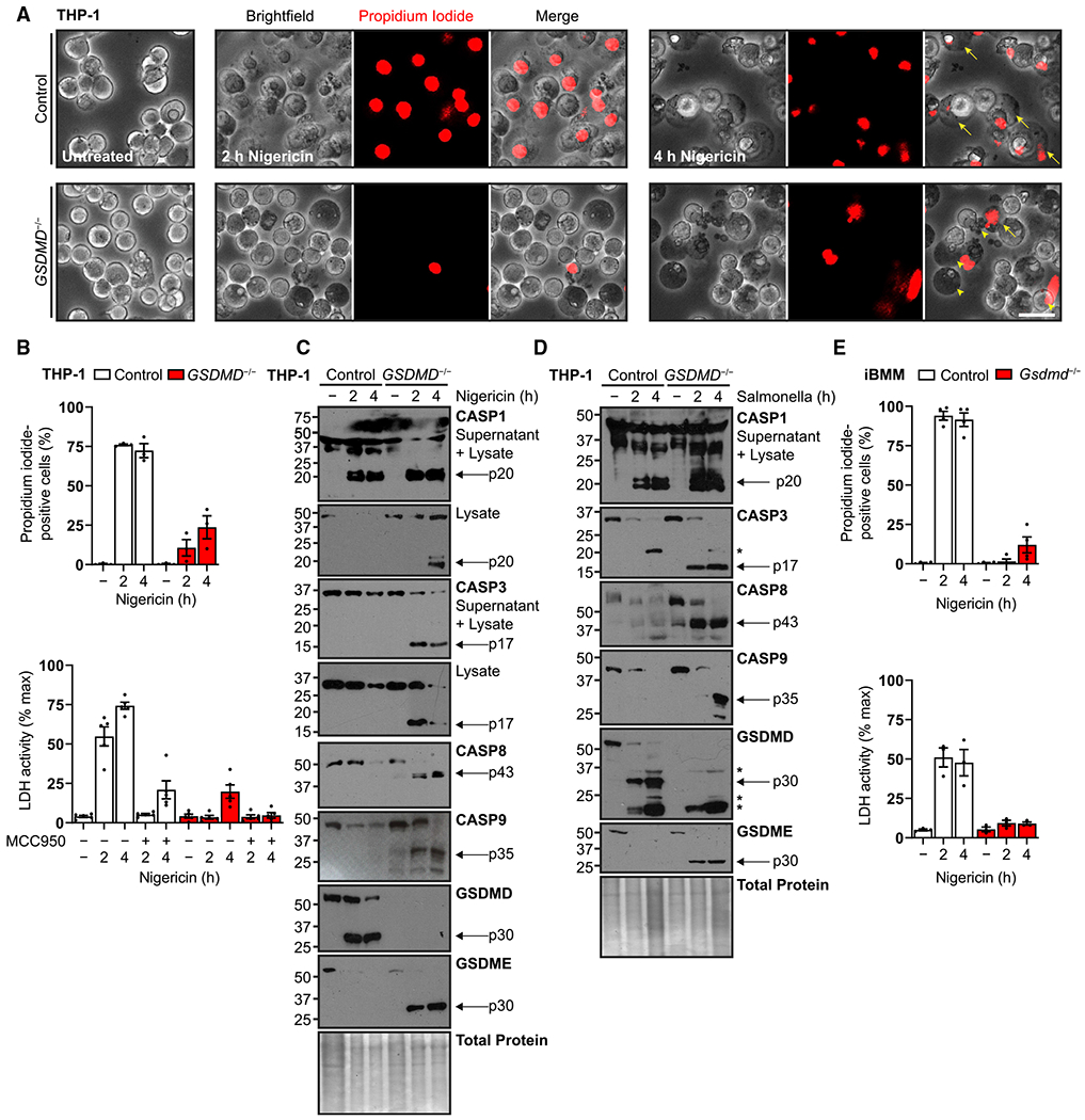
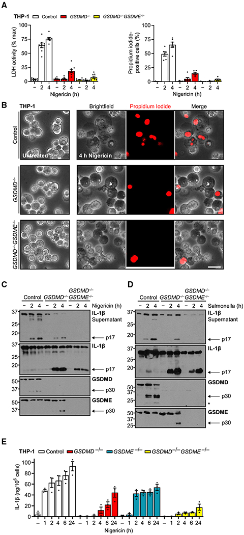
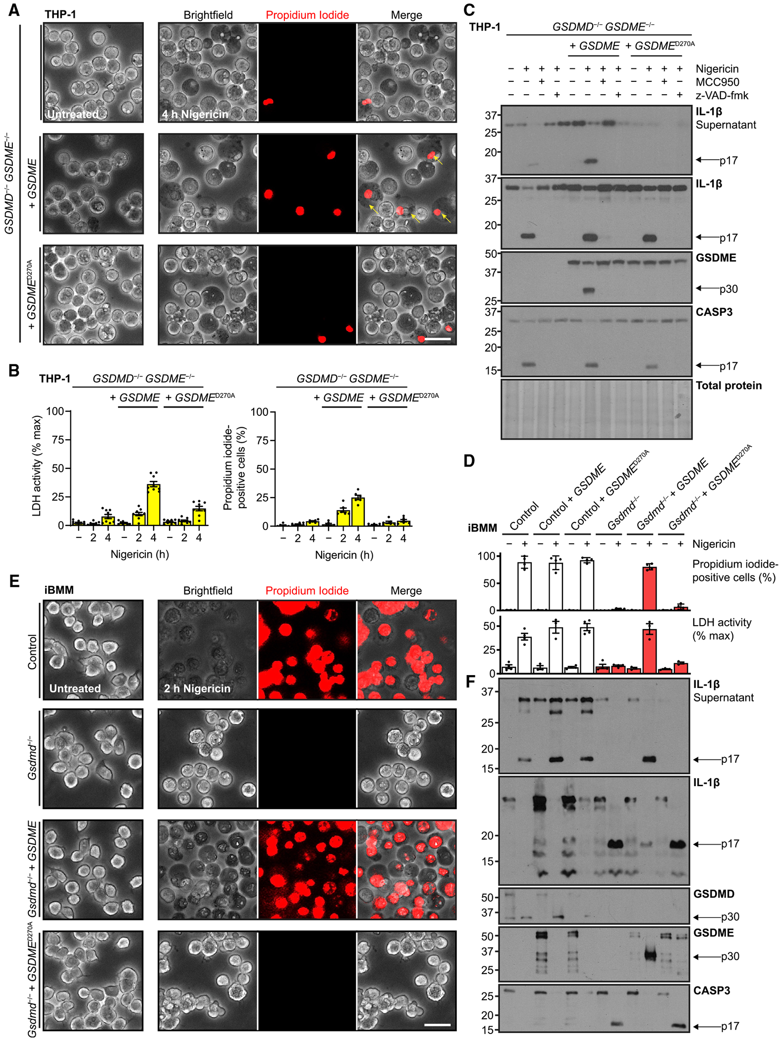
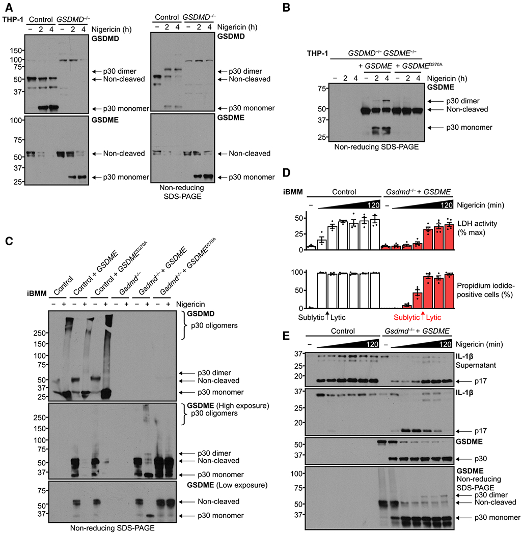
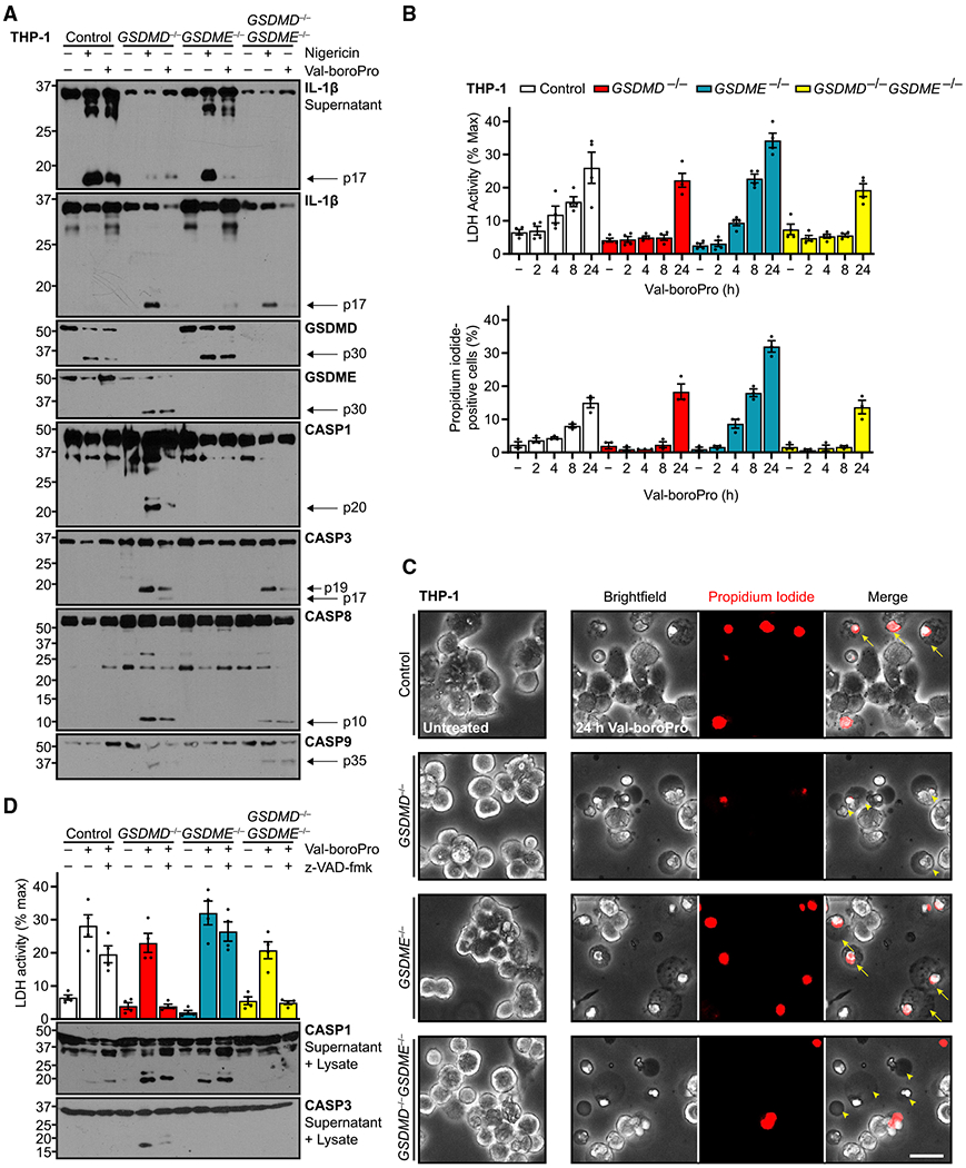
Cellular inflammasome activation causes caspase-1 cleavage of the pore-forming protein gasdermin D (GSDMD) with subsequent pyroptotic cell death and cytokine release. Here, we clarify the ambiguous role of the related family member gasdermin E (GSDME) in this process. Inflammasome stimulation in GSDMD-deficient cells led to apoptotic caspase cleavage of GSDME. Endogenous GSDME activation permitted sublytic, continuous interleukin-1β (IL-1β) release and membrane leakage, even in GSDMD-sufficient cells, whereas ectopic expression led to pyroptosis with GSDME oligomerization and complete liberation of IL-1β akin to GSDMD pyroptosis. We find that NLRP3 and NLRP1 inflammasomes ultimately rely concurrently on both gasdermins for IL-1β processing and release separately from their ability to induce cell lysis. Our study thus identifies GSDME as a conduit for IL-1β release independent of its ability to cause cell death.

Keywords: GSDMD; GSDME; IL-1; cell death; gasdermin D; gasdermin E; gasdermins; inflammasome; interleukin-1; pore-forming proteins.


Conflict of interest statement

Declaration of interests The authors declare no competing interests.

Figures

Figure 1.
Figure 1.. Inflammasome activation triggers apoptotic cascade and GSDME cleavage in GSDMD-deficient macrophages
(A) Representative micrographs of human PMA-differentiated (100 ng/mL, 16 h) wild-type and GSDMD−/− THP-1 cells treated with nigericin (20 μM) and incubated with cell-impermeable propidium iodide dye to visualize membrane permeability upon inflammasome activation. Arrows indicate classically swollen pyroptotic cells that are transparent, speckled, and positive for propidium iodide uptake. Arrowheads indicate atypical opaque, impermeable cells and apoptotic bodies. (B) Quantification of propidium iodide uptake and quantification of frank cell lysis by supernatant lactate dehydrogenase activity assay for similar experiments. For the latter, cells were also pre-treated with NLRP3 inhibitor MCC950 (10 μM, 0.5 h). (C and D) Immunoblots of similar cells treated with nigericin (20 μM) to activate the NLRP3 inflammasome or S. typhimurium (MOI = 10) for the NLRC4 inflammasome. Combined supernatant and lysate were assayed to detect caspase-1 fragments expelled during pyroptosis. (E) Quantification of propidium iodide uptake and quantification of cell lysis for wild-type and Gsdmd−/− murine immortalized bone marrow-derived macrophages (iBMMs) treated with S. typhimurium (MOI = 10) or primed with LPS (0.2 μg/mL, 4 h) before nigericin treatment (20 μM). Scale bar, 50 μm. Graph bars represent mean ± standard error of biological replicates. Graph points represent pooled technical replicates per biological replicate. Immunoblots are representative of at least six independent experiments. Asterisks indicate cross-reactive S. typhimurium protein bands. See also Figure S1.
Figure 2.
Figure 2.. Endogenous GSDME permits IL-1β release with limited secondary pyroptosis
(A and B) PMA-differentiated (100 ng/mL, 16 h) wild-type, GSDMD−/−, and GSDMD−/−GSDME−/− THP-1 cells treated with nigericin (20 μM) assessed for propidium iodide uptake, cell lysis through supernatant lactate dehydrogenase activity assay (A), and cell morphology by microscopy (B). (C and D) Immunoblots of these cells treated with nigericin or S. typhimurium (MOI = 10). (E) Mature IL-1β ELISA for supernatants of wild-type and indicated gasdermin knockout differentiated THP-1 treated with nigericin (20 μM). Immunoblots are representative of at least five independent experiments. Scale bar, 50 μm. Graph bars represent mean ± standard error of biological replicates. Graph points represent pooled technical replicates per biological replicate. See also Figure S2.
Figure 3.
Figure 3.. GSDME expression level determines secondary pyroptosis
(A) Micrographs of GSDMD−/−GSDME−/− THP-1 cells rescued with constitutively expressed wild-type or caspase-immune D270A GSDME, PMA differentiated (100 ng/mL, 16 h), and treated with nigericin (20 μM). (B) For similarly treated cells, quantification of propidium iodide uptake and quantification of cell lysis with supernatant lactate dehydrogenase activity assay. (C) Immunoblots of indicated PMA-differentiated cells pre-treated with NLRP3 inhibitor MCC950 (10 μM, 0.5 h) or pan-caspase and calpain inhibitor z-VAD-fmk (20 μM, 0.5 h) before exposure to nigericin (20 μM). (D–F) Identical analyses of murine control or Gsdmd−/− iBMMs with orthologous expression of human GSDME or caspase-immune variant D270A. Immunoblots are representative of at least four independent experiments. Scale bar, 50 μm. Graph bars represent mean ± standard error of biological replicates. Graph points represent pooled technical replicates per biological replicate.
Figure 4.
Figure 4.. Sublytic GSDME activity transitions to pore-dependent lysis
(A and B) Immunoblots of PMA-differentiated (100 ng/mL, 16h) human THP-1 cells with the indicated genotypes were treated with nigericin (20 μM). Where noted, lysates were processed under non-reducing conditions to assess higher-order self-association of GSDMD or GSDME p30 fragments as a readout for pore formation. (C–E) Murine immortalized bone marrow-derived macrophages were LPS primed (0.2 μg/mL, 4 h) and treated with nigericin (20 μM) for up to 2 h and similarly prepared to assess gasdermin self-association. (D) Quantification of propidium iodide uptake and quantification of frank cell lysis by supernatant lactate dehydrogenase activity assay for similar experiments. Arrows indicate transition from sublytic phase to full-blown cell lysis. Immunoblots are representative of at least three independent experiments. Graph bars represent mean ± standard error of biological replicates. Graph points represent pooled technical replicates per biological replicate.
Figure 5.
Figure 5.. Gasdermins segregate Val-boroPro pyroptosis from IL-1β release
Indicated genotypes of PMA-differentiated (100 ng/mL, 16 h) human THP-1 cells were treated with NLRP1 inflammasome activator Val-boroPro (2 μM). (A) Immunoblots of these cells also were treated with nigericin (20 μM, 3 h) as pyroptosis positive control. (B) Quantification of propidium iodide uptake and cell lysis through supernatant lactate dehydrogenase activity assay. (C) Micrographs of Val-boroPro treatment. Arrows indicate classically pyroptotic cells that are swollen, speckled, transparent, and permeable to propidium iodide. Arrowheads indicate atypical, opaque cells that are impermeable to propidium iodide and opaque. (D) Quantification of cell lysis induced with Val-boroPro with additional pretreatment with calpain and pan-caspase inhibitor z-VAD-fmk (20 μM, 0.5 h). Scale bar, 50 μm. Immunoblots are representative of at least three independent experiments. Graph bars represent mean ± standard error of biological replicates. Graph points represent pooled technical replicates per biological replicate.

Similar articles

Cited by

References

    1. Aglietti RA, Estevez A, Gupta A, Ramirez MG, Liu PS, Kayagaki N, Ciferri C, Dixit VM, and Dueber EC (2016). GsdmD p30 elicited by caspase-11 during pyroptosis forms pores in membranes. Proc. Natl. Acad. Sci. USA 113, 7858–7863. - PMC - PubMed
    1. Aizawa E, Karasawa T, Watanabe S, Komada T, Kimura H, Kamata R, Ito H, Hishida E, Yamada N, Kasahara T, et al. (2020). GSDME-dependent incomplete pyroptosis permits selective IL-1α release under caspase-1 inhibition. iScience 23, 101070. - PMC - PubMed
    1. Boucher D, Monteleone M, Coll RC, Chen KW, Ross CM, Teo JL, Gomez GA, Holley CL, Bierschenk D, Stacey KJ, et al. (2018). Caspase-1 self-cleavage is an intrinsic mechanism to terminate inflammasome activity. J. Exp. Med 215, 827–840. - PMC - PubMed
    1. Broz P, Pelegrín P, and Shao F (2020). The gasdermins, a protein family executing cell death and inflammation. Nat. Rev. Immunol 20, 143–157. - PubMed
    1. Bulek K, Zhao J, Liao Y, Rana N, Corridoni D, Antanaviciute A, Chen X, Wang H, Qian W, Miller-Little WA, et al. (2020). Epithelial-derived gasdermin D mediates nonlytic IL-1β release during experimental colitis. J. Clin. Invest 130, 4218–4234. - PMC - PubMed

Publication types

MeSH terms

LinkOut - more resources