. 2021 May 6;81(9):1905-1919.e12.
doi: 10.1016/j.molcel.2021.02.033. Epub 2021 Apr 13.

Loss of LUC7L2 and U1 snRNP subunits shifts energy metabolism from glycolysis to OXPHOS

Affiliations

Loss of LUC7L2 and U1 snRNP subunits shifts energy metabolism from glycolysis to OXPHOS

Alexis A Jourdain et al. Mol Cell. .

Abstract

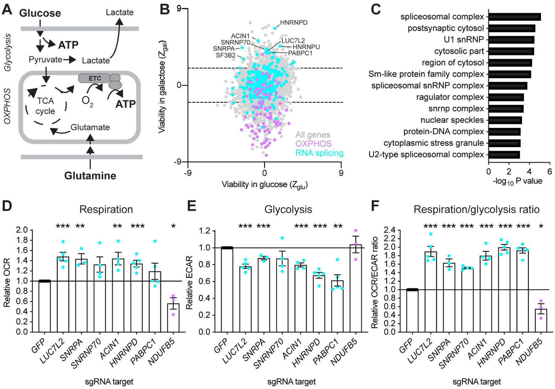
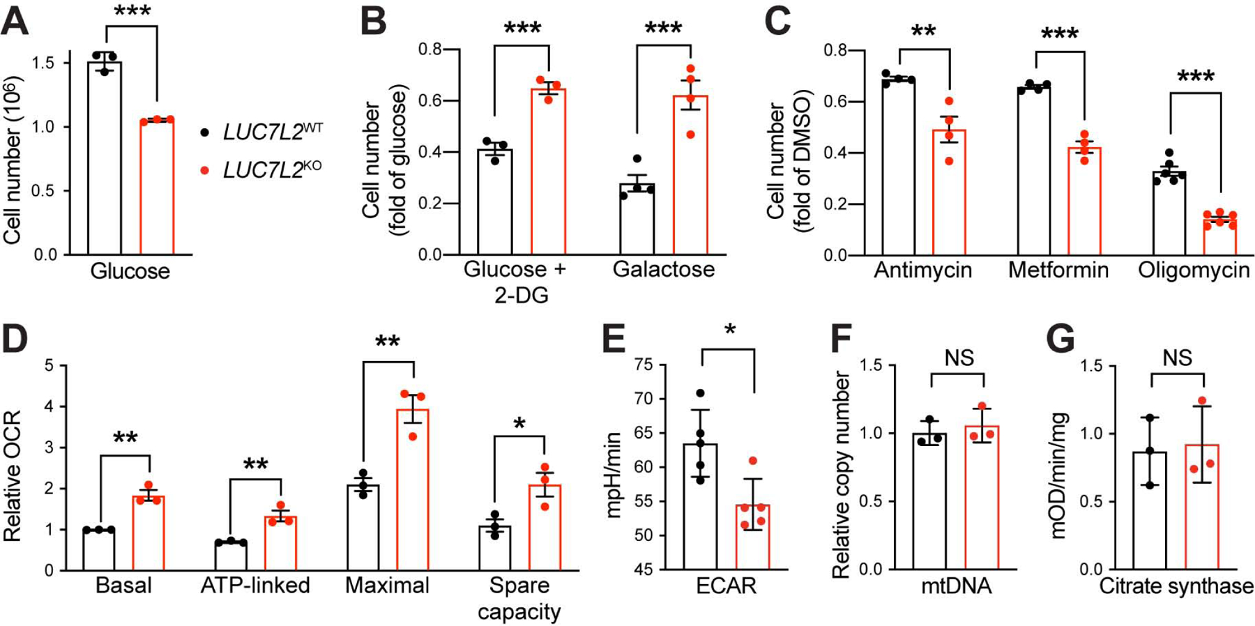
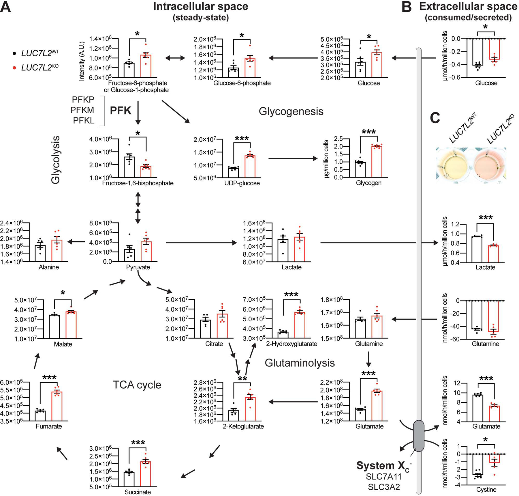
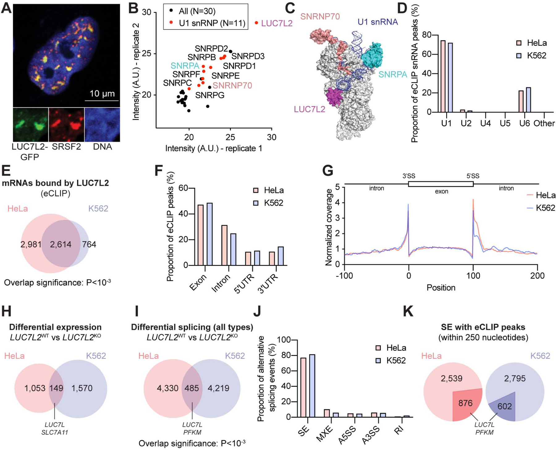
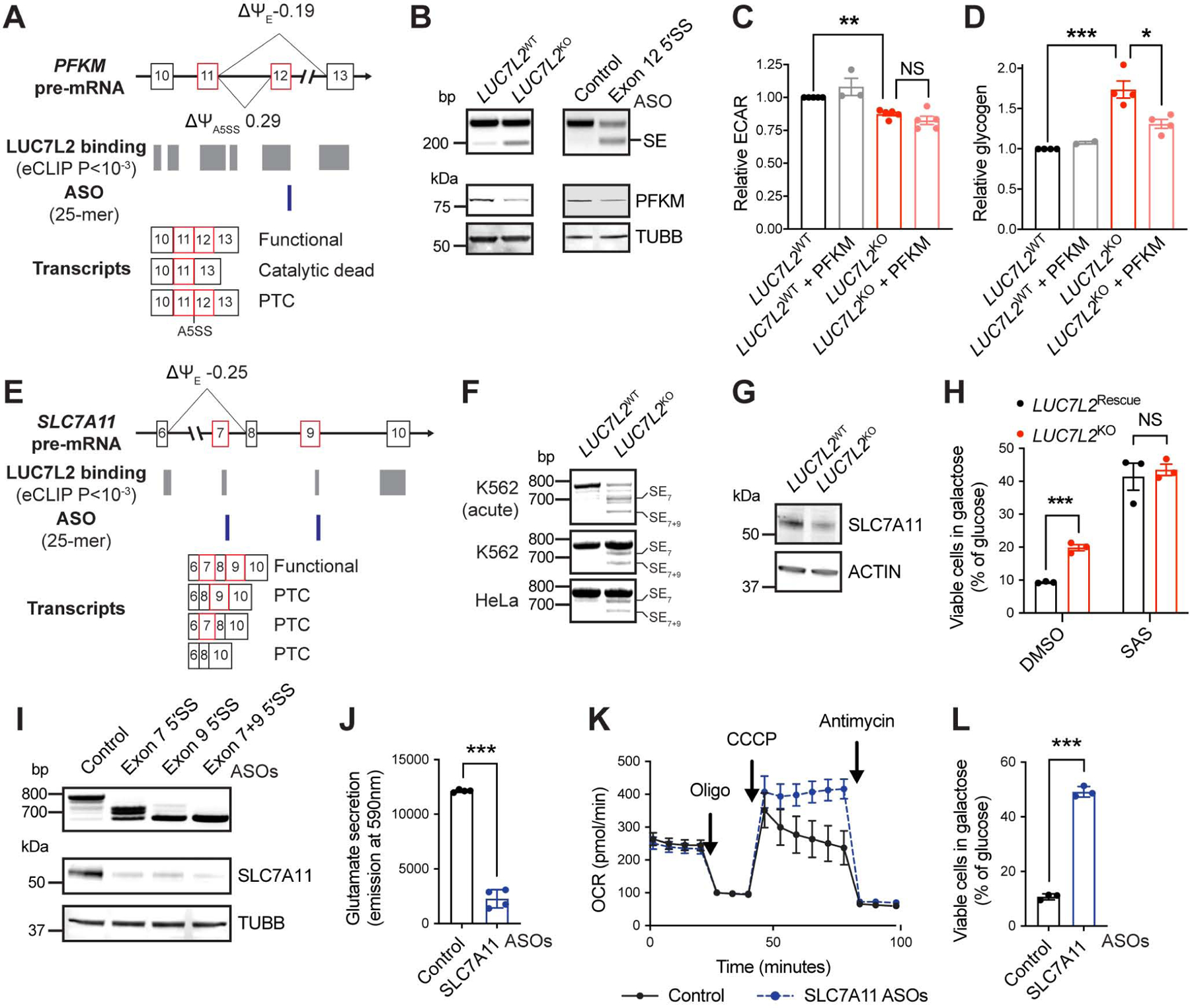
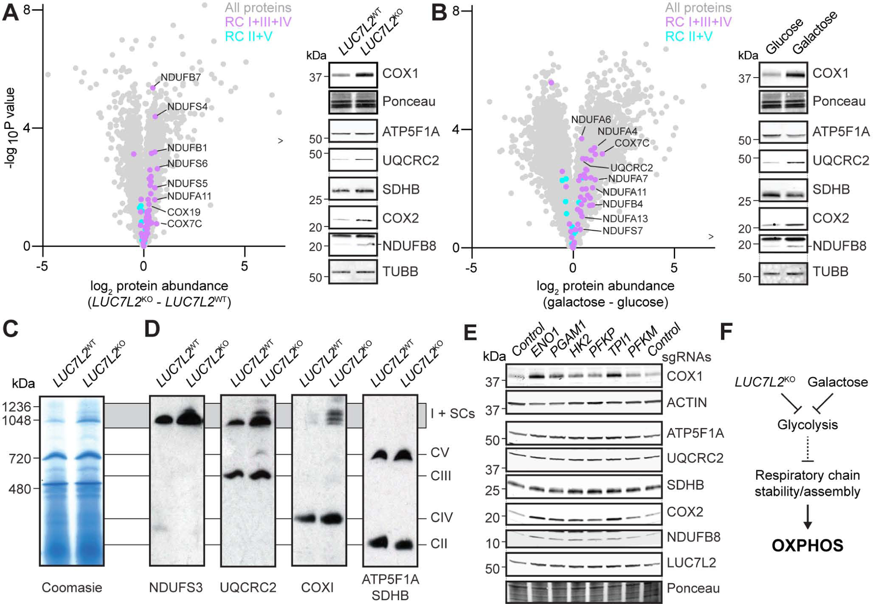
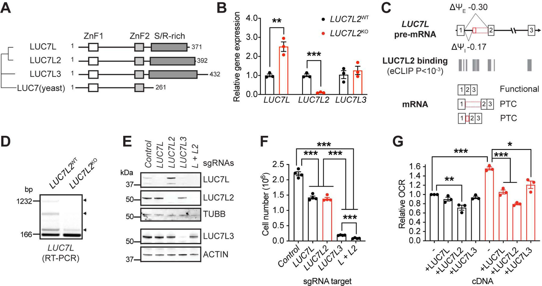
Oxidative phosphorylation (OXPHOS) and glycolysis are the two major pathways for ATP production. The reliance on each varies across tissues and cell states, and can influence susceptibility to disease. At present, the full set of molecular mechanisms governing the relative expression and balance of these two pathways is unknown. Here, we focus on genes whose loss leads to an increase in OXPHOS activity. Unexpectedly, this class of genes is enriched for components of the pre-mRNA splicing machinery, in particular for subunits of the U1 snRNP. Among them, we show that LUC7L2 represses OXPHOS and promotes glycolysis by multiple mechanisms, including (1) splicing of the glycolytic enzyme PFKM to suppress glycogen synthesis, (2) splicing of the cystine/glutamate antiporter SLC7A11 (xCT) to suppress glutamate oxidation, and (3) secondary repression of mitochondrial respiratory supercomplex formation. Our results connect LUC7L2 expression and, more generally, the U1 snRNP to cellular energy metabolism.

Keywords: 7q-; LUC7; MDS; Tarui disease; cancer; ferroptosis; myelodysplastic syndrome; phosphofructokinase; spliceosome; system X(c)(−).


Conflict of interest statement

Declaration of interests V.K.M. is a paid scientific advisor to 5AM Ventures and Janssen Pharmaceuticals. O.S.S. is a paid consultant for Proteinaceous. R.S. holds equity in BlueBird Bio. G.W.Y. is co-founder, member of the Board of Directors, on the scientific advisory board, equity holder, and paid consultant for Locanabio and Eclipse Bioinnovations. G.W.Y. is a visiting professor at the National University of Singapore. G.W.Y.’s interest(s) have been reviewed and approved by the University of California, San Diego in accordance with its conflict of interest policies. A.A.J. and V.K.M. are co-inventors on a US provisional patent application related to the work in this manuscript. The authors declare no other competing interests.

Figures

Figure 1:
Figure 1:. Identification of Pre-mRNA Splicing Components as Repressors of OXPHOS. See also Figure S1 and Table S1.
(A) Overview of the main ATP-generating pathways in human cells. OXPHOS: oxidative phosphorylation. ETC: electron transport chain. TCA: tricarboxylic acid cycle. (B) Gene-level analysis of a genome-wide CRISPR/Cas9 screen in glucose and galactose. Each dot represents an expressed, non-essential gene (n = 9,189). (C) Gene ontology analysis generated using a gene list ranked by viability in galactose against GO components. (D–F) Functional validation of the screening results. Basal whole-cell oxygen consumption rates (OCR), extracellular acidification rates (ECAR) and OCR/ECAR ratios were simultaneously measured after CRISPR/Cas9-mediated gene depletion in K562 cells grown in glucose-containing media. Data are shown as mean ± SEM (n≥3 independent experiments. *P<0.05, **P<0.01, ***P<0.001, t-test relative to control (GFP) sgRNA-treated cells. NDUFB5 is a control with known role in OXPHOS.
Figure 2:
Figure 2:. LUC7L2 Impacts Metabolic State-Dependent Cell Growth and Bioenergetics. See also Figure S2.
(A–C) Cell proliferation of LUC7L2KO K562 cells grown in (A) glucose and (B) treated with 2-deoxyglucose (2-DG) or when glucose was replaced by galactose or (C) glucose with OXPHOS inhibitors. (D) Respiratory parameters of LUC7L2KO cells as determined by oxygen consumption rate (OCR). (E) Basal glycolytic activity in LUC7L2KO cells as determined by extracellular acidification rate (ECAR). (F) Relative mtDNA abundance and (G) citrate synthase activity of LUC7L2KO cells. All data are shown as mean ±SEM (n≥3). *P< 0.05, **P<0.01, ***P<0.001, t-test relative to LUC7L2WT cells.
Figure 3:
Figure 3:. Metabolite Analysis in LUC7L2-Depleted Cells Reveals Crossovers at Phosphofructokinase and System Xc. See also Figure S3 and Table S2–3.
(A) Intracellular levels of metabolites in LUC7L2KO K562 cells as determined by LC-MS. (B) Extracellular levels of metabolites as determined by LC-MS analysis of the spent media from (A). Positive and negative values illustrate metabolite secretion and consumption by the cells, respectively. All data are shown as mean ±SEM (n = 5–8). *P<0.05, **P<0.01, ***P<0.001, t-test relative to LUC7L2WT. (C) Media acidification of LUC7L2KO K562 cells grown in glucose.
Figure 4:
Figure 4:. LUC7L2 Encodes a U1 snRNP Subunit Involved in Pre-mRNA Splicing. See also Figure S4 and Table S4–7.
(A) Confocal microscopy of a single nucleus from a HeLa cell expressing LUC7L2-GFP and immunolabeled with antibodies to SRSF2. (B) LUC7L2-interacting proteins as determined by IP-MS (n = 2). (C) Representation of LUC7, SNRPA and SNRNP70 on the yeast U1 snRNP (PDB 5UZ5) (Li et al., 2017). (D) Proportion of eCLIP peaks mapping to splicing snRNAs in HeLa and K562 cells (eCLIP n = 2, each). (E) Representation of the genes bound by LUC7L2 at P<10−4. (F) Proportion of LUC7L2 eCLIP peaks in pre-mRNAs at P<10−4. (G) Meta-analysis of LUC7L2 binding sites across shared eCLIP peaks at P<10−4. (H) Differential gene expression in LUC7L2KO cells (n = 3 for each cell type and each genotype) as determined by RNA deep-sequencing at FDR<10−4 and >|1.5| fold change. (I) Alternative splicing events seen in LUC7L2KO cells as determined by rMATS at FDR<0.1 and |Δψ|>0.05 (n = 3 for each cell type and each genotype). (J) Types of alternative splicing in LUC7L2KO cells with SE: skipped exon; MXE: mutually-exclusive exons; A5SS: alternative 5′ splice site; A3SS: alternative 3′ splice site; RI: retained intron. (K) Alternative events presenting an eCLIP peak at a 250-nucleotide distance from splicing events at P<10−2 (in darker shade).
Figure 5:
Figure 5:. Role of LUC7L2-Mediated PFKM and SLC7A11 Alternative Splicing in Energy Metabolism. See also Figure S5.
(A) Representation of PFKM exons 10–13, LUC7L2 binding sites as determined by eCLIP, antisense oligonucleotides (ASO) targeting sites and the expected transcripts. Ψ: percent spliced in reported by rMATS in K562 cells. E: exon. A negative Δψ value indicates exon skipping. PTC: premature termination codons. (B) RT-PCR (top) and immunoblot (bottom) of LUC7L2KO K562 cells (left) or HAP1 cells treated for 48h with ASO targeting the 5′SS of PFKM exon 12 (right). (C) Relative ECAR (n = 3–5) and (D) glycogen in LUC7L2KO K562 cells expressing control cDNAs (GFP) or PFKM cDNA (n = 2–4). (E) Representation of SLC7A11 exons 6–10 as in (A). (F) RT-PCR of LUC7L2KO K562 cells with primers amplifying transcripts corresponding to SLC7A11 exons 6–12. SE: skipped exon. (G) Immunoblot on LUC7L2KO K562 cells with antibodies to SLC7A11 and ACTIN. (H) Cell viability of LUC7L2Rescue (corresponds to LUC72L2KO expressing LUC7L2 cDNA) and LUC7L2KO HAP1 cells grown for 24h in galactose relative to glucose (n = 3). SAS: 500μM sulfasalazine. (I) RT-PCR (top) and immunoblot (bottom) of HAP1 cells treated for 48h with ASOs targeting the 5′SS of exon 7 and/or exon 9 of SLC7A11. (J) Media glutamate (n = 4), (K) representative seahorse trace (shown as mean ±SD), and (L) viability in galactose of HAP1 cells treated for 48h with the indicated ASOs (n = 3). All data are shown as mean ±SEM (unless otherwise stated) with * P<0.05, ** P<0.01, ***P<0.001, t-test relative to control.
Figure 6:
Figure 6:. Proteomic Analysis of LUC7L2KO and Galactose-Grown Cells Reveals Secondary Complexes I+III+IV Accumulation. See also Figure S6 and Table S8.
(A) Volcano plots and immunoblots of OXPHOS protein expression in LUC7L2KO and (B) in galactose-grown K562 cells. > indicates a protein not shown but reported in Table S8. (C) Blue-Native PAGE on a mitochondria-rich fraction isolated from LUC7L2KO K562 cells and stained with coomassie or (D) immunoblotted with the indicated antibodies. Parallel blots in which the same lysate was loaded were used to avoid antibodies cross-reactivity. SCs: supercomplexes. CI-V: complexes I to V. (E) Immunoblot on K562 cells expressing Cas9 and treated with sgRNAs targeting glycolytic enzymes with the indicated antibodies. (F) Model of the secondary regulation of the respiratory chain by LUC7L2 and galactose.
Figure 7:
Figure 7:. Pre-mRNA Splicing and Partial Redundancy Within the LUC7 Family. See also Figure S7.
(A) Phylogenetic tree of the LUC7 protein family. LUC7 is from S. cerevisiae. ZnF: Zinc-finger domain. S/R-rich: Serine and arginine-rich domain. (B) Quantitative PCR detecting LUC7 family transcripts in LUC7L2KO K562 cells (n = 3). (C) Representation of LUC7L exons 1–3, LUC7L2 binding sites as determined by eCLIP, and the expected transcripts. Ψ: percent spliced in reported by rMATS in K562 cells. I: intron. E: exon. (D) RT-PCR amplifying LUC7L exon 1 to exon 2. Arrowheads: retained entities in LUC7L. (E) Immunoblot of LUC7 proteins in cell lines expressing Cas9 and sgRNAs targeting the indicated genes using the indicated antibodies and (F) number of cells after 4 days of growth in glucose-containing media. (G) Oxygen consumption analysis of LUC7L2KO K562 cells expressing cDNAs of LUC7 family members. All data are shown as mean ±SEM with * P<0.05, ** P<0.01, ***P<0.001, t-test relative to control.

Comment in

Similar articles

Cited by

References

    1. ACIN-PEREZ R, BAYONA-BAFALUY MP, FERNANDEZ-SILVA P, MORENOLOSHUERTOS R, PEREZ-MARTOS A, BRUNO C, MORAES CT & ENRIQUEZ JA 2004. Respiratory complex III is required to maintain complex I in mammalian mitochondria. Mol Cell, 13, 805–15. - PMC - PubMed
    1. ARROYO JD, JOURDAIN AA, CALVO SE, BALLARANO CA, DOENCH JG, ROOT DE & MOOTHA VK 2016. A Genome-wide CRISPR Death Screen Identifies Genes Essential for Oxidative Phosphorylation. Cell Metab, 24, 875–885. - PMC - PubMed
    1. BADGLEY MA, KREMER DM, MAURER HC, DELGIORNO KE, LEE HJ, PUROHIT V, SAGALOVSKIY IR, MA A, KAPILIAN J, FIRL CEM, DECKER AR, SASTRA SA, PALERMO CF, ANDRADE LR, SAJJAKULNUKIT P, ZHANG L, TOLSTYKA ZP, HIRSCHHORN T, LAMB C, LIU T, GU W, SEELEY ES, STONE E, GEORGIOU G, MANOR U, IUGA A, WAHL GM, STOCKWELL BR, LYSSIOTIS CA & OLIVE KP 2020. Cysteine depletion induces pancreatic tumor ferroptosis in mice. Science, 368, 85–89. - PMC - PubMed
    1. BAO XR, ONG SE, GOLDBERGER O, PENG J, SHARMA R, THOMPSON DA, VAFAI SB, COX AG, MARUTANI E, ICHINOSE F, GOESSLING W, REGEV A, CARR SA, CLISH CB & MOOTHA VK 2016. Mitochondrial dysfunction remodels one-carbon metabolism in human cells. Elife, 5. - PMC - PubMed
    1. BERG MG, SINGH LN, YOUNIS I, LIU Q, PINTO AM, KAIDA D, ZHANG Z, CHO S, SHERRILL-MIX S, WAN L & DREYFUSS G 2012. U1 snRNP determines mRNA length and regulates isoform expression. Cell, 150, 53–64. - PMC - PubMed

Publication types

MeSH terms