. 2021 May 13;184(10):2715-2732.e23.
doi: 10.1016/j.cell.2021.03.032. Epub 2021 Apr 13.

Reducing acetylated tau is neuroprotective in brain injury

Affiliations

Reducing acetylated tau is neuroprotective in brain injury

Min-Kyoo Shin et al. Cell. .

Abstract

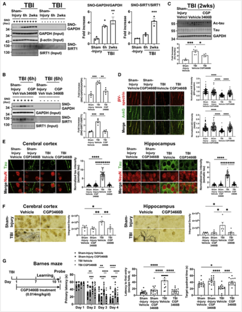
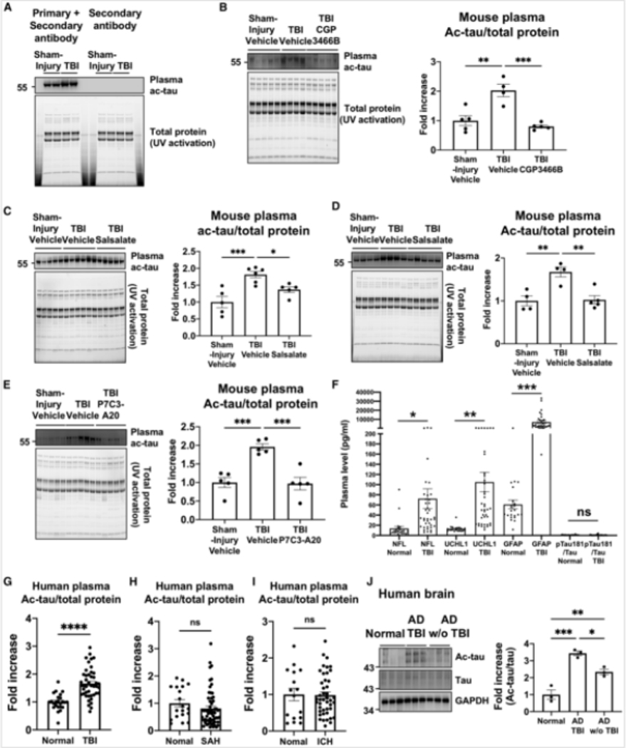
Traumatic brain injury (TBI) is the largest non-genetic, non-aging related risk factor for Alzheimer's disease (AD). We report here that TBI induces tau acetylation (ac-tau) at sites acetylated also in human AD brain. This is mediated by S-nitrosylated-GAPDH, which simultaneously inactivates Sirtuin1 deacetylase and activates p300/CBP acetyltransferase, increasing neuronal ac-tau. Subsequent tau mislocalization causes neurodegeneration and neurobehavioral impairment, and ac-tau accumulates in the blood. Blocking GAPDH S-nitrosylation, inhibiting p300/CBP, or stimulating Sirtuin1 all protect mice from neurodegeneration, neurobehavioral impairment, and blood and brain accumulation of ac-tau after TBI. Ac-tau is thus a therapeutic target and potential blood biomarker of TBI that may represent pathologic convergence between TBI and AD. Increased ac-tau in human AD brain is further augmented in AD patients with history of TBI, and patients receiving the p300/CBP inhibitors salsalate or diflunisal exhibit decreased incidence of AD and clinically diagnosed TBI.

Keywords: Alzheimer’s disease; P7C3; acetylation; congenital muscular dystrophy; diflunisal; neurodegeneration; neuroprotection; omigapil; salsalate; tau; traumatic brain injury.


Conflict of interest statement

Declaration of interests A.A.P. is an inventor on patents related to P7C3. L.G. is a founder of Aeton Therapeutics. No other authors declare competing interests.

Figures

Figure 1
Figure 1. Neuronal tau acetylation after TBI induces axon initial segment degradation and pathologic tau mislocalization
(A) Quantified western blot shows increased ac-tau 6 h-2 weeks after TBI in cerebral cortex and hippocampus. Each group n = 3, **p <0.01, ***p <0.001, ****p < 0.0001 versus sham-injury group, one-way ANOVA and Dunnett multiple comparisons test. (B) Quantified western blot shows increased ac-tau in neurons (NeuN+, GFAP), but not glia (NeuN, GFAP+), of the cerebral cortex, with greater tau expression in neurons. Each lane consists of pooled brain tissue from 3 animals, **p <0.01 versus sham-injury group, Student’s t test. (C) Quantified western blot shows TBI intensity-dependent increase in ac-tau. Each group n = 3–4, **p < 0.01, ****p < 0.0001 versus sham-injury group, one-way ANOVA and Dunnett multiple comparisons test. PSI, pounds per square inch of explosive pressure. (D) Quantified western blot shows reduced AIS proteins AnkG and βIV-spectrin after TBI, consistent with AIS degradation. Each group n = 3, *p < 0.05, **P < 0.01, *** p < 0.001 versus sham-injury group, one-way ANOVA and Dunnett multiple comparisons test. (E) Immunohistochemical staining for tau and the neuronal marker NeuN shows normal axonal localization of tau 2 weeks after sham-injury, and pathological tau mislocalization into the somatodendritic companment 2 weeks after TBI (scale bar, 5 μm). Images are representative of 3 animals per group. (F) Blast-injury induces increased cleaved caspase-3 levels and LDH release in TauKQ (mimicking acetylated lysine) overexpressing cells compared to TauWT (Wild type) and TauKR (nonacetylatable tau mutant) transfected cells (***P < 0.001, ****p < 0.0001 versus TauWT, TauKR transfected cells, one-way ANOVA; and Tukey’s post hoc analysis). (G) TauKQhigh mice have axonal degeneration in cerebral cortex, hippocampus, and hypothalamus, which is absent from nontransgenic littermates (*p < 0.05, Student’s t test). See also Figures S1 and S2.
Figure 2
Figure 2. SNO-GAPDH mediates the post-TBI p300/CBP acetyltransferase activation and Sirtl deacetylase inhibition that leads to accumulated ac-tau, AIS degradation, tau mislocalization, neurodegeneration, and cognitive deficits
(A) Western blot and its quantification show significantly increased S-nitrosylation (SNO) of GAPDH and Sirtl in cerebral cortex after TBI (n = 3 per group, *p < 0.05, **p < 0.01, ***p < 0.001 versus sham-injury group, one-way ANOVA; and Dunnett multiple comparisons test). “Ascorbate (Asc) - negative control” shows specificity of signal in the SNO-resin-assisted capture technique. (B) Western blot and its quantification show that treatment of CGP3466B inhibits S-nitrosylation of GAPDH and Sirtl at 0.014 mg/kg. Each group n = 4, **p < 0.01, ***p < 0.001 versus TBI+Vehicle group, one-way ANOVA and Tukey’s post hoc analysis. Asc + represents SNO, and Asc - represents control. (C) Western blot and its quantification show that 0.014 mg/kg CGP3466B reduces ac-tau in cerebral cortex after TBI. Each group n = 4–7,*p < 0.05, ***p < 0.001 versus TBI+vehicle group, one-way ANOVA; and Tukey’s post hoc analysis. (D) CGP3466B protects mice from post-TBI AIS degradation in the cerebral cortex_(scale bar, 5 4μm). (E) CGP3466B protects mice from post-TBI tau mislocalization (scale bar, 5 μm). Lower magnification pictures of the field from which these pictures were derived are shown in Figure S8C. (F) CGP3466B protects mice from post-TBI axonal degeneration, as evidenced by silver staining of degenerating axons (scale bar, 5 μm). In (D)-(F), each group n = 3–5, *p < 0.05, **P < 0.01, ****P < 0.0001 versus TBI+vehicIe group, one-way ANOVA; and Tukey’s post hoc analysis. (G) CGP3466B protects mice from post-TBI impaired cognition in both leaming and memory phases of the Bames maze task (*p < 0.05, **P < 0.01, ***P < 0.001, ****p < 0.0001 versus TBI-+vehicle group, repeated-measures two-way ANOVA (learning) and one-way ANOVA (memory) and Tukey ‘s post hoc analysis. See also Figures S3 and S4.
Figure 3
Figure 3. Low-dose salsalate-mediated inhibition of p300/CBP acetyltransferase protects mice from post-TBI-induced elevated ac-tau, AIS degradation, tau mislocalization, neurodegeneration, and cognitive deficits
(A) Low-dose salsalate dose-dependently reduces post-TBI elevations in ac-tau in the brain (n = 3, *p < 0.05, **p < 0.01, ***p <0.001 versus TBI-+Vehicle group, one-way ANOVA; and Tukey’s post hoc analysis). (B) Low-dose salsalate protects mice from post-TBI AIS degradation (scale bar, 5 μm). (C) Low-dose salsalate protects mice from post-TBI tau mislocalization (scale bar, 5 μm). (D) Low-dose salsalate protects mice from post-TBI axonal degeneration (scale bar, 5 μm). In (BHD), each group n = 3, *p < 0.05, **p < 0.01, ***p <0.001, ****p < 0.0001 versus TBI+Vehicle group, one-way ANOVA and Tukey’s post hoc analysis. (E) Low-dose salsalate protects mice from post-TBI impairments in motor (foot slip assay) and cognitive (learning and memory in the Bames maze) behavioral assays. (*p < 0.05, **p < 0.01, ***p<0.001, ****p < 0.0001 versus TBI+vehicle group, repeated-measures two-way ANOVA (learning) and one-way ANOVA (memory) and Tukey’s post hoc analysis. See also Figures S5 and S6.
Figure 4
Figure 4. WldS mice are protected from post-TBI-induced elevated ac-tau, AIS degradation, and tau mislocalization
(A) WldS mice are resistant to post-TBI elevations in ac-tau in the brain (for each group n =4, *p < 0.05, **p < 0.01 versus WT+TBI group, one-way ANOVA; and Tukey’s post hoc analysis). (B) WldS mice are resistant to post-TBI AIS degradation (scale bar, 5 μm). (C) WldS mice are resistant to post-TBI tau mislocalization (scale bar, 5 μm). In (B)-(C), each group n = 3, ****p < 0.0001 versus WT+TBI group, one-way ANOVA; and Tukey’s post hoc analysis. See also Figure S5.
Figure 5
Figure 5. P7C3-A20 treatment protects mice from post-TBI-induced elevated ac-tau, AIS degradation, and tau mislocalization
(A) treatment rescued normal NAD+ levels after TBI, which was blocked by co-administration ofFK866 (each group n = 3, **p < 0.01, ***p < 0.001 one-way ANOVA and Tukey’s post hoc analysis). (B) P7C3-A20 treatment protects mice from post-TBI elevations in ac-tau in the brain (each group n = 3–4, *p < 0.05, **p< 0.01, ***p <0.001 ****p <0.0001, one-way ANOVA; and Tukey’s post hoc analysis). This protective effect is blocked by treatment with the NAMPT inhibitor FK866 or the Silti Inhibitor EX527. (C) P7C3-A20 treatment protects mice from post-TBI AIS degradation (n = 3 per group, ****p < 0.0001, one-way ANOVA; and Tukey’s post hoc analysis). This protective effect is blocked by treatment with the NAMPT inhibitor FK866 or the Sirtl Inhibitor EX527. (D) P7C3-A20 treatment protects mice from post-TBI tau mislocalization (each group n = 3, ****p < 0.0001, one-way ANOVA, and Tukey’s post hoc analysis, scale bar, 5 μm). This protective effect is blocked by treatment with the NAMPT inhibitor FK866 or the Sirtl Inhibitor EX527. (E) SNO-GAPDH is not affected by P7C3-A20 (western blot, each group n = 3, *p < 0.05, **p < 0.01, one-way ANOVA and Tukey’s post hoc analysis). “Ascorbate (Asc) - negative control” shows specificity of signal in the SNO-resin-assisted capture technique. See also Figure S5.
Figure 6
Figure 6. Elevated blood plasma ac-tau is a blood biomarker of TBI in mice and humans
(A) Western blot shows that TBI increases plasma ac-tau. Because tau and immunoglobulin G (IgG) have closely similar molecular weights, secondary antibody alone served as a control to ensure there was no cross reactivity with any residual IgG after IgG depletion. (B) Western blot and its quantification show that CGP3466B significantly reduced plasma ac-tau levels after TBI. Each group n = 4–5 with each lane representing a separated animal, **p < 0.01, ***p < 0.001 versus TBI+vehicle group, one-way ANOVA; and Tukey’s post hoc analysis. (C) Western blot and its quantification show that salsalate reduces plasma ac-tau levels. Each group n = 54 with each lane representing a separated animal, *p < 0.05, ***< 0.001 versus TBI+vehicle group, one-way ANOVA; and Tukey’s post hoc analysis. (D) A repeat experiment in an independent cohort of animals confirmed the results shown in (C). Each lane represents a separate animal. For both (C) and (D), **p <0.01, TBI+vehicle group, one-way ANOVA; and Tukey’s post hoc analysis. (E) Western blot and its quantification show that P7C3-A20 significantly reduced plasma ac-tau levels after TBL Each group n = 5 with each lane representing a separated animal, ***p < 0.001 versus TBI+Nehicle group, one-way ANOVA; and Tukey’s post hoc analysis. (F) Plasma Nil, UCHLI, and GFAP, but not pTauI 81/Tau, levels are higher in TBI cohorts at 24 h after injury, compared to controls (*p <0.05, **P <0.01,***P <0.001). (G) The mean level of ac-tau was significantly higher in the TBI cohort at 24 h in comparison to the controls (1.8 ± 0.58 versus 1.16 ± 0.5, ****p <0.0001). (H) The mean level of ac-tau in the subarachnoid hemorrhage (SAH) cohort at 24 h was no different from controls. (I) The mean level of ac-tau in the intracranial hemorrhage (ICH) cohort at 24 h was no different from controls. (J) Western blot and its quantification show that AD patients with TBI history have higher ac-tau levels than AD patients without TBI exposure (*p <0.05, **p <0.01, ***p < 0.001, one-way ANOVA; and Tukey’s post hoc analysis). See also Figures S6 and S7 and Tables S1 and S2.
Figure 7
Figure 7. Diflunisal usage is associated with decreased incidence of TBI and AD in people and with inhibition of ac-tau after TBI in mice
(A) Longitudinal analyses reveal that salsalate and diflunisal usages reduce risk of traumatic brain injury (TBI) in all patient data from the IBM MarketScan Medicare Supplemental Database. The un-stratified Kaplan-Meier curves, conducted propensity score stratified (n strata = 10) log-rank test and Cox model, and hazard ratio and 95% confidence interval for two cohort studies, were illustrated for both (A) and (B). Two cohott studies were conducted including: (I) salsalate users and aspirin users, and (2) diflunisal users and aspirin users. Using propensity score stratified survival analyses by adjusting the initiation time of drugs, enrollment history, age and gender, and disease comorbidities (diabetes, or hypertension, or coronary anery disease). Propensity score stratified Cox-proportional hazards models were used to conduct statistical inference for the hazard ratios. (B) Longitudinal analyses reveal that salsalate and diflunisal usage in the same group as (A) is also associated with reduced incidence of AD in people. (C) Subgroup analyses after excluding patients with type 2 diabetes, hypertension, or coronary artety disease (known risk factors for AD) further confirms that salsalate or diflunisal usage is associated with decreased incidence of AD. (D) LC-MS/MS analysis shows modest penetration of diflunisal into mouse brain but the plasma:brain ratio is decreased when protein binding is taken into account and free drug levels are compared. Drug levels in plasma and brain were determined by LC-MS\MS analysis after mice were administered three different concentrations of diflunisal and euthanized 60 or 180 min later, followed by collection of blood and perfusion with saline, prior to harvesting brain tissue. Rapid equilibrium dialysis was used to determine binding of diflunisal in mouse plasma and brain homogenate. “P” and “B” denote plasma and brain, respectively. (E) Diflunisal treatment dose-dependently reduces post-TBI elevations in ac-tau in the brain. Each group n = 4–5, ***P < 0.001, ****P < 0.0001 versus TBI+vehicIe group, one-way ANOVA; and Tukey’s post hoc analysis. (F) Western blot and its quantification show that diflunisal dose-dependently reduced plasma ac-tau levels after TBI. Each group n = 4–5 With each lane representing a separate animal, *P< 0.05, ***P <0.001 ***p < 0.0001 versus TBI+NehicIe group, one-way ANOVA; and Tukey’s post hoc analysis. (G) There is a significant correlation between brain and plasma ac-tau levels after brain injury (data from C and D; R = 0.857, p < 0.0001). See also Table S3 and S4

Comment in

Similar articles

Cited by

References

    1. Allen RS, Motz CT, Feola A, Chesler KC, Haider R, Ramachandra Rao S, Skelton LA, Fliesler SJ, Pardue MT, 2018. Long-term functional and structural consequences of primary blast overpressure to the eye. J. Neurotrauma 35, 2104–2116. - PMC - PubMed
    1. Arun P, Abu-Taleb R, Oguntayo S, Tanaka M, Wang Y, Valiyaveettil M, Long JB, Zhang Y, Nambiar MP, 2013. Distinct patterns of expression of traumatic brain injury biomarkers after blast exposure: role of compromised cell membrane integrity. Neurosci. Lett. 552, 87–91. - PubMed
    1. Bolós M, Llorens-Martín M, Perea JR, Jurado-Arjona J, Rábano A, Hernández F, Avila J, 2017. Absence ofCX3CR1 impairs the intemalization of Tau by microglia. Mol. Neurodegener. 12, 59. - PMC - PubMed
    1. Cao G, Xing J, Xiao X, Liou AKF, Gao Y, Yin X-M, Clark RSB, Graham SH, Chen J, 2007. Critical role of calpain I in mitochondrial release of apoptosis-inducing factor in ischemic neuronal injury. J. Neurosci. 27, 9278–9293. - PMC - PubMed
    1. Centers for Disease Control and Prevention, 2015. Report to Congress on Traumatic Brain Injury in the United States: Epidemiology and Rehabilitation. National Center for Injury Prevention and Control: Division of Unintentional Injury Prevention.

Publication types

MeSH terms