

The study of screw placement parameters for Ogawa type I acromial fractures by 3D simulation
- PMID: 33853640
- PMCID: PMC8045168
- DOI: 10.1186/s13018-021-02416-3
The study of screw placement parameters for Ogawa type I acromial fractures by 3D simulation
Abstract
Background: Acromial fractures are rare and there is no consensus on fixation, but an increasing number of studies have reported using two screws to fix Ogawa type I acromial fractures. The objective of this study was to obtain the ideal length, diameter, insertion point, and angle of the screw using a novel 3D simulation.
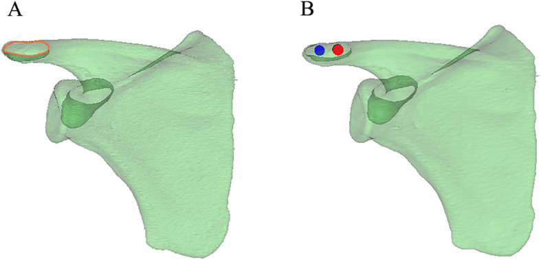
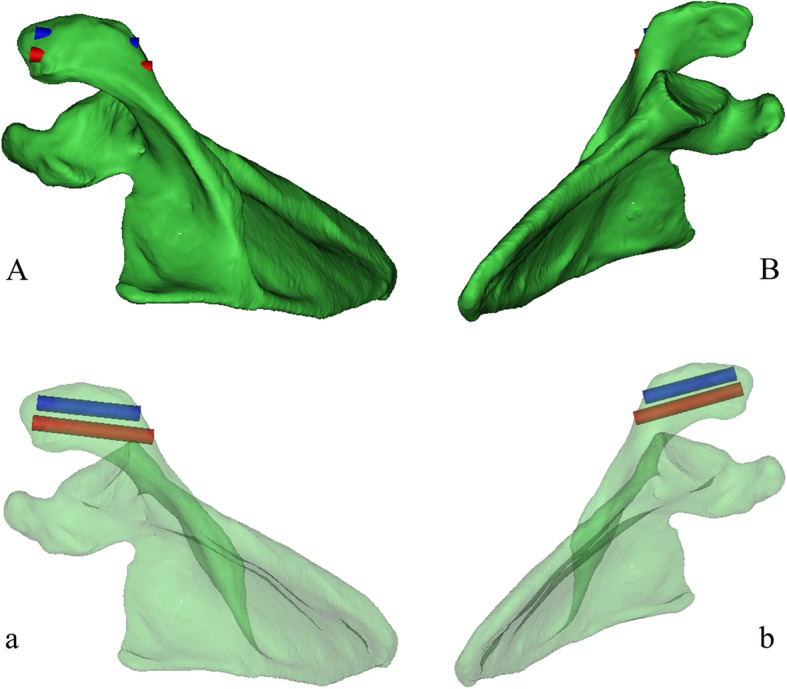
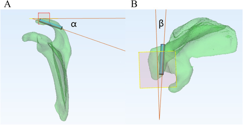
Methods: The scapular CT data of 100 individuals were obtained to reconstruct 3D models. The transparency of the 3D model was then downgraded along the axial perspective (the view perpendicular to the cross section of the acromion axis) to find the maximum translucent area. Two virtual screws were placed at the anterior edge of the acromion until they penetrated the posterior cortical bon. The largest diameters and lengths of the screw were measured, and the direction and insertion point of the screw were observed.
Results: The mean maximum lengths of the medial and lateral screws were 43.33 ± 6.17 mm and 39.23 ± 6.01 mm, respectively. The mean maximum diameters of the medial and lateral screws were 4.71 ± 1.23 mm and 4.97 ± 1.07 mm, respectively. Differences in screw length, diameter, and insertion point between males and females were found. The differences in screw angle between sexes were not statistically significant.
Conclusions: Based on a 3D model test, we recommend the size, entry points, and angles of screws for Ogawa type I acromial fractures, providing valuable guidance for clinical work. More accurate screw parameters can be obtained preoperatively by establishing an individualized 3D model.
Keywords: 3D technology; Acromion; Axial perspective; Screw fixation.
Conflict of interest statement
The authors declare that they have no conflict of interest.
Figures


Similar articles
-
A guideline for screw fixation of coracoid process base fracture by 3D simulation.J Orthop Surg Res. 2021 Jan 14;16(1):58. doi: 10.1186/s13018-021-02203-0. J Orthop Surg Res. 2021. PMID: 33446228 Free PMC article.
-
Axial perspective to find the largest intraosseous space available for percutaneous screw fixation of fractures of the acetabular anterior column.Int J Comput Assist Radiol Surg. 2015 Aug;10(8):1347-53. doi: 10.1007/s11548-015-1149-6. Epub 2015 Jan 9. Int J Comput Assist Radiol Surg. 2015. PMID: 25572704
-
Definition of a safe zone for antegrade lag screw fixation of fracture of posterior column of the acetabulum by 3D technology.Injury. 2016 Mar;47(3):702-6. doi: 10.1016/j.injury.2016.01.026. Epub 2016 Feb 1. Injury. 2016. PMID: 26867979
-
2D versus 3D fluoroscopy-based navigation in posterior pelvic fixation: review of the literature on current technology.Int J Comput Assist Radiol Surg. 2017 Jan;12(1):69-76. doi: 10.1007/s11548-016-1465-5. Epub 2016 Aug 8. Int J Comput Assist Radiol Surg. 2017. PMID: 27503119 Review.
-
Open reduction internal fixation after displacement of a previously nondisplaced acromial fracture in a multiply injured patient: case report and review of literature.J Orthop Trauma. 2001 Jun-Jul;15(5):369-73. doi: 10.1097/00005131-200106000-00013. J Orthop Trauma. 2001. PMID: 11433145 Review.
Cited by
-
Ipsilateral fractures of the acromion and coracoid processes of the scapula.Chin J Traumatol. 2024 Mar;27(2):121-124. doi: 10.1016/j.cjtee.2023.04.003. Epub 2023 Apr 11. Chin J Traumatol. 2024. PMID: 37210253 Free PMC article.
References
-
- Goss TP. The scapula: coracoid, acromial, and avulsion fractures. Am J Orthop (Belle Mead NJ) 1996;25:106–115. - PubMed
MeSH terms
LinkOut - more resources
Full Text Sources
Other Literature Sources
Medical
