

Accuracy of CT for measuring femoral neck anteversion in children with developmental dislocation of the hip verified using 3D printing technology
- PMID: 33853657
- PMCID: PMC8045201
- DOI: 10.1186/s13018-021-02400-x
Accuracy of CT for measuring femoral neck anteversion in children with developmental dislocation of the hip verified using 3D printing technology
Abstract
Background: Accurate femoral neck anteversion angle (FNA) measurement is of great significance in the diagnosis and treatment of developmental dysplasia of the hip (DDH) in children. The FNA measurement still remains a controversy. We aimed to verify the accuracy of our CT method by 3D printing technology and to evaluate its clinical value.
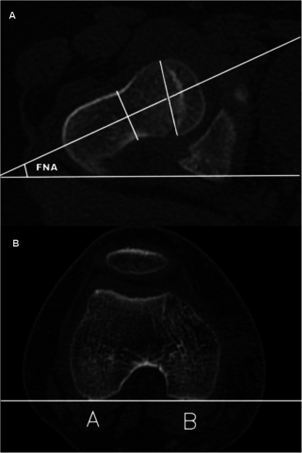
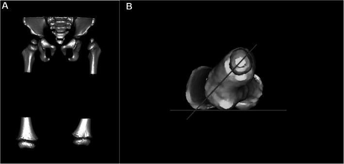
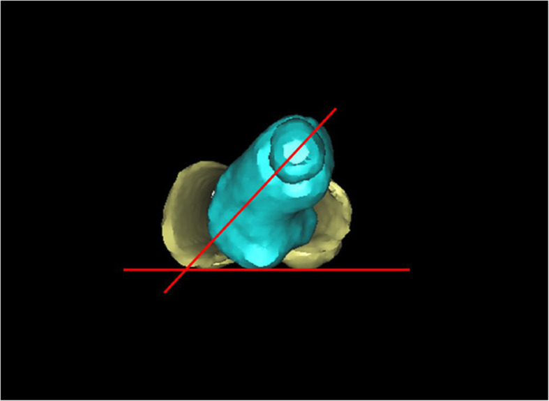
Methods: Sixty-eight children with unilateral DDH were enrolled, and their FNA was measured using 2D-CT and 3D-CT, respectively, by three observers. This procedure was repeated 3 months later. The above measurement outcomes were then compared with the results in the 3D-printed femur (3D-PF) model. The FNA in the 3D-PF model was measured by three observers (two radiologists and one orthopedist; all were professors) collectively through electronic angle instrument.
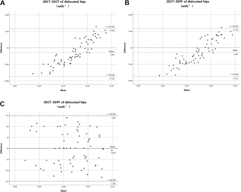
Results: The primary measurement of FNA at the affected hips by 2D-CT was 44.0 ± 6.1, 49.5 ± 8.9, and 52.8 ± 7.9°, respectively. On the 3D-CT, it was 47.6 ± 5.4, 49.3 ± 6.8, and 48.6 ± 6.2°. Three months later, the FNA on 2D-CT was 49.3 ± 10.5, 42.8 ± 7.4, and 45.1 ± 9.3°, and it was 48.0 ± 6.5, 48.9 ± 7.2, and 49.0 ± 5.7° on 3D-CT, respectively. The FNA in the 3D-PF model at the affected and unaffected hips was 48.5 ± 6.6 and 36.9 ± 13.1°. There were significant differences between 2D-CT and 3D-PF measurements, but no significant difference was found between 3D-CT and 3D-PF measurements. The results by 2D-CT showed significant differences among groups and between the groups. However, the results by 3D-CT had no significant differences among groups or between the groups.
Conclusion: The results of our study showed that 3D-CT is a more precise, and reproducible method for FNA measurement in DDH. The FNA at the affected hips is 11.6° larger than the unaffected in DDH children aged 3-8 years.
Keywords: 3D printing technology; Anteversion angle; CT measurement; Femoral neck; Hip joint.
Conflict of interest statement
The authors declare that they have no conflicts of interest.
Figures
References
-
- Klein C, Fontanarosa A, Khouri N, Bellity J, Padovani JP, Glorion C, Wicart P. Anterior and lateral overcoverage after triple pelvic osteotomy in childhood for developmental dislocation of the hip with acetabular dysplasia: Frequency, features, and medium-term clinical impact [J] Orthop Traumatol Surg Res. 2018;104(3):383–387. doi: 10.1016/j.otsr.2017.12.020. - DOI - PubMed
-
- Billing L. Roentgen examination of the proximal femur end in children and adolescents; a standardized technique also suitable for determination of the collum-, anteversion-, and epiphyseal angles; a study of slipped epiphysis and coxa plana [J] Acta Radiol Suppl. 1954;110:1–80. - PubMed
MeSH terms
LinkOut - more resources
Full Text Sources
Other Literature Sources
Medical
Research Materials
