. 2021 Apr 14;6(2):e00250-21.
doi: 10.1128/mSphere.00250-21.

Term Human Placental Trophoblasts Express SARS-CoV-2 Entry Factors ACE2, TMPRSS2, and Furin

Affiliations

Term Human Placental Trophoblasts Express SARS-CoV-2 Entry Factors ACE2, TMPRSS2, and Furin

Yingshi Ouyang et al. mSphere. .

Abstract

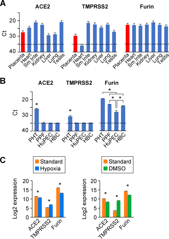
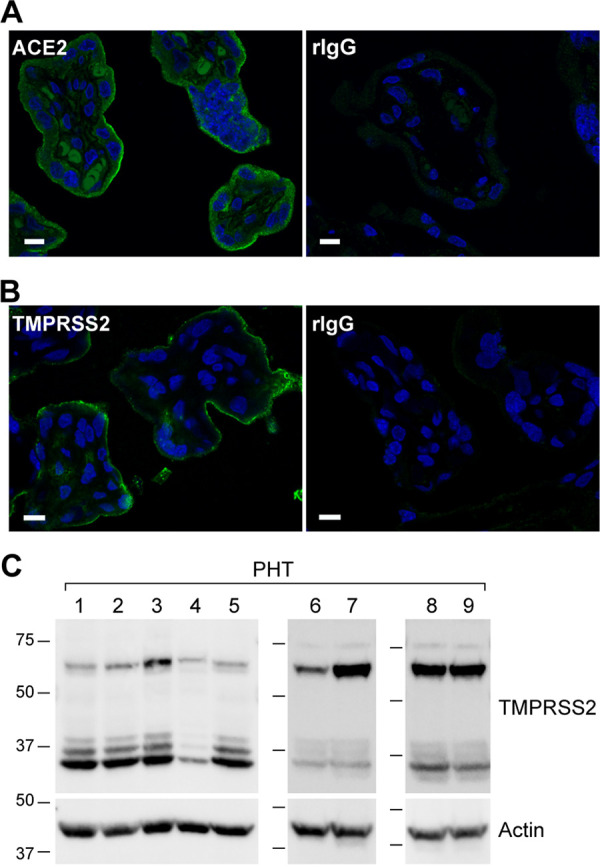
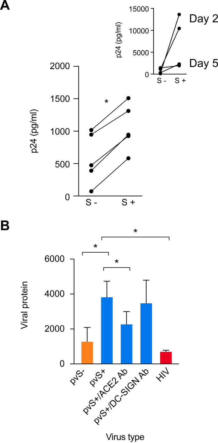
The coronavirus disease 2019 (COVID-19) pandemic, caused by severe acute respiratory syndrome coronavirus 2 (SARS-CoV-2), has had a massive impact on human lives worldwide. While the airborne SARS-CoV-2 primarily affects the lungs, viremia is not uncommon. As placental trophoblasts are directly bathed in maternal blood, they are vulnerable to SARS-CoV-2. Intriguingly, the human fetus is largely spared from SARS-CoV-2 infection. We tested whether the human placenta expresses the main SARS-CoV-2 entry factors angiotensin-converting enzyme 2 (ACE2), transmembrane protease serine 2 (TMPRSS2), and furin and showed that ACE2 and TMPRSS2 are expressed in the trophoblast rather than in other placental villous cells. While furin is expressed in the main placental villous cell types, we surveyed, trophoblasts exhibit the highest expression. In line with the expression of these entry factors, we demonstrated that a SARS-CoV-2 pseudovirus could enter primary human trophoblasts. Mechanisms underlying placental defense against SARS-CoV-2 infection likely involve postentry processing, which may be germane for mitigating interventions against SARS-CoV-2.IMPORTANCE Pregnant women worldwide have been affected by COVID-19. As the virus is commonly spread to various organs via the bloodstream and because human placental trophoblasts are directly bathed in maternal blood, feto-placental infection by SARS-CoV-2 seems likely. However, despite the heightened risk to pregnant women, thus far the transmission risk of COVID-19 to the feto-placental unit seems extremely low. This has been recently attributed to a negligible expression of SARS-CoV-2 entry factors in the human placenta. We therefore sought to explore the expression of the entry factors ACE2 and TMPRSS2 in the different cell types of human placental villi. Using a combination of transcriptome sequencing (RNA-seq), real-time quantitative PCR (RT-qPCR), in situ hybridization, and immunofluorescence, we found that trophoblasts, but not the other main villous cell types, express ACE2 and TMPRSS2, with a broad expression of furin. Correspondingly, we also showed that primary human trophoblasts are permissive to entry of SARS-CoV-2 pseudovirus particles.

Keywords: ACE2; SARS-CoV-2; TMPRSS2; furin; placenta; trophoblast.


Figures

FIG 1
FIG 1
The expression landscape of ACE2, TMPRSS2, and furin in the human placenta and placental cells. (A) The expression of the processing proteins in human organs (Sm Inte, small intestine). Transcripts of ACE2, TMPRSS2, and furin were detected by RT-qPCR using the FirstChoice Human Total RNA Survey Panel. Expression levels are shown by threshold cycle (Ct) values in various human organs. n = 3. (B) The expression of the processing proteins in primary human placental villous cells. Data are derived from cultures of the major primary human placenta cells, including trophoblasts (PHT), fibroblasts (PPF), endothelial cells (HuPEC), and Hofbauer cells (HBC). Expression (mRNA), detected by RT-qPCR, is shown by Ct values. n = 3. *, P < 0.01, ANOVA with post hoc Bonferroni test. For panels A and B, the dashed line represents the assay threshold for the presence of each transcript. Note that lower Ct values indicate higher expression. (C) The expression of the processing proteins in cytotrophoblasts versus syncytiotrophoblasts. Standard/hypoxic conditions (left) and culture in the presence of 1.5% DMSO (right) were performed and analyzed as described in Materials and Methods. RNA-seq data (n = 5) are expressed as log2 value. *, P < 0.01.
FIG 2
FIG 2
In situ hybridization for the expression of ACE2 and TMPRSS2 mRNA in human placental villi. Paraffin-embedded human placental villi sections were processed as detailed in Materials and Methods. RNAscope probes targeting ACE2 (A), TMPRSS2 (B), or control probe (C) were used according to the manufacturer’s instructions. The hybridized probe signal was amplified using the preamplifier reagent followed by the addition of Opal fluorophores. Both ACE2 (A) and TMPRSS2 (B) show notable punctae (white arrow) in the outermost syncytiotrophoblast layer of the placental villi, compared to the negative control (C). The panels on the right are a magnification of the white squares in panels A to C. The panels represent three independent experiments. Bars, 20 μm.
FIG 3
FIG 3
Immunofluorescence localization of ACE2 and TMPRSS2 proteins in the trophoblasts of human placental villi. (A and B) The human placenta villous sections were immunostained with antibodies to ACE2 (A) and TMPRSS2 (B). The concentrations of each pair of specific and control, nonspecific IgG (rIgG), were identical. All image acquisition parameters and intensity scales were identical for each pair of specific and nonspecific antibodies. The panels represent three independent experiments. Bars, 10 μm. (C) Western blot of TMPRSS2 in primary human trophoblasts (PHTs). Whole-cell lysates of PHT cells (60 μg), derived from nine independent placentas, were resolved on SDS-PAGs and transferred to PVDF membranes that were blotted in three independent Western immunoblots using a mouse anti-TMPRSS2 antibody as detailed in Materials and Methods. Note that TMPRSS2 is expressed as a full-length zymogen protein (∼70 kDa) and is processed by proteolytic cleavage (89, 90), producing three activated mature forms with a smaller size (∼37 kDa), derived from two isoforms.
FIG 4
FIG 4
The expression of ACE2 and early endosomal EEA.1 in human placental villi and cultured PHT cells. Placenta crysections were immunolabeled for ACE2 (green) and EEA.1 (red, marker of early/sorting endosomes), and z stacks of x-y confocal images were acquired. (A) Merged image is shown. (B to D) High-magnification ACE2 and EEA.1 images of a cross section of an individual terminal villus, corresponding to the area marked by the square outlined by a white dashed line in panel A. (E to G) Fixed PHT cells were permeabilized and costained for ACE2 and EEA.1 antibodies. x-z images in panel E show a single cross section of the three-dimensional (3-D) image. Arrows in panels E to G show examples of ACE2 localization in EEA.1-containing endosomes. Individual x-y confocal images of 3-D image z stacks are shown. Arrows point at endosomes. (H and I) Representative examples of live-cell images of PHT cells incubated with the Alexa Fluor 488-conjugated ACE2 antibody for 90 min at 37°C. The arrowheads in panel H show ACE2 localization on the plasma membrane, and the arrows in panel I denote the ACE2 cytoplasmic punctae following endocytosis. The panels represent three independent experiments. Bars, 10 μm.
FIG 5
FIG 5
The ACE2-dependent entry of SARS-CoV-2 pseudovirus into cultured PHT cells. (A) PHT (five donors) and 293T-ACE2 cells inoculated with SARS-CoV-2 protein pseudotyped lentiviruses containing SMNE proteins (spike, membrane, nucleocapsid, and envelope) or MNE proteins, as detailed in Materials and Methods. Cells, harvested on day 2 postinoculation with the virus, were washed three times with PBS and trypsinized to remove adherent virus, and the cell pellet was lysed to release intracellular p24. Shown is p24 content (in picograms per milliliter) in cell lysates. *, P < 0.01 (paired t test). (Inset) 293T cells expressing ACE2, assessed at 2 days or 5 days (n = 2 for each) and serving as a positive control. (B) PHT were preincubated or not with 20 μg/ml of anti-ACE-2 or anti-DC-SIGN antibody (Ab) and then inoculated with pvSARS-CoV-2 S+. The PHT cells were also inoculated with pseudovirus lacking S protein (pvS-) or with inactivated HIV-1 as negative controls. Cells were harvested on day 2 postinoculation, and p24 content (in picograms per milliliter) was measured in cell lysates, as detailed in Materials and Methods. *, P < 0.05 (ANOVA and Tukey test, n = 5).

Similar articles

Cited by

References

    1. Sungnak W, Huang N, Bécavin C, Berg M, Queen R, Litvinukova M, Talavera-López C, Maatz H, Reichart D, Sampaziotis F, Worlock KB, Yoshida M, Barnes JL, HCA Lung Biological Network . 2020. SARS-CoV-2 entry factors are highly expressed in nasal epithelial cells together with innate immune genes. Nat Med 26:681–687. doi:10.1038/s41591-020-0868-6. - DOI - PMC - PubMed
    1. Bhaskar S, Sinha A, Banach M, Mittoo S, Weissert R, Kass JS, Rajagopal S, Pai AR, Kutty S. 2020. Cytokine storm in COVID-19-immunopathological mechanisms, clinical considerations, and therapeutic approaches: the REPROGRAM Consortium Position Paper. Front Immunol 11:1648. doi:10.3389/fimmu.2020.01648. - DOI - PMC - PubMed
    1. Brosnihan KB, Neves LA, Joyner J, Averill DB, Chappell MC, Sarao R, Penninger J, Ferrario CM. 2003. Enhanced renal immunocytochemical expression of ANG-(1–7) and ACE2 during pregnancy. Hypertension 42:749–753. doi:10.1161/01.HYP.0000085220.53285.11. - DOI - PubMed
    1. Brosnihan KB, Neves LA, Anton L, Joyner J, Valdes G, Merrill DC. 2004. Enhanced expression of Ang-(1-7) during pregnancy. Braz J Med Biol Res 37:1255–1262. doi:10.1590/s0100-879x2004000800017. - DOI - PubMed
    1. Valdés G, Neves LA, Anton L, Corthorn J, Chacón C, Germain AM, Merrill DC, Ferrario CM, Sarao R, Penninger J, Brosnihan KB. 2006. Distribution of angiotensin-(1-7) and ACE2 in human placentas of normal and pathological pregnancies. Placenta 27:200–207. doi:10.1016/j.placenta.2005.02.015. - DOI - PubMed

Publication types