. 2021 Apr 14;13(589):eabe9805.
doi: 10.1126/scitranslmed.abe9805.

Imaging Enterobacterales infections in patients using pathogen-specific positron emission tomography

Affiliations

Imaging Enterobacterales infections in patients using pathogen-specific positron emission tomography

Alvaro A Ordonez et al. Sci Transl Med. .

Abstract

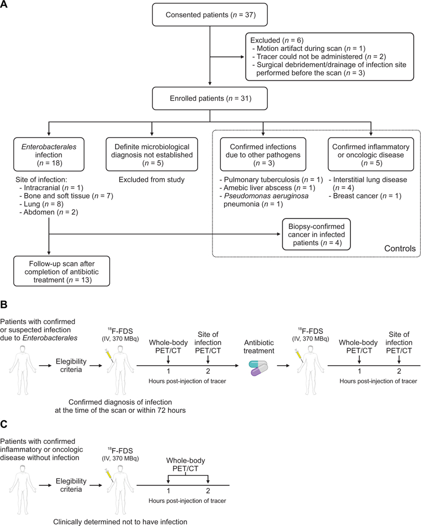
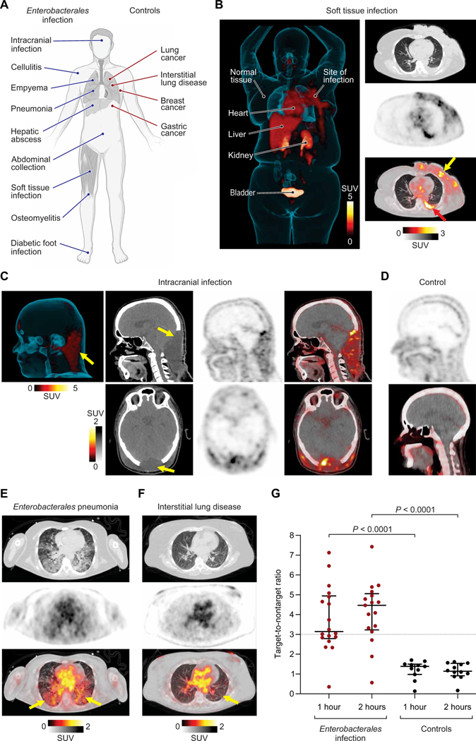
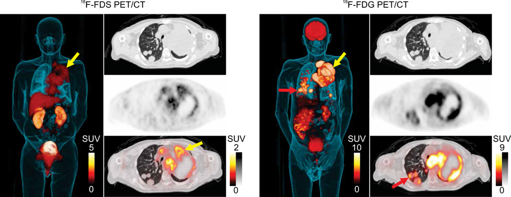
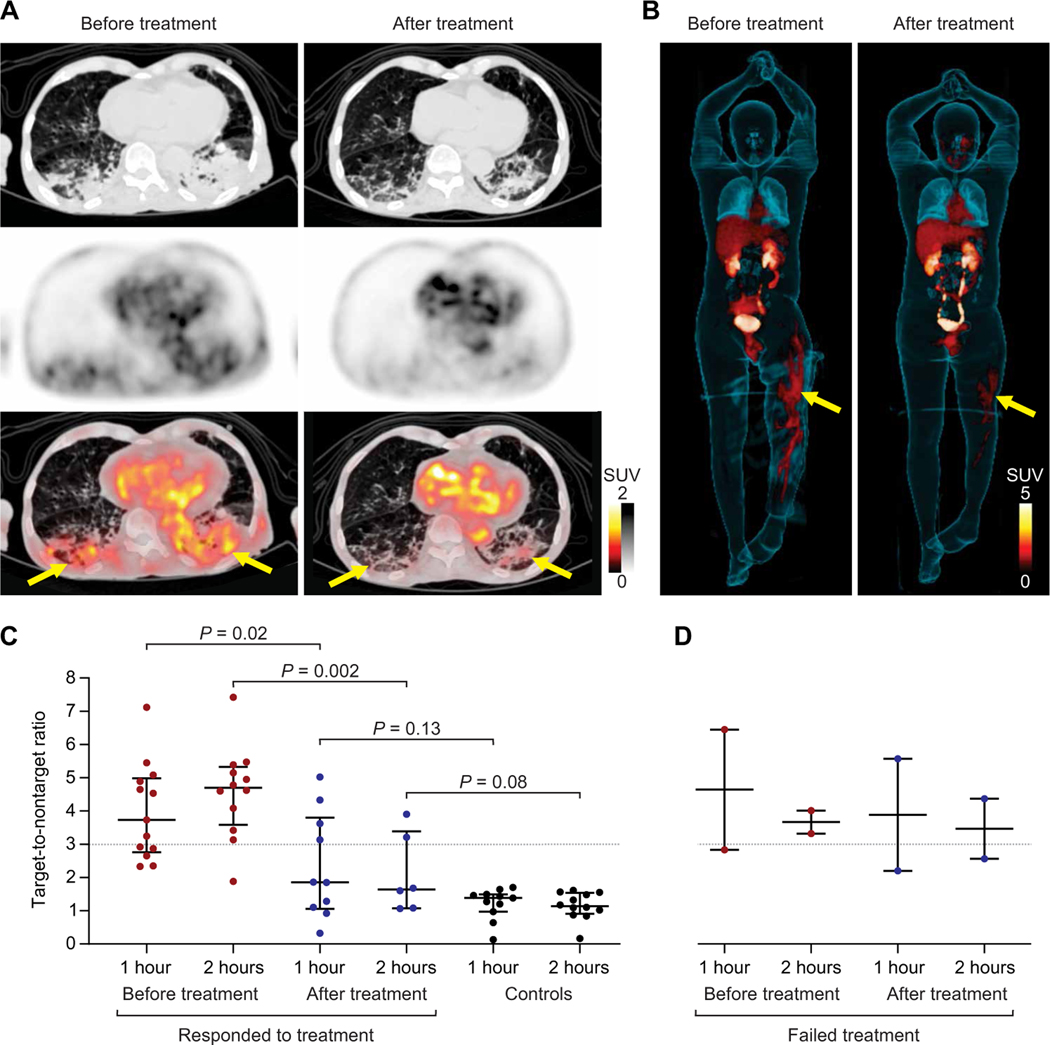
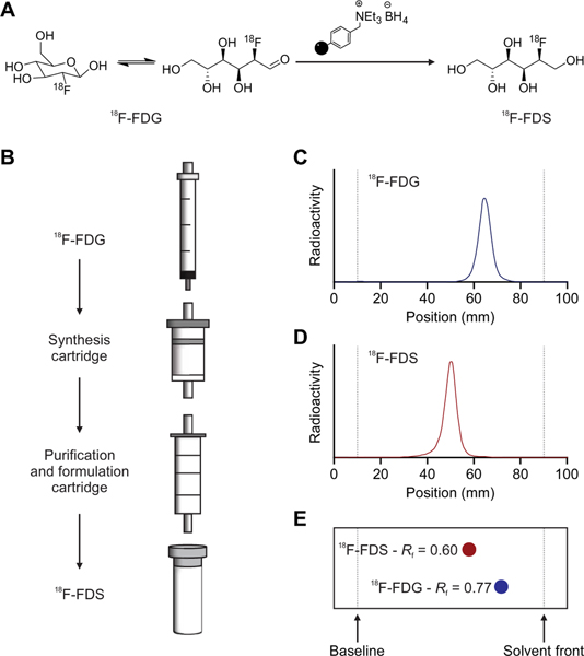
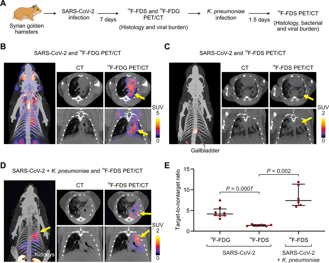
Enterobacterales represent the largest group of bacterial pathogens in humans and are responsible for severe, deep-seated infections, often resulting in sepsis or death. They are also a prominent cause of multidrug-resistant (MDR) infections, and some species are recognized as biothreat pathogens. Tools for noninvasive, whole-body analysis that can localize a pathogen with specificity are needed, but no such technology currently exists. We previously demonstrated that positron emission tomography (PET) with 2-deoxy-2-[18F]fluoro-d-sorbitol (18F-FDS) can selectively detect Enterobacterales infections in murine models. Here, we demonstrate that uptake of 18F-FDS by bacteria occurs via a metabolically conserved sorbitol-specific pathway with rapid in vitro 18F-FDS uptake noted in clinical strains, including MDR isolates. Whole-body 18F-FDS PET/computerized tomography (CT) in 26 prospectively enrolled patients with either microbiologically confirmed Enterobacterales infection or other pathologies demonstrated that 18F-FDS PET/CT was safe, could rapidly detect and localize Enterobacterales infections due to drug-susceptible or MDR strains, and differentiated them from sterile inflammation or cancerous lesions. Repeat imaging in the same patients monitored antibiotic efficacy with decreases in PET signal correlating with clinical improvement. To facilitate the use of 18F-FDS, we developed a self-contained, solid-phase cartridge to rapidly (<10 min) formulate ready-to-use 18F-FDS from commercially available 2-deoxy-2-[18F]fluoro-d-glucose (18F-FDG) at room temperature. In a hamster model, 18F-FDS PET/CT also differentiated severe acute respiratory syndrome coronavirus 2 (SARS-CoV-2) pneumonia from secondary Klebsiella pneumoniae pneumonia-a leading cause of complications in hospitalized patients with COVID-19. These data support 18F-FDS as an innovative and readily available, pathogen-specific PET technology with clinical applications.


Figures

Fig. 1.
Fig. 1.. Clinical study design.
(A) Patients with microbiologically confirmed or high suspicion for Enterobacterales infection and control patients with other pathologies were prospectively enrolled. For all infections, microbiological confirmation was required from the clinical/biopsy sample obtained from the infection site. Definite microbiological diagnosis was not established in five patients who were excluded from the study. Oncologic diagnosis was made via tissue biopsies, and sterile inflammatory pathologies were determined clinically and/or with tissue biopsies. Four patients with Enterobacterales infections had coexisting biopsy-proven oncologic disease. Patients meeting the eligibility criteria underwent 18F-FDS PET/CT at 1 and 2 hours after tracer injection. (B) Patients with microbiologically confirmed or high suspicion for Enterobacterales infection were imaged. A subset of patients with microbiologically confirmed Enterobacterales infection was imaged again after completion of antibiotic treatment. (C) Patients with confirmed inflammatory or oncologic diseases without infection were also enrolled and served as controls.
Fig. 2.
Fig. 2.. 18F-FDS PET can selectively detect and localize Enterobacterales infection at several body sites.
(A) Patients with microbiologically confirmed Enterobacterales infection (blue lines) or control patients with other pathologies (red lines) were prospectively enrolled and underwent PET/CT at 1 and 2 hours after intravenous administration of 18F-FDS. (B) Three-dimensional (3D) maximum intensity projection (MIP), transverse CT (top right), PET (middle right), and overlaid PET/CT (bottom right) from a 67-year-old female with Klebsiella aerogenes cellulitis of the left breast. The breast infection site is marked by a yellow arrow, and a microbiologically confirmed pleural effusion is also noted (red arrow). Signal is also noted in the heart (blood pool), liver, kidneys, and the urinary bladder, but no signal is noted in the brain. (C) From left to right, 3D MIP (left), CT, PET, and overlaid PET/CT from a sagittal view (top) and transverse view (bottom), where 18F-FDS PET signal is observed in a 38-year-old male with an intracranial infection due to an MDR bacteria. ESBL-producing K. pneumoniae was isolated from the site of infection (yellow arrow) and cerebrospinal fluid. (D) Sagittal 18F-FDS PET (top) and overlaid PET/CT (bottom) of the healthy brain of a control patient with low background signal. (E) Transverse CT (top), PET (middle), and overlaid PET/CT (bottom), where 18F-FDS PET signal is observed in the infected lung areas (yellow arrows) in a 51-year-old female patient with K. pneumoniae pneumonia. (F) Transverse CT (top), PET (middle), and overlaid PET/CT (bottom) show lack of 18F-FDS PET signal in the affected lung areas (yellow arrow) of a 54-year-old male with interstitial lung disease (control patient). (G) Target-to-nontarget ratio at 1 and 2 hours after 18F-FDS injection for all 26 patients. Data are represented as median and IQR. Statistical comparisons were performed using a two-tailed Mann-Whitney U test. SUV, standardized uptake value.
Fig. 3.
Fig. 3.. Patient with Enterobacterales infection and coexisting biopsy-proven oncologic disease.
3D MIP, transverse CT, PET, and overlaid PET/CT are shown from a 67-year-old male with squamous cell carcinoma of the lung and K. pneumoniae pneumonia that underwent 18F-FDG PET for clinical reasons 12 days after the study-related 18F-FDS PET. 18F-FDG avid pulmonary lesions are noted in red arrows; 18F-FDS PET signal is noted selectively in the infected tissues in yellow arrow. 18F-FDG PET signal is noted in infected lesions and in the cancerous pulmonary lesions (red arrows). Also, note the 18F-FDG PET signal in the brain, which is absent for 18F-FDS PET.
Fig. 4.
Fig. 4.. 18F-FDS PET can monitor antibiotic treatments.
Thirteen patients with microbiologically confirmed Enterobacterales infections underwent repeat 18F-FDS PET/CT after completion of antibiotic treatments. Transverse CT, PET, and overlaid PET/CT from (A) a 91-year-old male with MDR, ESBL-producing K. pneumoniae pneumonia before and after appropriate treatment. The yellow arrows indicate the sites of infection. (B) 3D MIP from a 33-year-old male with MDR, ESBL-producing E. coli osteomyelitis (yellow arrows) before and after inadequate treatment. The extent of 18F-FDS PET signal decreased, but the intensity did not change. (C and D) Target-to-nontarget tissue ratio at 1 and 2 hours after 18F-FDS injection for all 13 patients before and after completion of antibiotic treatments. Ratios from control patients are shown as reference. (C) Eleven patients who clinically responded to treatment and (D) two patients with treatment failure are shown. Data are represented as median and IQR. Statistical comparisons were performed using a two-tailed Mann-Whitney U test.
Fig. 5.
Fig. 5.. Solid-phase cartridge system to synthesize 18F-FDS.
(A) 18F-FDS synthesis from 18F-FDG reduction at room temperature using a solid-supported borohydride source in under 10 min. (B) Schematic representation of kit-based synthesis and formulation of 18F-FDS, which can be achieved by eluting a solution of 18F-FDG through a solid-phase synthesis cartridge followed by a purification and formulation cartridge (Chromabond Set V). Radiochemical purity (>90%, n = 3) was assessed by radio-TLC, which shows that 18F-FDG (C) and 18F-FDS (D) have different retention times and that these are identical to those of cold FDG and FDS (E).
Fig. 6.
Fig. 6.. 18F-FDS and SARS-CoV-2 versus K. pneumoniae infections in hamsters.
(A) Syrian golden hamsters were infected intranasally with SARS-CoV-2 and, 7 days later, imaged with 18F-FDS or 18F-FDG PET/CT. A group of animals was euthanized at that time point for histology and quantification of viral burden (fig. S7). A subset of animals was subsequently coinfected with K. pneumoniae and imaged with 18F-FDS PET 36 hours later. (B) 3D MIP, transverse (top) and coronal (bottom) CT, and overlaid PET/CT for 18F-FDG in SARS-CoV-2–infected animals. The affected lung parenchyma is noted by yellow arrows. (C) 3D MIP, transverse (top) and coronal (bottom) CT, and overlaid PET/CT for 18F-FDS in SARS-CoV-2–infected animals, where minimal signal is observed in the affected lung areas (yellow arrows). (D) 3D MIP, transverse (top) and coronal (bottom) CT, and overlaid PET/CT for 18F-FDS in SARS-CoV-2– and K. pneumoniae–infected animals. Yellow arrows indicate the affected lung areas. (E) Target-to-nontarget ratio 18F-FDS or 18F-FDG injection for all animals (n = 6 animals per group of 18F-FDS and n = 8 animals for 18F-FDG). Data are represented as median and IQR. Statistical comparisons were performed using a two-tailed Mann-Whitney U test.

References

    1. Donnenberg MS, Mandell, Douglas, and Bennett’s Principles and Practice of Infectious Diseases, Gerald JEB Mandell L, Dolin R, Eds. (Elsevier Inc., 2010), chap. 218, pp. 2815–2833.
    1. Guentzel MN, in Medical Microbiology, Baron S, Ed. (University of Texas Medical Branch at Galveston, 1996), chap. 26. - PubMed
    1. Centers for Disease Control and Prevention, Antibiotic Resistance Threats in the United States (Centers for Disease Control and Prevention, 2019); https://cdc.gov/drugresistance/pdf/threats-report/2019-ar-threats-report....
    1. Sabino S, Soares S, Ramos F, Moretti M, Zavascki AP, Rigatto MH, A cohort study of the impact of carbapenem-resistant Enterobacteriaceae infections on mortality of patients presenting with sepsis. mSphere 4, e00052–19 (2019). - PMC - PubMed
    1. Dhesi Z, Enne VI, Brealey D, Livermore DM, High J, Russell C, Colles A, Kandil H, Mack D, Martin D, Page V, Parker R, Roulston K, Singh S, Wey E, Swart AM, Stirling S, Barber JA, O’Grady J, Gant VA, Organisms causing secondary pneumonias in COVID-19 patients at 5 UK ICUs as detected with the FilmArray test. medRxiv 10.1101/2020.2006.2022.20131573, (2020). - DOI

Publication types

MeSH terms

Substances