. 2021 Apr 14;41(15):3400-3417.
doi: 10.1523/JNEUROSCI.2470-20.2020.

Perinatal Fentanyl Exposure Leads to Long-Lasting Impairments in Somatosensory Circuit Function and Behavior

Affiliations

Perinatal Fentanyl Exposure Leads to Long-Lasting Impairments in Somatosensory Circuit Function and Behavior

Jason B Alipio et al. J Neurosci. .

Abstract

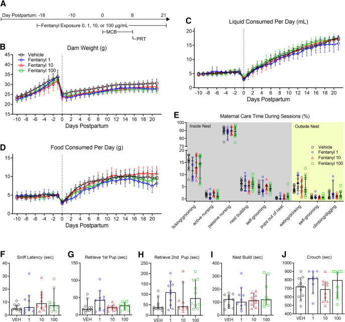
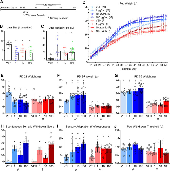
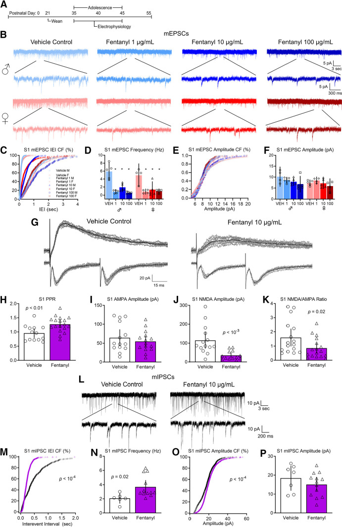
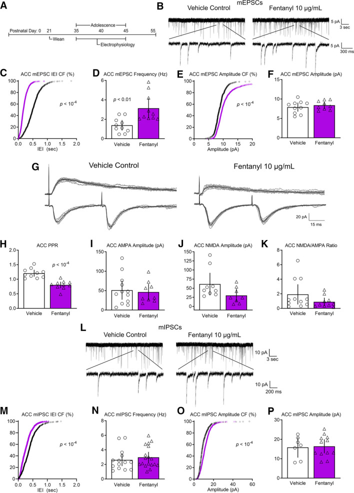
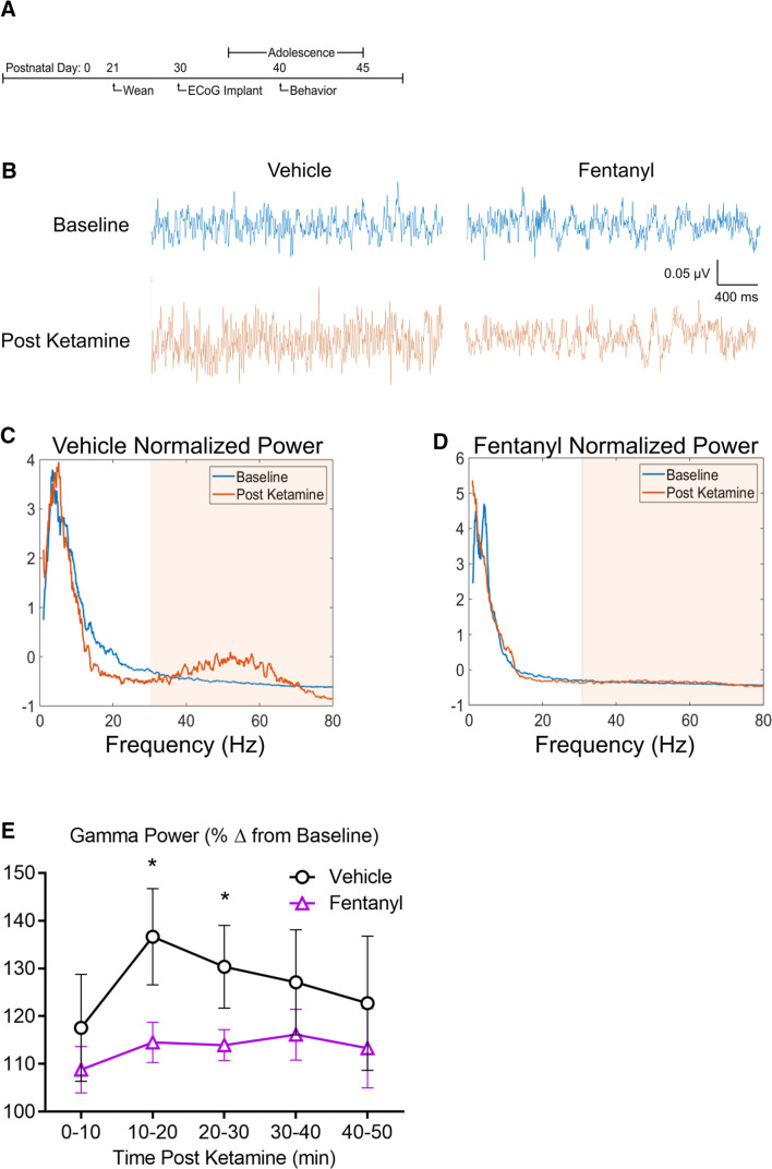
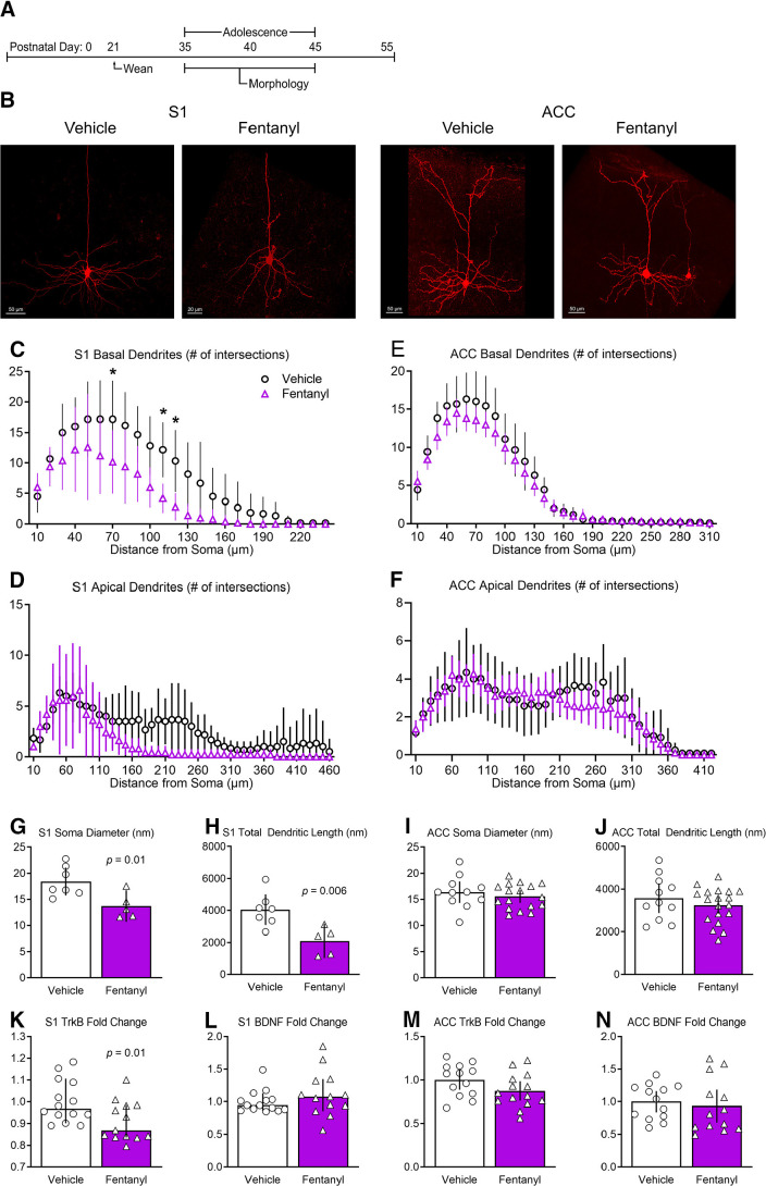
One consequence of the opioid epidemic are lasting neurodevelopmental sequelae afflicting adolescents exposed to opioids in the womb. A translationally relevant and developmentally accurate preclinical model is needed to understand the behavioral, circuit, network, and molecular abnormalities resulting from this exposure. By employing a novel preclinical model of perinatal fentanyl exposure, our data reveal that fentanyl has several dose-dependent, developmental consequences to somatosensory function and behavior. Newborn male and female mice exhibit signs of withdrawal and sensory-related deficits that extend at least to adolescence. As fentanyl exposure does not affect dams' health or maternal behavior, these effects result from the direct actions of perinatal fentanyl on the pups' developing brain. At adolescence, exposed mice exhibit reduced adaptation to sensory stimuli, and a corresponding impairment in primary somatosensory (S1) function. In vitro electrophysiology demonstrates a long-lasting reduction in S1 synaptic excitation, evidenced by decreases in release probability, NMDA receptor-mediated postsynaptic currents, and frequency of miniature excitatory postsynaptic currents (mEPSCs), as well as increased frequency of miniature inhibitory postsynaptic currents (mIPSCs). In contrast, anterior cingulate cortical neurons exhibit an opposite phenotype, with increased synaptic excitation. Consistent with these changes, electrocorticograms (ECoGs) reveal suppressed ketamine-evoked γ oscillations. Morphologic analysis of S1 pyramidal neurons indicate reduced dendritic complexity, dendritic length, and soma size. Further, exposed mice exhibited abnormal cortical mRNA expression of key receptors involved in synaptic transmission and neuronal growth and development, changes that were consistent with the electrophysiological and morphologic changes. These findings demonstrate the lasting sequelae of perinatal fentanyl exposure on sensory processing and function.SIGNIFICANCE STATEMENT This is the first study to show that exposure to fentanyl in the womb results in behavioral, circuitry, and synaptic effects that last at least to adolescence. We also show, for the first time, that this exposure has different, lasting effects on synapses in different cortical areas.

Keywords: Developmental Biology; opioid; somatosensory; thalamocortical.


Figures

Figure 1.
Figure 1.
Perinatal fentanyl exposure does not influence dam health or maternal care. A, Timeline depicting fentanyl exposure on dams, maternal care behavior, and pup retrieval test. There was no difference in dam weight (B), liquid consumption (C), or food consumption (D) across all concentrations of fentanyl tested. E, Fentanyl exposure did not adversely affect passive maternal care behaviors during PD1–PD7. FJ, There were no differences between exposure groups in latencies to sniff, retrieve pups, start nest building, or crouch over pups. Data depict means for parametric or medians for non-parametric comparisons with 95% confidence intervals.
Figure 2.
Figure 2.
Perinatal fentanyl exposure results in aberrant effects at birth, withdrawal behavior, and impaired sensory function. A, Timeline depicting withdrawal behavior 24 h after weaning and sensory behavior test during adolescence. Exposure resulted in smaller litter size (B) and a higher litter mortality rate (C). D, Mice exposed to fentanyl perinatally exhibited abnormal weight during early development. E, At weaning (PD21), exposed male mice weighed less than controls. F, At adolescence (PD35), exposed male mice weighed more than controls and males weighed more than females at each concentration of fentanyl tested. G, By adulthood (PD55), males weighed more than females, but there were no differences between fentanyl exposure groups. H, Perinatal fentanyl exposure induces spontaneous somatic withdrawal behavior 24 h after cessation, at all concentrations of fentanyl tested. I, Perinatal fentanyl exposure impaired sensory adaptation to continuous application of tactile stimuli (I) but did not influence paw withdrawal threshold (J). Data depict means with 95% confidence intervals. *p < 0.05 compared to vehicle control, †p < 0.05 compared to opposite sex condition.
Figure 3.
Figure 3.
Perinatal fentanyl exposure impairs synaptic transmission in somatosensory cortical neurons. A, Timeline depicting slice electrophysiology recordings in S1 layer 5 neurons of adolescent mice. B, Example traces of mEPSCs. C, Cumulative frequency plot of the interevent intervals of mEPSCs are shifted to the right in fentanyl-exposed mice compared with controls. D, Grouped data reflect decreased mEPSC frequency in fentanyl-exposed mice. There were no differences in the cumulative frequency of the mEPSC amplitude (E) nor in the grouped data for mEPSC amplitude (F). G, Example traces of evoked paired pulse and NMDAR-mediated response. Perinatal fentanyl exposure results in increased paired pulse ratio in fentanyl-exposed mice (H). There were no differences in AMPAR-mediated response amplitude (I). J, There was decreased NMDAR-mediated response amplitude, which is also reflected in the NMDA/AMPA ratio (K). L, Example traces of mIPSCs. M, Cumulative frequency plot of the interevent intervals of mIPSCs are shifted to the left in fentanyl-exposed mice compared with controls. N, Grouped data reflect increased mIPSC frequency in fentanyl-exposed mice. O, Cumulative frequency plot of the mIPSC amplitude was shifted to the right. P, There were no differences in grouped data of mIPSC amplitude. Data depict means for parametric or medians for non-parametric comparisons with 95% confidence intervals.
Figure 4.
Figure 4.
Perinatal fentanyl exposure impairs synaptic transmission in anterior cingulate cortical neurons. A, Timeline depicting slice electrophysiology recordings in ACC layer 5 neurons of adolescent mice. B, Example traces of mEPSCs. C, Cumulative frequency plot of the interevent intervals of mEPSCs are shifted to the left in fentanyl-exposed mice compared with controls. D, Grouped data reflect increased mEPSC frequency in fentanyl-exposed mice. E, Cumulative frequency of the mEPSC amplitude was shifted to the right in fentanyl-exposed mice. F, There were no differences in grouped data of mEPSC amplitudes. G, Example traces of evoked paired pulse and NMDAR-mediated responses. H, Perinatal fentanyl exposure results in decreased paired pulse ratio in fentanyl-exposed mice. There were no differences in AMPAR-mediated response amplitudes (I), NMDAR-mediated response amplitudes (J), or the NMDA/AMPA ratio (K). L, Example traces of mIPSCs. M, Cumulative frequency plot of the interevent intervals of mIPSCs are shifted to the left in fentanyl-exposed mice compared with controls. N, There were no differences in grouped data of mIPSC frequency. O, Cumulative frequency plot of the mIPSC amplitude was shifted to the right. P, There were no differences in grouped data of mIPSC amplitude. Data depict means for parametric or medians for non-parametric comparisons with 95% confidence intervals.
Figure 5.
Figure 5.
Perinatal fentanyl exposure reduces ketamine-evoked cortical oscillations. A, Timeline depicting transmitter implant and in vivo ECoG recordings. B, Example ECoG traces of cortical oscillations recorded from fentanyl or vehicle control adolescent mice, before and after intraperitoneal injection of subanesthetic dose of ketamine (10 µg/ml). Normalized power of cortical oscillations in control (C) and perinatal fentanyl exposed (D) adolescent mice. E, Grouped data of γ power across 10-min time bins after ketamine injection. Data depict means with 95% confidence intervals. *p < 0.05.
Figure 6.
Figure 6.
Perinatal fentanyl exposure reduces morphology of basal dendrites of pyramidal neurons in S1. A, Timeline depicting morphologic assays of pyramidal neurons. B, Example of biocytin-filled layer 5 pyramidal neurons in S1 and ACC. Sholl analysis reveals that perinatal fentanyl exposure results in reduced branching of S1 basal (C) but not apical dendrites (D). There were no differences in branching of ACC basal (E) or apical dendrites (F). Fentanyl-exposed mice exhibited, in S1, decreased soma diameter (G) and decreased total dendritic length (H). Fentanyl-exposed mice also had decreased mRNA expression of TrkB receptors (K), with no difference in expression of BDNF (L). In ACC, there were no differences in soma diameter (I), total dendritic length (J), mRNA expression of TrkB receptors (M), or BDNF (N). Data depict means for parametric or medians for non-parametric comparisons with 95% confidence intervals. *p < 0.05.

References

    1. Adaikkan C, Tsai LH (2020) Gamma entrainment: impact on neurocircuits, glia, and therapeutic opportunities. Trends Neurosci 43:24–41. 10.1016/j.tins.2019.11.001 - DOI - PubMed
    1. Alipio JB, Brockett AT, Fox ME, Tennyson SS, deBettencourt CA, El-Metwally D, Francis NA, Kanold PO, Lobo MK, Roesch MR, Keller A (2020) Enduring consequences of perinatal fentanyl exposure in mice. Addict Biol. Advance online publication. Retrieved March 18, 2020. doi: 10.1111/adb.12895. - DOI - PMC - PubMed
    1. Angevine JB, Sidman RL (1961) Autoradiographic study of cell migration during histogenesis of cerebral cortex in the mouse. Nature 192:766–768. 10.1038/192766b0 - DOI - PubMed
    1. Ayres AJ (1964) Tactile functions. Their relation to hyperactive and perceptual motor behavior. Am J Occup Ther 18:6–11. - PubMed
    1. Bakhireva LN, Holbrook BD, Shrestha S, Leyva Y, Ashley M, Cano S, Lowe J, Stephen JM, Leeman L (2019) Association between prenatal opioid exposure, neonatal opioid withdrawal syndrome, and neurodevelopmental and behavioral outcomes at 5-8 months of age. Early Hum Dev 128:69–76. 10.1016/j.earlhumdev.2018.10.010 - DOI - PMC - PubMed

Publication types

MeSH terms

LinkOut - more resources