. 2021 Apr 14;12(4):405.
doi: 10.1038/s41419-021-03666-z.

Sirt1 deacetylates and stabilizes p62 to promote hepato-carcinogenesis

Affiliations

Sirt1 deacetylates and stabilizes p62 to promote hepato-carcinogenesis

Lifeng Feng et al. Cell Death Dis. .

Abstract

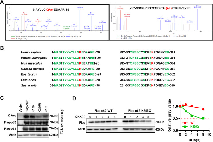
p62/SQSTM1 is frequently up-regulated in many cancers including hepatocellular carcinoma. Highly expressed p62 promotes hepato-carcinogenesis by activating many signaling pathways including Nrf2, mTORC1, and NFκB signaling. However, the underlying mechanism for p62 up-regulation in hepatocellular carcinoma remains largely unclear. Herein, we confirmed that p62 was up-regulated in hepatocellular carcinoma and its higher expression was associated with shorter overall survival in patients. The knockdown of p62 in hepatocellular carcinoma cells decreased cell growth in vitro and in vivo. Intriguingly, p62 protein stability could be reduced by its acetylation at lysine 295, which was regulated by deacetylase Sirt1 and acetyltransferase GCN5. Acetylated p62 increased its association with the E3 ligase Keap1, which facilitated its poly-ubiquitination-dependent proteasomal degradation. Moreover, Sirt1 was up-regulated to deacetylate and stabilize p62 in hepatocellular carcinoma. Additionally, Hepatocyte Sirt1 conditional knockout mice developed much fewer liver tumors after Diethynitrosamine treatment, which could be reversed by the re-introduction of exogenous p62. Taken together, Sirt1 deacetylates p62 at lysine 295 to disturb Keap1-mediated p62 poly-ubiquitination, thus up-regulating p62 expression to promote hepato-carcinogenesis. Therefore, targeting Sirt1 or p62 is a reasonable strategy for the treatment of hepatocellular carcinoma.


Conflict of interest statement

The authors declare no competing interests.

Figures

Fig. 1
Fig. 1. High expression of p62 promotes hepato-carcinogenesis.
A Expression of p62 protein in 12 pairs of fresh HCC tissues and adjacent non-tumor tissues was analyzed by western blot. B p62 protein expression in 6 pairs of fresh liver cancer tissues and adjacent non-tumor tissues of DEN-induced mice liver cancer model was analyzed by western blot. C The impact of p62 protein expression on overall survival (OS) was analyzed by Kaplan–Meier survival curve analysis (n(Low) = 46, n(High) = 47, Cox-regression analysis, p < 0.05, Hazard Ration = 2.347, 95% CI of ratio 1.340–4.109). D Relative cell vitality of PLC/PRF/5, Huh7, and SK-Hep1 cells transient transfected with SQSTM1(p62) or negative control siRNAs was measured with MTS assay (n = 6, T-test, p < 0.05). E Growth curve, F representative image, and G Tumor weight of xenografts with stable p62-silenced (shp62) or scramble control (shNC) PLC/PRF/5 cells(n = 5, T-test, p < 0.05).
Fig. 2
Fig. 2. Acetylation at lysine 295 reduces p62 stabilization.
A The acetylation site of Flag-p62 was analyzed by mass spectrometry (MS). The MS results showed that Lysine 13 (K13), 295 (K295) of p62 might be acetylated. B Conservation of p62 K13 and K295 in various species was aligned. C Acetylation of p62 wild type (WT) or p62 mutants (K13R or K295R) expressed in HEK293T cells were detected via western blot. D Wild type (WT) of p62 or p62(K295Q) mutants were overexpressed in HEK293T cells, followed cycloheximide (CHX) treatment with indicated time points, and the half-life of the WT and K295Q was detected via western blot, and the relative expression was measured by ImageJ software.
Fig. 3
Fig. 3. p62 is deacetylated by Sirt1 at K295.
A Exogenous Flag-p62 in HEK293T cells treated with deacetylase inhibitors Nicotinamide (NAM, 5 mM, 6 h) or Trichostatin A (TSA, 1 μm, 12 h) was immunoprecipitated with anti-Flag, and p62 acetylation was analyzed using an anti-acetyl-Lysine (K-Ace) antibody via western blot. B Exogenous Flag-p62 in HEK293T cells after Sirt1 knockdown with siRNAs transfection was immunoprecipitated with anti-Flag, and the acetylation of p62 was analyzed using an anti-acetyl-Lysine (K-Ace) antibody via western blot. C Acetylation of exogenous Flag-p62 in HEK293T cells with or without co-overexpression of HA-Sirt1 or HA-Sirt1H363Y was detected by western blot. D Co-IP was performed to detect the interaction of full-length Flag-p62 or deletion (△LB (Lim-binding domain deletion, from amino acids 167–220)) with Sirt1 in HEK293T cells using anti-Flag, Empty vector was used as the negative control. E Co-IP was performed to detect the interaction of endogenous Sirt1 with p62 in PLC/PRF/5 cells using anti-p62, blank IgG was used as the negative control. F Acetylation of exogenous Flag-p62(WT) or mutant Flag-p62(K295R) in PLC/PRF/5 cells with or without Sirt1 knockdown and MG132 incubation was detected by western blot. G, H The protein expression of p62 and the phosphorylation of mTOR (p-mTOR) in PLC/PRF/5, Huh7 and SK-Hep1 cells with or without Sirt1 knockdown was analyzed by western blot.
Fig. 4
Fig. 4. Sirt1 inhibits GCN5-mediated p62 acetylation.
A The expression of p62 protein and the phosphorylation of mTORC1 in PLC/PRF/5 and SK-Hep1 cells with GCN5 siRNAs and Sirt1 siRNA co-transfection were analyzed by western blot. B Acetylation of Flag-p62 in HEK293T cells co-transfected with Myc-GCN5 and Myc-Sirt1 or empty vector were analyzed by anti-Flag immunoprecipitation, and probed with anti-K-ace. C Co-IP was performed to detect the interaction of Flag-p62 with Myc-GCN5 in HEK293T cells with anti-Flag. D Acetylation of the mutants of Flag-p62, WT, or K295R in HEK293T cells co-transfected with Myc-GCN5 were analyzed by anti-Flag immunoprecipitation, and probed with anti-K-ace. E Co-IP was performed to detect the interaction of Flag-p62 or Flag-p62 △LB with Myc-GCN5 in HEK293T cells. Flag-p62 or Flag-p62△LB and Myc-GCN5 co-transfection were analyzed with anti-Flag co-IP. And the acetylation of Flag-p62 or Flag-p62 △LB Flag-p62 in HEK293T with Myc-GCN5 overexpression was probed with anti-K-ace. F Co-IP was performed to detect the interaction of Flag-p62 and Myc-GCN5 with or without overexpression of HA-Sirt1 with anti-Myc.
Fig. 5
Fig. 5. Sirt1 stabilizes p62 via inhibiting its acetylation.
A The protein and mRNA expression of p62 in HCCLM3 cells with or without Sirt1 overexpression was analyzed by western blot or RT-PCR. B The protein and mRNA expression of p62 in of Sirt1fl/fl MEF cells with or without GFP-Cre adenovirus infection were analyzed by western blot or RT-PCR. C The mRNA expression of p62 in PLC/PRF/5 and Huh7 cells with or without Sirt1 knowdown via siRNAs was analyzed by RT-PCR. D Exogenous Flag-p62 protein expression in PLC/PRF/5 cells with or without Sirt1 knockdown were analyzed by western blot. E The half-life of p62 in PLC/PRF/5 with or without Sirt1 knowdown via siRNAs was analyzed by western blot and the half-life of p62 in PLC/PRF/5 with or without Sirt1 knowdown via siRNAs was determined by ImageJ software. F Exogenous Flag-p62(WT) or p62 mutants (K295R) protein expression in PLC/PRF/5 and SK-Hep1 cells with or without Sirt1 knockdown were analyzed by western blot. G The half-life of p62 in PLC/PRF/5 with or without Sirt1 inhibitor (EX527, 20 μM, 24 h) treatment was analyzed by western blot and the half-life of p62 in PLC/PRF/5 with or without Sirt1 inhibitoor treatment was determined by ImageJ software.
Fig. 6
Fig. 6. Sirt1 suppresses Keap1-mediated ubiquitination of p62.
A p62 protein level in PLC/PRF/5 and SK-Hep1 cells treated with CQ(50 μM, 16 h) or MG132(5 μM, 16 h) after Sirt1 knockdown were detected by western blot. B Ni-NTA beads pull down was applied for ubiquitylation analysis of Flag-p62 in HEK293T cells co-transfected with Flag-p62, HA-sirt1 and His-ubiquitin (His-Ub) or empty vector, and anti-Flag antibody was used to detect ubiquitinated p62. C Ubiquitylation of WT or K295Q in HEK293T cells co-transfected with His-Ub or empty vector were analyzed by Ni-NTA beads pull down. D p62 protein level and the phosphorylation of mTORC1 in PLC/PRF/5 and SK-Hep1 cells with Keap1 siRNAs and Sirt1 siRNA co-transfection were analyzed by western blot. E Co-IP was performed to detect the interaction of WT or K295R with Keap1 in 293T cells by anti-Flag. F Ubiquitylation of Flag-p62 WT or K295R in HEK293T cells co-transfected with Keap1 and His-Ub or empty vector were analyzed by Ni-NTA beads pull down.
Fig. 7
Fig. 7. Sirt1 promotes hepatocellular carcinogenesis via up-regulating p62 expression.
A Represented photos of p62 and Sirt1 expression in HCC tissues were shown, cases were grouped based on median expression and the correlation was analyzed (n = 93, χ2 test, p < 0.05 represents statistically significant). B Patients were grouped as above in A, and the overall survival of co-high (both Sirt1 and p62 high expressed) and co-low (both Sirt1 and p62 low expressed) groups of patients was analyzed via Kaplan–Meier survival curve analysis (n = 71, Cox-regression analysis, p < 0.05, Hazard Ration = 2.347, 95% CI of ratio 1.340–4.109). C The average tumor numbers (>1 mm) per mouse and the average liver weight/body weight ratio of three groups (wild-type mice without DEN treatment (WT-Control, n = 5), DEN-treated wild-type mice (Sirt1fl/fl, Alb-cre−/−, WT-DEN, n = 15) and DEN treated Sirt1 hepatocyte-specific knockout mice (Sirt1fl/fl, Alb-cre+/+, CKO-DEN, n = 11) were showed respectively. D The representative liver images, E the average tumor numbers (>1 mm) per mouse and the average liver weight/body weight ratio in CKO mice with p62 re-introduced (n = 5) or not (n = 5) were showed respectively. F The expressions of Sirt1, Flag-p62, and p62 downstream targets, p-mTOR were detected by western blot in liver tumors.
Fig. 8
Fig. 8. The model of the regulation mechanism of Sirt1-p62 axis.
Type III deacetylase Sirt1 interacted with p62 to decrease its acetylation mediated via GCN5 at K295 and disturbed Keap1 mediated ubiquitination-dependent degradation of p62. Thus, Sirt1 up-regulation resulted into p62 accumulation, and contributed to hepatocellular carcinogenesis.

Similar articles

Cited by

References

    1. Villanueva A. Hepatocellular carcinoma. N. Engl. J. Med. 2019;380:1450–1462. doi: 10.1056/NEJMra1713263. - DOI - PubMed
    1. Ishizawa T, et al. Neither multiple tumors nor portal hypertension are surgical contraindications for hepatocellular carcinoma. Gastroenterology. 2008;134:1908–1916. doi: 10.1053/j.gastro.2008.02.091. - DOI - PubMed
    1. Siegel RL, Miller KD, Jemal A. Cancer statistics, 2020. CA Cancer J. Clin. 2020;70:7–30. doi: 10.3322/caac.21590. - DOI - PubMed
    1. Moscat J, Karin M, Diaz-Meco MT. p62 in cancer: signaling adaptor beyond autophagy. Cell. 2016;167:606–609. doi: 10.1016/j.cell.2016.09.030. - DOI - PMC - PubMed
    1. Feng LF, et al. YY1-MIR372-SQSTM1 regulatory axis in autophagy. Autophagy. 2014;10:1442–1453. doi: 10.4161/auto.29486. - DOI - PMC - PubMed

Publication types

MeSH terms