Review
. 2021 Apr;53(4):517-527.
doi: 10.1038/s12276-021-00590-2. Epub 2021 Apr 14.

The p53-caspase-2 axis in the cell cycle and DNA damage response

Affiliations
Review

The p53-caspase-2 axis in the cell cycle and DNA damage response

Yoon Lim et al. Exp Mol Med. 2021 Apr.

Abstract

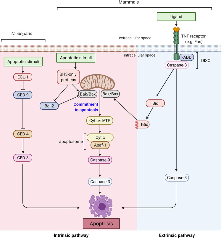
Caspase-2 was discovered almost three decades ago. It was one of the first two mammalian homologs of CED-3, the other being interleukin 1β-converting enzyme (ICE/caspase-1). Despite high similarity with CED-3 and its fly and mammalian counterparts (DRONC and caspase-9, respectively), the function of caspase-2 in apoptosis has remained enigmatic. A number of recent studies suggest that caspase-2 plays an important role in the regulation of p53 in response to cellular stress and DNA damage to prevent the proliferation and accumulation of damaged or aberrant cells. Here, we review these recent observations and their implications in caspase-2-mediated cellular death, senescence, and tumor suppression.


Conflict of interest statement

The authors declare no competing interests.

Figures

Fig. 1
Fig. 1. Conserved apoptotic pathways.
Core apoptotic machinery in C. elegans and in mammals that drives caspase activation is shown. In mammals, there are two main apoptosis pathways. In the intrinsic pathway, various cellular insults result in MOMP and cytochrome c release from mitochondria, which drives apoptosome-dependent caspase-3 activation. In the extrinsic pathway, TNF family members activate caspase-8 via DISC. Activated caspase-8 and caspase-9 mediate the processing and activation of effector caspases, of which caspase-3 is the most prominent. The figure was created with BioRender.com.
Fig. 2
Fig. 2. Caspase-2 structure and activation.
In the primary structure, the location of zymogen cleavage sites, CARD, p19, p12, nuclear localization signal (NLS), and the catalytic Cys residue are shown. The putative ubiquitination and phosphorylation sites are also indicated. Various biochemical steps that lead to mature dimeric caspase-2 are shown. A ribbon diagram displays the dimeric structure of caspase-2 with two substrate binding pockets, including catalytic Cys (PDB ID: 3R6G). The figure was created with BioRender.com.
Fig. 3
Fig. 3. Caspase-2 functions in cell cycle arrest and apoptosis.
a Schematic showing that the cell cycle arrest and apoptotic functions of caspase are mediated via cleavage of MDM2 and BID following mitotic catastrophe, respectively. Figure created with BioRender.com. b An example of MDM2 cleavage by caspase-2 (Casp2) following treatment of WT or caspase-2-deficient (KO) U2OS cells with or without the AURKB inhibitor ZM447439 (ZM). c Panels showing examples of increased multinucleated (arrow) caspase-2-deficient U2OS cells following AURKB inhibition. Scale bar, 50 µm. d Examples of caspase-2-mediated BID cleavage (appearance of truncated BID, tBID) following treatment with a PLK1 inhibitor, BI2536 (BI), which induces mitotic arrest. fl.; full-length, cl.; cleaved, p33 and p18; cleaved caspase-2. The data and images in bd were generated by Yoon Lim.
Fig. 4
Fig. 4. Different physiological outcomes mediated by caspase-2-dependent p53 stabilization.
A proposed scheme showing that PIDDosome-mediated caspase-2 activation, MDM2 cleavage and p53 stabilization serves as a checkpoint under conditions of sustained DNA damage to induce cell cycle arrest or apoptosis. The figure was created with BioRender.com.

References

    1. Galluzzi L, et al. Molecular mechanisms of cell death: recommendations of the Nomenclature Committee on Cell Death 2018. Cell Death Differ. 2018;25:486–541. - PMC - PubMed
    1. Shalini S, Dorstyn L, Dawar S, Kumar S. Old, new and emerging functions of caspases. Cell Death Differ. 2015;22:526–539. - PMC - PubMed
    1. Vigneswara, V. & Ahmed, Z. The role of caspase-2 in regulating cell fate. Cells10.3390/cells9051259 (2020). - PMC - PubMed
    1. Kumar S. Caspase function in programmed cell death. Cell Death Differ. 2007;14:32–43. - PubMed
    1. Dorstyn L, Akey CW, Kumar S. New insights into apoptosome structure and function. Cell Death Differ. 2018;25:1194–1208. - PMC - PubMed

Publication types