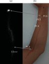
In Vivo Visualization of the Pericardium Meridian with Fluorescent Dyes
- PMID: 33854554
- PMCID: PMC8021474
- DOI: 10.1155/2021/5581227
In Vivo Visualization of the Pericardium Meridian with Fluorescent Dyes
Abstract
The anatomical basis of acupuncture meridians continues to be enigmatic. Although much attention has been placed on potential correlations with inter/intramuscular fascia or lower electrical impedance, animal studies performed in the past 40 years have shown that tracer dyes-specifically Tc-99m pertechnetate-injected at strategic skin points generate linear migrations closely aligning with acupuncture meridians. To evaluate whether this phenomenon is also observable in humans, we injected two fluorescent dyes-fluorescein sodium and indocyanine green (ICG)-into the dermal layer both at acupuncture points (PC5, PC6, and PC7) and a nonacupoint control. Fifteen healthy volunteers were enrolled in this study. Of the 19 trials of fluorescein injected at PC6, 15 (79%) were associated with slow diffusion of the dye proximally along a path matching closely with the pericardium meridian. Furthermore, the dye emerged and coalesced proximally at exactly acupoint PC3. Injections of ICG at the acupoints PC5, PC6, or PC7 showed a similar trajectory close to the injection site but diverged when migrating proximally, failing converge on acupoint PC3. Injections of either dye at an adjacent PC6-control did not generate any notable linear pathway. Both ultrasound imaging and vein-locating device did not reveal any corresponding vessels (arterial or venous) at the visualized tracer pathway but did demonstrate correlations with intermuscular fascia.
Copyright © 2021 Tongju Li et al.
Conflict of interest statement
The authors declare that they have no conflicts of interest.
Figures


Similar articles
-
Cortical functional networks of transcutaneous electrical stimulation at acupoints on the pericardial meridian.Neuropsychologia. 2023 Oct 10;189:108669. doi: 10.1016/j.neuropsychologia.2023.108669. Epub 2023 Aug 28. Neuropsychologia. 2023. PMID: 37648106
-
Application of Biophysical Properties of Meridians in the Visualization of Pericardium Meridian.J Acupunct Meridian Stud. 2023 Jun 30;16(3):101-108. doi: 10.51507/j.jams.2023.16.3.101. J Acupunct Meridian Stud. 2023. PMID: 37381032
-
[Experimental observation on the relation between pericardium meridian and cardiac function].Zhen Ci Yan Jiu. 1993;18(2):143-8. Zhen Ci Yan Jiu. 1993. PMID: 8070043 Chinese.
-
Stimuli-induced NOergic Molecules and Neuropeptides Mediated Axon Reflexes Contribute to Tracers along Meridian Pathways.Curr Top Med Chem. 2024;24(5):393-400. doi: 10.2174/0115680266260220240108114337. Curr Top Med Chem. 2024. PMID: 38243932 Free PMC article. Review.
-
[Review on skin temperature of acupoints].Zhongguo Zhen Jiu. 2017 Jan 12;37(1):109-114. doi: 10.13703/j.0255-2930.2017.01.029. Zhongguo Zhen Jiu. 2017. PMID: 29231335 Review. Chinese.
Cited by
-
Mitochondria as the Essence of Yang Qi in the Human Body.Phenomics. 2022 Jun 16;2(5):336-348. doi: 10.1007/s43657-022-00060-3. eCollection 2022 Oct. Phenomics. 2022. PMID: 36939762 Free PMC article. Review.
-
Proteomic Study Between Interstitial Channels Along Meridians and Adjacent Areas in Mini-Pigs.Biomolecules. 2025 Jun 1;15(6):804. doi: 10.3390/biom15060804. Biomolecules. 2025. PMID: 40563444 Free PMC article.
-
Research status and prospects of acupuncture for autism spectrum disorders.Front Psychiatry. 2023 May 25;14:942069. doi: 10.3389/fpsyt.2023.942069. eCollection 2023. Front Psychiatry. 2023. PMID: 37304438 Free PMC article. Review.
-
Portable devices for periodic monitoring of bioelectrical impedance along meridian pathways in healthy individuals.Biomed Eng Online. 2025 Jan 17;24(1):3. doi: 10.1186/s12938-025-01335-2. Biomed Eng Online. 2025. PMID: 39825427 Free PMC article.
-
The Science of Tai Chi and Qigong as Whole Person Health-Part I: Rationale and State of the Science.J Integr Complement Med. 2025 Jun;31(6):499-520. doi: 10.1089/jicm.2024.0957. Epub 2025 Mar 17. J Integr Complement Med. 2025. PMID: 40091656 Review.
References
LinkOut - more resources
Full Text Sources
Other Literature Sources
Miscellaneous

