

Evaluation of jaw bone density and morphology in bruxers using panoramic radiography
- PMID: 33854718
- PMCID: PMC8025186
- DOI: 10.1016/j.jds.2020.09.008
Evaluation of jaw bone density and morphology in bruxers using panoramic radiography
Abstract
Background/purpose: Bruxism affects the stomatognathic system and causes tissue damage by the excessive jaw movements. The purpose of this study was to evaluate the effects of sleep bruxism on jaw bone density, mineralisation and morphology by comparing bruxers and non-bruxers.
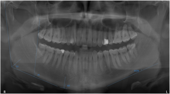
Materials and methods: 60 bruxers and 60 non-bruxers (control) patients were included in the analysis. Cortical width at the gonion (GI), at the mental foramen (MI), at the antegonion (AI), the panoramic mandibular index (PMI), the mandibular cortical index (MCI) and antegonial notch depth (AND) were measured bilaterally on 120 panoramic radiographs. The measurements were evaluated for repeatability, correlation with age, gender and correlation between the variables.
Results: A significant association was observed between cortical shape (MCI) and bruxism status (p = 0.012). The MI was significantly different between the bruxers and non-bruxers (p = 0.006). There was a significant but weak correlation between the MI value and age in bruxers and the control (p = 0.003, p = 0.04). The AI was not associated with bruxism status and did not vary by age or gender (p > 0.05). The AND was higher in bruxers than non-bruxers (p = 0.001). Male bruxers had a significantly higher AND value than female bruxers (p = 0.001). The GI was higher in male bruxers (p = 0.001).
Conclusion: Defects in the endosteal margin of the cortex and cortical thickening in the mental region were detected in bruxer patients. Furthermore, AND was increased in bruxers. Tiny bone peaks accompanied the cortical thickening seen in the gonial region. Male bruxer patients had higher GI and AND values than female bruxers.
Keywords: Bone mineral density; Bruxism; Mandible; Panoramic radiography.
© 2020 Association for Dental Sciences of the Republic of China. Publishing services by Elsevier B.V.
Conflict of interest statement
The authors have no conflicts of interest relevant to this article.
Figures


Similar articles
-
Morphological evaluation of gonial and antegonial regions in bruxers on panoramic radiographic images.BMC Oral Health. 2023 Jul 7;23(1):457. doi: 10.1186/s12903-023-03162-0. BMC Oral Health. 2023. PMID: 37420199 Free PMC article.
-
Evaluation of Bone Quality in Patients with Bruxism.Curr Med Imaging. 2024;20:e15734056299979. doi: 10.2174/0115734056299979240927101222. Curr Med Imaging. 2024. PMID: 39629572
-
Evaluation of mandibular bone density in bruxers: the value of panoramic radiographs.Oral Radiol. 2023 Jan;39(1):117-124. doi: 10.1007/s11282-022-00612-3. Epub 2022 Apr 19. Oral Radiol. 2023. PMID: 35438407
-
Evaluation of the effect of bruxism on mandibular cortical bone using radiomorphometric indices on panoramic radiographs.Niger J Clin Pract. 2021 Nov;24(11):1742-1748. doi: 10.4103/njcp.njcp_71_21. Niger J Clin Pract. 2021. PMID: 34782517
-
Relationship between bruxism and mandibular bone modifications based on medical imaging: a scoping review.BMC Oral Health. 2023 Jul 14;23(1):483. doi: 10.1186/s12903-023-03209-2. BMC Oral Health. 2023. PMID: 37452323 Free PMC article.
Cited by
-
Guided Bone Regeneration in a Periodontally Compromised Individual with Autogenous Tooth Bone Graft: A Radiomics Analysis.J Funct Biomater. 2023 Apr 14;14(4):220. doi: 10.3390/jfb14040220. J Funct Biomater. 2023. PMID: 37103310 Free PMC article.
-
3D comparative evaluation and correlation of condylar morphology in bruxers and non-bruxers.F1000Res. 2023 Sep 1;12:1099. doi: 10.12688/f1000research.133773.1. eCollection 2023. F1000Res. 2023. PMID: 39267819 Free PMC article.
-
A new perspective for radiologic findings of bruxism on dental panoramic radiography.Oral Radiol. 2023 Jul;39(3):544-552. doi: 10.1007/s11282-022-00667-2. Epub 2022 Dec 12. Oral Radiol. 2023. PMID: 36504381
-
Evaluation of fractal analysis and radiomorphometric measurements of mandibular bone structure in bruxism and non-bruxism paediatric patients.Oral Radiol. 2025 Jan;41(1):60-68. doi: 10.1007/s11282-024-00776-0. Epub 2024 Oct 8. Oral Radiol. 2025. PMID: 39375282
-
Bruxism assessment combining fractal analysis, clinical evaluation, and self-reports: a case-control study.BMC Oral Health. 2025 May 30;25(1):851. doi: 10.1186/s12903-025-06234-5. BMC Oral Health. 2025. PMID: 40448020 Free PMC article.
References
-
- Jensen R., Rasmussen B.K., Pedersen B. Prevalence of oromandibular dysfunction in a general population. J Orofac Pain. 1993;7:175–182. - PubMed
-
- Santos-Silva R., Bittencourt L.R., Pires M.L. Increasing trends of sleep complaints in the city of Sao Paulo, Brazil. Sleep Med. 2010;11:520–524. - PubMed
-
- Bayar G.R., Tutuncu R., Acikel C. Psychopathological profile of patients with different forms of bruxism. Clin Oral Invest. 2012;16:305–311. - PubMed
-
- Fernandes G., Franco A.L., Siqueira J.T. Sleep bruxism increases the risk for painful temporomandibular disorder, depression and non-specific physical symptoms. J Oral Rehabil. 2012;39:538–544. - PubMed
LinkOut - more resources
Full Text Sources
Other Literature Sources
