Review
. 2021 Mar 29;10(1):1900508.
doi: 10.1080/2162402X.2021.1900508.

Tertiary lymphoid structures and B lymphocytes in cancer prognosis and response to immunotherapies

Affiliations
Review

Tertiary lymphoid structures and B lymphocytes in cancer prognosis and response to immunotherapies

Jacquelot N et al. Oncoimmunology. .

Abstract

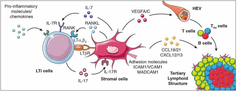
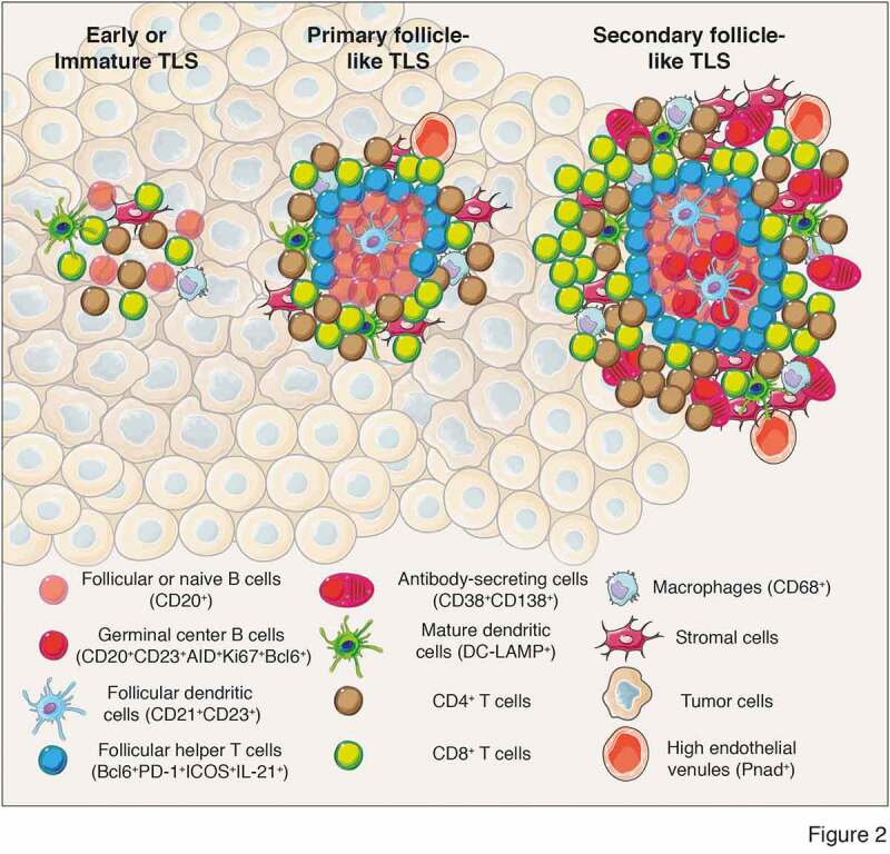
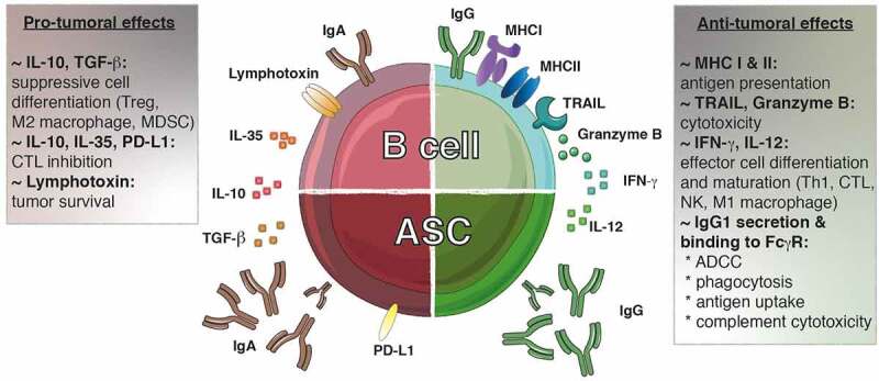
Tertiary lymphoid structures (TLS) are ectopic cellular aggregates that resemble secondary lymphoid organs in their composition and structural organization. In contrast to secondary lymphoid organs, TLS are not imprinted during embryogenesis but are formed in non-lymphoid tissues in response to local inflammation. TLS structures exhibiting a variable degree of maturation are found in solid tumors. They are composed of various immune cell types including dendritic cells and antigen-specific B and T lymphocytes, that together, actively drive the immune response against tumor development and progression. This review highlights the successive steps leading to tumor TLS formation and its association with clinical outcomes. We discuss the role played by tumor-infiltrating B lymphocytes and plasma cells, their prognostic value in solid tumors and immunotherapeutic responses and their potential for future targeting.

Keywords: Tertiary lymphoid structures; b lymphocytes; cancer; immunotherapy; plasma cells.


Conflict of interest statement

No potential conflict of interest was reported by the authors.

Figures

Figure 1.
Figure 1.
Cellular and molecular signals that control TLS formation. The local accumulation of pro-inflammatory molecules and chemokines promotes the recruitment of LTi cells to the inflammed site promoting their interaction with stromal cells to initiate TLS genesis and cytokine (IL-7, IL-17, RANKL, and LTα1β2) and cytokine receptor (IL-7R, IL-17R, RANK and LTβR) expression. When LTi cells are absent, other immune cells such as macrophages, B lymphocytes and Th17 cells can also interact with stromal cells to induce TLS formation. This interaction culminates in the production of chemokines (CCL19, CCL21, CXCL12 and CXCL13), pro-angiogenic molecules (vascular endothelial growth factors VEGFA and VEGFC) and the expression of adhesion molecules which facilitate the recruitment of additional immune cell types, their retention and organization into the nascent TLS. LTi cells, lymphoid tissue-inducer cells; Th17 cells, T helper cells secreting IL-17; LT, lymphotoxin; RANK, receptor activator of nuclear factor-κB; ICAM, intercellular adhesion molecule 1; VCAM1, Vascular adhesion molecule 1; MADCAM, mucosal vascular addressin cell adhesion molecule 1; VEGFC, vascular endothelial growth factor A/C; IL, interleukin; CCL19: C-C motif chemokine ligand 19; CXCL13, C-X-C motif chemokine ligand 13; HEV, high endothelial venules
Figure 2.
Figure 2.
Different levels of TLS maturation and their composition. Tumor-associated TLS are heterogenous and range from poorly-organized cellular aggregates (Early or immature TLS) to well-organized structures forming primary follicles or secondary follicles containing germinal centers surrounded by specific vessels called high endothelial venules (PNad+). Their cellular composition include stromal cells, innate and adaptive immune cells. In most of the analyses performed, cellular composition has been determined using immunofluorescence or immunochemistry analyses and relies on the expression of cell-specific markers to identify the cell types that form TLS. Mature secondary follicle-like TLS harbor a germinal center composed of proliferating mature germinal center B lymphocytes (CD20+CD23+AID+Ki67+Bcl6+) and follicular dendritic cells (CD21+CD23+) surrounded by naïve or follicular B cells (CD20+) and bordered by follicular helper T cells (Bcl6+PD-1+ICOS+IL-21+). In addition, TLS are formed of CD4+ and CD8+ T cells (CD3+), plasma cells (CD38+CD138+), mature dendritic cells (DC-LAMP+) and macrophages (CD68+). CD, cluster of differenciation; DC-LAMP, dendritic cell lysosomal associated membrane glycoprotein; PD-1, programmed cell death 1; ICOS, Inducible costimulator; AID, activation-induced deaminase; Bcl6, B cell lymphoma 6 protein; PNad, peripheral node addressin
Figure 3.
Figure 3.
The dichotomy of tumor-infiltrating B lymphocytes. Both pro- and anti-tumoral roles can be attributed to B lymphocytes. IgG1+ B cells promote the anti-tumoral response by presenting antigens to T cells and secreting cytokines (IFN-γ, IL-12) that polarizing the response toward an optimal Th1/CTL composition. These B cells can exert direct cytotoxic functions through the expression of TRAIL and granzyme B. Furthermore, the IgG1 antibodies secreted by the ASC can bind to the FcγR at the surface of NK cells, macrophages and dendritic cells allowing induction of ADCC, phagocytosis and antigen uptake, respectively. Furthermore, IgG1 antibodies fix complement to trigger its cytotoxic cascade. In contrast, IgA+ cells are associated with the secretion of inhibitory cytokines (IL-10, IL-35, TGF-β) that create a suppressive environment favoring the emergence of Treg, M2 macrophages and MDSC while repressing the function of the effector cells. In addition, the expression of lymphotoxin by B cells supports tumor cell survival. ADCC, antibody-dependent cell-mediated cytotoxicity; ASC, antibody secreting cell; CTL, cytotoxic T lymphocyte; FcγR, Fc gamma receptor; Ig, immunoglobulin; IFN-γ, interferon gamma; MDSC, myeloid-derived suppressor cells; MHC: major histocompatibility complex; PD-L1: progammed cell death ligand 1; TGF-β, transforming growth factor-beta; TRAIL, tumor-necrosis-factor related apoptosis-inducing ligand; Treg, regulatory T cell

References

    1. Fridman WH, Zitvogel L, Sautes-Fridman C, Kroemer G.. The immune contexture in cancer prognosis and treatment. Nat Rev Clin Oncol. 2017;14(12):717–16. doi:10.1038/nrclinonc.2017.101. - DOI - PubMed
    1. Vesely MD, Kershaw MH, Schreiber RD, Smyth MJ. Natural innate and adaptive immunity to cancer. Annu Rev Immunol. 2011;29(1):235–271. doi:10.1146/annurev-immunol-031210-101324. - DOI - PubMed
    1. Petitprez F. B cells are associated with survival and immunotherapy response in sarcoma. Nature. 2020;577(7791):556–560. doi:10.1038/s41586-019-1906-8. - DOI - PubMed
    1. Helmink BA. B cells and tertiary lymphoid structures promote immunotherapy response. Nature. 2020;577(7791):549–555. doi:10.1038/s41586-019-1922-8. - DOI - PMC - PubMed
    1. Cabrita R. Tertiary lymphoid structures improve immunotherapy and survival in melanoma. Nature. 2020;577(7791):561–565. doi:10.1038/s41586-019-1914-8. - DOI - PubMed

Publication types

LinkOut - more resources