

Primary Central Nervous System Vasculitis as an Unusual Cause of Intracerebral Hemorrhage: A Case Report
- PMID: 33854857
- PMCID: PMC8038648
- DOI: 10.7759/cureus.13847
Primary Central Nervous System Vasculitis as an Unusual Cause of Intracerebral Hemorrhage: A Case Report
Abstract
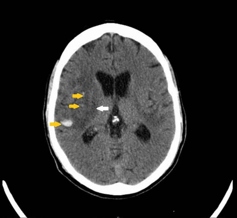
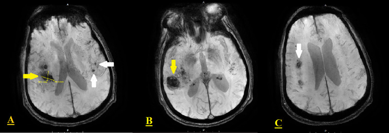
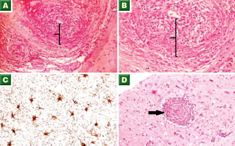
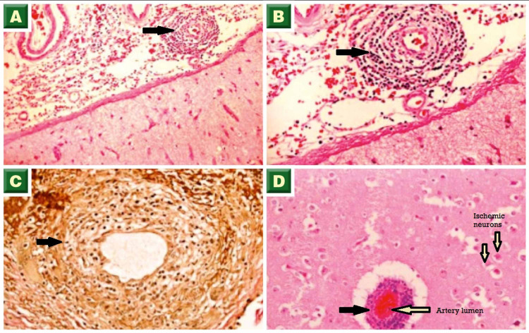
A 64-year-old male with a history of transverse myelitis presented to the hospital with a decreased level of consciousness of one day's duration. CT of the head revealed intracranial hemorrhage measuring 2 x 1.2 cm in the right temporal lobe and multiple small hemorrhages in the left hemisphere, suggestive of vasculitis. Initial vasculitis workup was negative for antinuclear antibody (ANA), complement component 3 (C3), and antineutrophil cytoplasmic antibodies: P-ANCA, C-ANCA. Syphilis, hepatitis B and C, West Nile virus antibody [immunoglobulin G (IgG) and immunoglobulin M (IgM)], herpes simplex virus (HSV) polymerase chain reaction (PCR), and HIV 1 and 2 were also negative. In view of the CT scan findings suggestive of vasculitis and the vague presentation of primary central nervous system vasculitis (PCNSV), a brain biopsy was performed. It revealed angiocentric granulomatous inflammation with focal vessel disruption and associated parenchymal hemorrhage, consistent with a diagnosis of granulomatous vasculitis. The patient received levetiracetam, multiple high doses of steroids, and six cycles of cyclophosphamide for a six-month duration. After induction, he has remained in remission without any maintenance therapy until now (eight years post-presentation).
Keywords: cns angiitis; induction therapy; intracerebral hemorrhage; primary cns vasculitis.
Copyright © 2021, Borcheni et al.
Conflict of interest statement
The authors have declared that no competing interests exist.
Figures
Similar articles
-
Hepatitis B virus induced cytoplasmic antineutrophil cytoplasmic antibody-mediated vasculitis causing subarachnoid hemorrhage, acute transverse myelitis, and nephropathy: a case report.J Med Case Rep. 2017 Apr 3;11(1):91. doi: 10.1186/s13256-017-1255-x. J Med Case Rep. 2017. PMID: 28366165 Free PMC article.
-
Primary and secondary central nervous system vasculitis: clinical manifestations, laboratory findings, neuroimaging, and treatment analysis.Clin Rheumatol. 2015 Apr;34(4):729-38. doi: 10.1007/s10067-014-2831-8. Epub 2014 Nov 27. Clin Rheumatol. 2015. PMID: 25425493
-
A fatal case of propylthiouracil-induced ANCA-associated vasculitis resulting in rapidly progressive glomerulonephritis, acute hepatic failure, and cerebral angiitis.Clin Nephrol. 2015 May;83(5):309-14. doi: 10.5414/CN108322. Clin Nephrol. 2015. PMID: 25208313
-
[Small vessel-childhood primary angiitis of the central nervous system: a case report and literature review].Zhonghua Er Ke Za Zhi. 2018 Feb 2;56(2):142-147. doi: 10.3760/cma.j.issn.0578-1310.2018.02.014. Zhonghua Er Ke Za Zhi. 2018. PMID: 29429204 Review. Chinese.
-
[Central nervous system involvement in patients with HCV-related cryoglobulinemia: literature review and a case report].Reumatismo. 2002 Apr-Jun;54(2):150-5. doi: 10.4081/reumatismo.2002.150. Reumatismo. 2002. PMID: 12105684 Review. Italian.
Cited by
-
Subarachnoid Hemorrhage, CNS Vasculitis and Stroke as a Sequela of Q Fever Infection.Neurohospitalist. 2025 Jul 18:19418744251361313. doi: 10.1177/19418744251361313. Online ahead of print. Neurohospitalist. 2025. PMID: 40688476 Free PMC article.
-
Different Types of Vasculitis Complicated by Heparin-Induced Thrombocytopenia-Analysis of Four Cases and Literature Review.J Clin Med. 2023 Sep 24;12(19):6176. doi: 10.3390/jcm12196176. J Clin Med. 2023. PMID: 37834820 Free PMC article. Review.
-
Primary Angiitis of the Central Nervous System in Adults: A Comprehensive Review of 76 Biopsy-Proven Case Reports.J Inflamm Res. 2023 Nov 7;16:5083-5094. doi: 10.2147/JIR.S434126. eCollection 2023. J Inflamm Res. 2023. PMID: 37953861 Free PMC article. Review.
References
-
- Godasi R, Pang G, Chauhan S, Bollu PC. Treasure Island, FL: StatPearls Publishing; 2020. Primary Central Nervous System Vasculitis; p. 29494083. - PubMed
-
- Involvement of CD45RO+ T lymphocyte infiltration in a patient with primary angiitis of the central nervous system restricted to small vessels. Iwase T, Ojika K, Mitake S, et al. Eur Neurol. 2001;45:184–185. - PubMed
-
- Primary central nervous system vasculitis: pathology and mechanisms. Giannini C, Salvarani C, Hunder G, Brown RD. Acta Neuropathol. 2012;123:759–772. - PubMed
Publication types
LinkOut - more resources
Full Text Sources
Other Literature Sources
Miscellaneous