Review
. 2021 Feb 12;8(7):2002545.
doi: 10.1002/advs.202002545. eCollection 2021 Apr.

Tailor-Made Nanomaterials for Diagnosis and Therapy of Pancreatic Ductal Adenocarcinoma

Affiliations
Review

Tailor-Made Nanomaterials for Diagnosis and Therapy of Pancreatic Ductal Adenocarcinoma

Xi Hu et al. Adv Sci (Weinh). .

Abstract

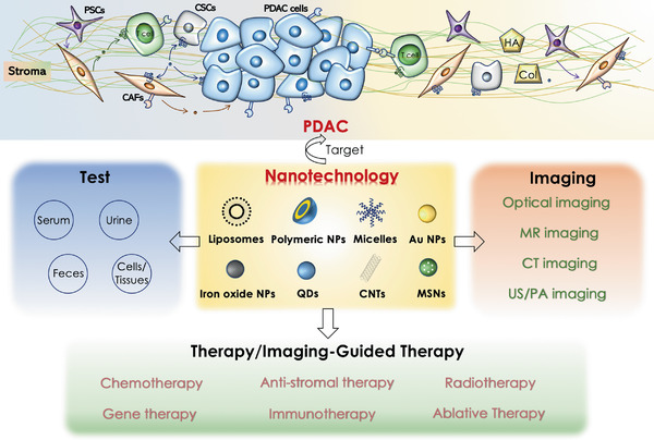
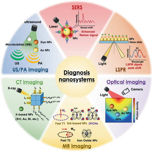
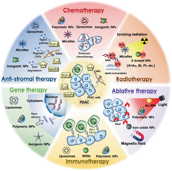
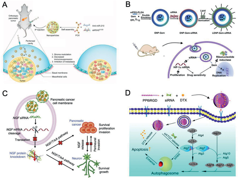
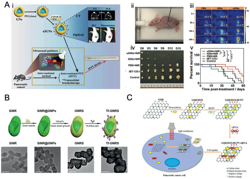
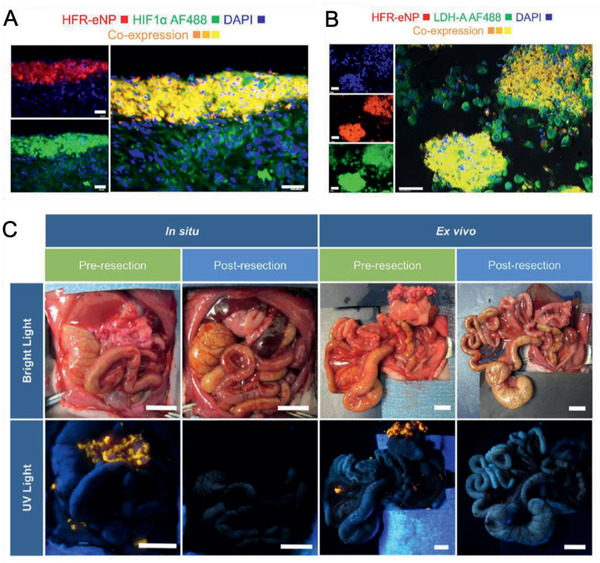
Pancreatic ductal adenocarcinoma (PDAC) is one of the deadliest cancers worldwide due to its aggressiveness and the challenge to early diagnosis and treatment. In recent decades, nanomaterials have received increasing attention for diagnosis and therapy of PDAC. However, these designs are mainly focused on the macroscopic tumor therapeutic effect, while the crucial nano-bio interactions in the heterogeneous microenvironment of PDAC remain poorly understood. As a result, the majority of potent nanomedicines show limited performance in ameliorating PDAC in clinical translation. Therefore, exploiting the unique nature of the PDAC by detecting potential biomarkers together with a deep understanding of nano-bio interactions that occur in the tumor microenvironment is pivotal to the design of PDAC-tailored effective nanomedicine. This review will introduce tailor-made nanomaterials-enabled laboratory tests and advanced noninvasive imaging technologies for early and accurate diagnosis of PDAC. Moreover, the fabrication of a myriad of tailor-made nanomaterials for various PDAC therapeutic modalities will be reviewed. Furthermore, much preferred theranostic multifunctional nanomaterials for imaging-guided therapies of PDAC will be elaborated. Lastly, the prospects of these nanomaterials in terms of clinical translation and potential breakthroughs will be briefly discussed.

Keywords: diagnosis; imaging‐guided therapy; nanomaterials; pancreatic ductal adenocarcinoma; therapy.


Conflict of interest statement

The authors declare no conflict of interest.

Figures

Figure 1
Figure 1
The development of tailor‐made nanomaterials in laboratory tests, medical imaging, therapy, and imaging‐guided therapy of heterogeneous PDAC. Liposomes, micelles, polymer NPs, and inorganic NPs can be easily modificated or loaded with therapeutic/imaging agents. Au NPs, QDs, and CNTs with intrinsic optical properties can be used for biosensing and biological imaging. Moreover, iron oxide NPs and high‐Z element NPs (e.g., Au NPs) can be utilized for MR imaging and CT imaging, respectively.
Figure 2
Figure 2
Illustration for various nanomaterials in the diagnosis of PDAC, including tailor‐made nanomaterials‐based in vitro detection technologies (e.g., SERS, LSPR) and various nanomaterials‐enabled in vivo imaging modalities (e.g., optical imaging, MR imaging, CT imaging, and US/PA imaging).
Figure 3
Figure 3
PDAC‐tailored nanomaterials for laboratory tests. A) Schematic illustration of the fabrication of the glass substrate‐bound Au nanoprisms to prepare LSPR‐based microRNA sensor for miR‐10b detection in plasma samples.Reproduced with permission.[ 36 ] Copyright 2015, American Chemical Society. B) Schematic illustration of the construction of chip‐based exosome‐PEARL SERS immunosensor. Reproduced with permission.[ 41 ] Copyright 2018, Royal Society of Chemistry. C) Illustration of fabrication of 1) SPIDE/CNO‐GO‐Ab and 2) SPIDE/GO‐Ab and CA19‐9 detection process. Reproduced with permission.[ 44 ] Copyright 2019, Elsevier. D) Apoferritin‐based modular nanoplatform (e.g., QDs, Au, or magnetic nanoprobes) for the detection of claudin 4. Reproduced with permission.[ 46 ] Copyright 2013, American Chemical Society.
Figure 4
Figure 4
PDAC‐tailored nanomaterials for optical imaging. A) Synthesis of bMSN@Cy 7.5‐FA NPs. B) Fluorescence imaging of tumor metastasis sites after i.v. injection of bMSN@Cy 7.5‐FA NPs. Reproduced with permission.[ 57 ] Copyright 2018, Elsevier. C) The construction of HSA‐GEM/IR780 nanocomplexes. D) Fluorescence imaging of nude mice bearing tumors after i.v. injection of HSA‐GEM/780 nanocomplexes. Reproduced with permission.[ 60 ] Copyright 2017, Elsevier.
Figure 5
Figure 5
PDAC‐tailored nanomaterials for multimodal imaging. A–D) The TEM image of A) CD326‐grafted UPGs micelles and B) normalized upconversion luminescence spectra (inset: full‐color photographs) under excitation of a 980 nm laser beam. C) The in vivo fluorescence images and D) T1‐weighted MR images of BxPC3 subcutaneous tumor models after i.v. injection of CD326‐grafted UPGs micelles. Reproduced with permission.[ 91 ] Copyright 2018, Springer Nature. E) The TEM image and F) AFM 3D topographic image of Gd‐Au‐NC‐GPC‐1. G) The in vivo fluorescence images and H) T1‐weighted MR images of COLO‐357 subcutaneous tumor models after i.v. injection of Gd‐Au‐NC‐GPC‐1. Reproduced with permission.[ 92 ] Copyright 2018, Dove Medical Press, Ltd.
Figure 6
Figure 6
Schematic illustration of various tailor‐made nanomaterials (e.g., liposomes, micelles, polymeric nanomaterials, protein‐based nanomaterials, and inorganic nanomaterials) and their therapeutic modalities (e.g., chemotherapy, anti‐stromal therapy, radiotherapy, gene therapy, immunotherapy, and ablative therapies) for PDAC.
Figure 7
Figure 7
PDAC‐tailored hybrid nanomaterials for chemotherapy. A) Schematic illustration of size‐switchable DGL/GEM@PP/GA hybrid NPs for deep tumor cell killing. Reproduced with permission.[ 132 ] Copyright 2019, American Chemical Society. B) The synthesis of thermosensitive liposomes (TSL/HSAPE) for enhanced drug penetration and therapeutic efficacy against PDAC. Reproduced with permission.[ 133 ] Copyright 2017, American Chemical Society. C) Thermosensitive gel composed of PTX‐loaded mPEG‐PLGA‐PLL‐cRGD NPs to reverse drug resistance and realize sustained drug release for PDAC interventional therapy. Reproduced with permission.[ 134 ] Copyright 2015, American Chemical Society.
Figure 8
Figure 8
PDAC‐tailored nanomaterials for anti‐stromal therapy. A) Schematic illustration of PFD loaded MMP‐2 responsive peptide hybrid liposome (MRPL‐PFD) to downregulate ECM and enhance drug penetration for improved PDAC therapy. B) Rhd penetration in PSCs/Miapaca‐2 subcutaneous tumor models after the treatment for 2 weeks. Blue, DAPI. Red: Rhd. Dotted lines: border between edge and core of tumor tissue. Scale bar, 100 µm. Reproduced with permission.[ 152 ] Copyright 2017, American Chemical Society. C) Schematic illustration of fraxinellone‐loaded CGKRK‐modified NPs (Frax‐NP‐CGKRK) to attenuate the dense stroma and improve tumor blood perfusion. D) B‐mode, maximum intensity persistence (MIP) mode, and pseudocolor parametric mode images of Panc‐1/NIH3T3 subcutaneous tumor models treated with PBS, Frax, and Frax‐NP‐CGKRK. Reproduced with permission.[ 154 ] Copyright 2019, Wiley‐VCH.
Figure 9
Figure 9
PDAC‐tailored nanomaterials for gene therapy. A) Scheme of PCX/(siKRAS + miR‐210) NPs for stromal modulation and metastatic PDAC therapy. Reproduced with permission.[ 178 ] Copyright 2020, American Chemical Society. B) Schematic diagram of LENPs for codelivery of si‐HIF1a and GEM for synergistic antitumor therapy. Reproduced with permission.[ 181 ] Copyright 2015, Elsevier. C) Schematic representation of GNC‐siRNA nanocomplexes for NGF silencing and PDAC therapy. Reproduced with permission.[ 183 ] Copyright 2017, Springer Nature. D) Schematic illustration of PP6iRGD/DTX/siAtg7 micelles for improved DTX therapeutic outcomes. Reproduced with permission.[ 185 ] Copyright 2019, American Chemical Society.
Figure 10
Figure 10
PDAC‐tailored nanomaterials for PTT. A) Schematic illustration of i) uIGN, ii) photoimage using a self‐developed IPTT device, iii) infrared thermal images for IPTT of PDAC, iv) photoimages of the resected tumors and v) percent survival in different treatment groups at 3 d interval. Reproduced with permission.[ 233 ] Copyright 2017, Wiley‐VCH. B) Synthesis illustration of Tf‐GNRS and the corresponding TEM images. Scale bar, 100 nm. Reproduced with permission.[ 234 ] Copyright 2017, American Chemical Society. C) Schematic illustration of GQD/DOX/BCPV/siRNA nanocomplexes for synergistic PTT and chemotherapy of PDAC. Reproduced with permission.[ 179 ] Copyright 2019, American Chemical Society.
Figure 11
Figure 11
PDAC‐tailored nanomaterials for surgical resection. A) Colocalization of HRF‐eNPs and HIF1α (a marker for hypoxia). Scale bar, 40 µm. B) Colocalization of HFR‐eNPs and LDH‐A (a marker for tumor acidification). Scale bar, 40 µm. C) Bright light and corresponding UV light images before and after HFR‐eNPs guided cytoreductive surgery in a xenograft Panc‐1‐CSCs rat model. Scale bar, 1 cm. Reproduced with permission.[ 283 ] Copyright 2017, American Chemical Society.

Similar articles

Cited by

References

    1. Riquelme E., Zhang Y., Zhang L., Montiel M., Zoltan M., Dong W., Quesada P., Sahin I., Chandra V., San Lucas A., Scheet P., Xu H., Hanash S. M., Feng L., Burks J. K., Do K.‐A., Peterson C. B., Nejman D., Tzeng C.‐W. D., Kim M. P., Sears C. L., Ajami N., Petrosino J., Wood L. D., Maitra A., Straussman R., Katz M., White J. R., Jenq R., Wargo J., McAllister F., Cell 2019, 178, 795. - PMC - PubMed
    1. Lee J.‐S., Rhee T.‐M., Pietrasz D., Bachet J.‐B., Laurent‐Puig P., Kong S.‐Y., Takai E., Yachida S., Shibata T., Lee J. W., Park H.‐c., Zang D. Y., Jeon K., Lee J., Kim M., Kim H.‐S., Kang H. J., Lee Y. K., Sci. Rep. 2019, 9, 16971. - PMC - PubMed
    1. Kamisawa T., Wood L. D., Itoi T., Takaori K., Lancet 2016, 388, 73. - PubMed
    1. Tummers W. S., Willmann J. K., Bonsing B. A., Vahrmeijer A. L., Gambhir S. S., Swijnenburg R. J., Pancreas 2018, 47, 675. - PMC - PubMed
    1. Kim D. W., Kim S. Y., Kim H. K., Kim S. W., Shin S. W., Kim J. S., Park K., Lee M. Y., Heo D. S., Ann. Oncol. 2007, 18, 2009. - PubMed

Publication types

MeSH terms