

Photoacoustic Computed Tomography of Breast Cancer in Response to Neoadjuvant Chemotherapy
- PMID: 33854889
- PMCID: PMC8025032
- DOI: 10.1002/advs.202003396
Photoacoustic Computed Tomography of Breast Cancer in Response to Neoadjuvant Chemotherapy
Abstract
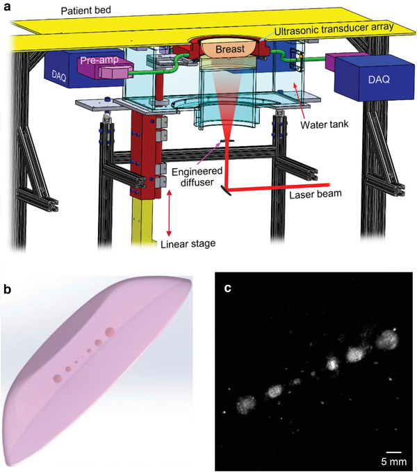
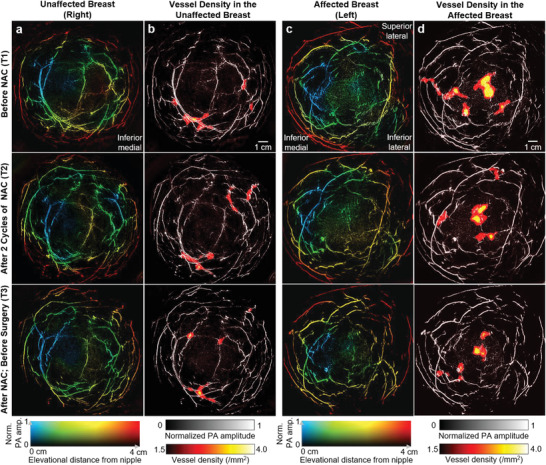
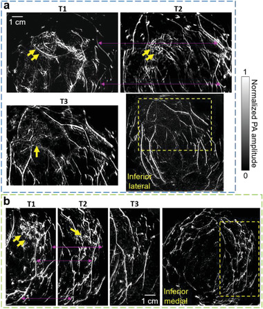
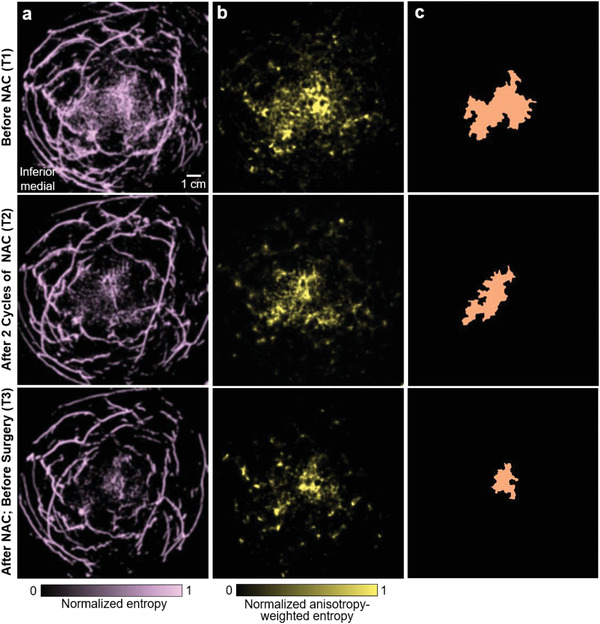
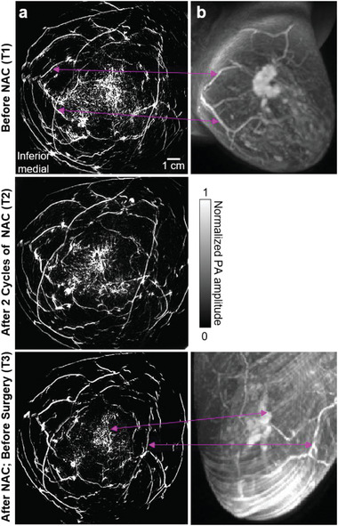
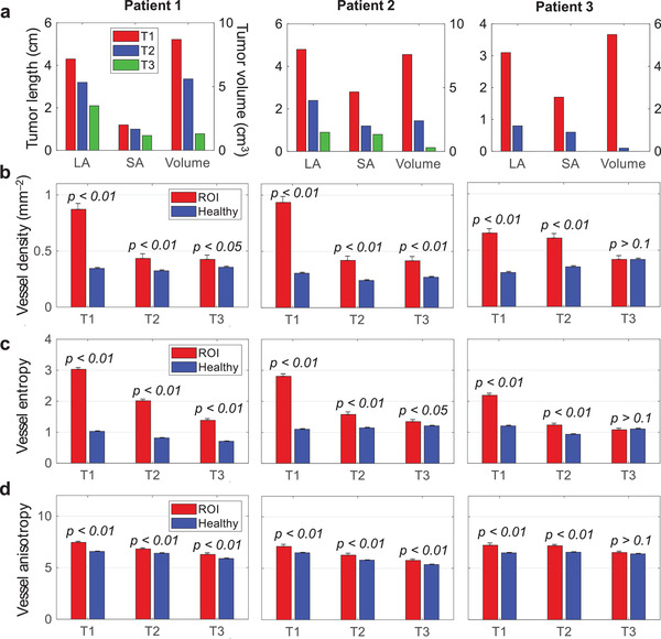
Neoadjuvant chemotherapy (NAC) has contributed to improving breast cancer outcomes, and it would ideally reduce the need for definitive breast surgery in patients who have no residual cancer after NAC treatment. However, there is no reliable noninvasive imaging modality accepted as the routine method to assess response to NAC. Because of the inability to detect complete response, post-NAC surgery remains the standard of care. To overcome this limitation, a single-breath-hold photoacoustic computed tomography (SBH-PACT) system is developed to provide contrast similar to that of contrast-enhanced magnetic resonance imaging, but with much higher spatial and temporal resolution and without injection of contrast chemicals. SBH-PACT images breast cancer patients at three time points: before, during, and after NAC. The analysis of tumor size, blood vascular density, and irregularity in the distribution and morphology of the blood vessels on SBH-PACT accurately identifies response to NAC as confirmed by the histopathological diagnosis. SBH-PACT shows its near-term potential as a diagnostic tool for assessing breast cancer response to systemic treatment by noninvasively measuring the changes in cancer-associated angiogenesis. Further development of SBH-PACT may also enable serial imaging, rather than the use of current invasive biopsies, to diagnose and follow indeterminate breast lesions.
Keywords: breast cancer; neoadjuvant chemotherapy; photoacoustic computed tomography; response to treatment; tumor‐associated microvasculature.
© 2021 The Authors. Advanced Science published by Wiley‐VCH GmbH.
Conflict of interest statement
L.V.W. has a financial interest in Microphotoacoustics, Inc., CalPACT, LLC, and Union Photoacoustic Technologies, Ltd., which, however, did not support this work.
Figures
References
-
- Pal S. K., Miller M. J., Agarwal N., Chang S. M., Mac Gregor M. C., Cohen E., Cole S., Dale W., Diefenbach C. S. M., Disis M. L., J. Clin. Oncol. 2019, 37, 834. - PubMed
-
- Andre F., Ismaila N., Henry N. L., Somerfield M. R., Bast R. C., Barlow W., Collyar D. E., Hammond M. E., Kuderer N. M., Liu M. C., J. Clin. Oncol. 2019, 37, 1956. - PubMed
-
- Cardoso F., van't Veer L. J., Bogaerts J., Slaets L., Viale G., Delaloge S., Pierga J.‐Y., Brain E., Causeret S., DeLorenzi M., N. Engl. J. Med. 2016, 375, 717. - PubMed
-
- Glück S., De Snoo F., Peeters J., Stork‐Sloots L., Somlo G., Breast Cancer Res. Treat. 2013, 139, 759. - PubMed
Publication types
MeSH terms
Grants and funding
LinkOut - more resources
Full Text Sources
Other Literature Sources
Medical
Miscellaneous