

Incidence of pedicle breach following open and minimally invasive spinal instrumentation: A postoperative CT analysis of 513 pedicle screws applied under fluoroscopic guidance
- PMID: 33854918
- PMCID: PMC7608845
- DOI: 10.37796/2211-8039.1016
Incidence of pedicle breach following open and minimally invasive spinal instrumentation: A postoperative CT analysis of 513 pedicle screws applied under fluoroscopic guidance
Abstract
Background: Even though pedicle screw application is a common procedure, and in-spite of spine surgeons being proficient with the technique, mal-positioning of pedicle screws can still occur. We intend to determine by postoperative CT analysis, the incidence of pedicle screw breach in the thoracolumbar spine despite satisfactory intraoperative placement confirmed by fluoroscopy.
Materials and methods: Consecutive patients diagnosed with thoracolumbar fractures who underwent open or minimally invasive posterior stabilization under fluoroscopic guidance were retrospectively reviewed. Postoperative CT scans of patients were analysed to determine the incidence of pedicle breach despite satisfactory intraoperative placement, and also to determine the factors that can predict a breach during intraoperative assessment.
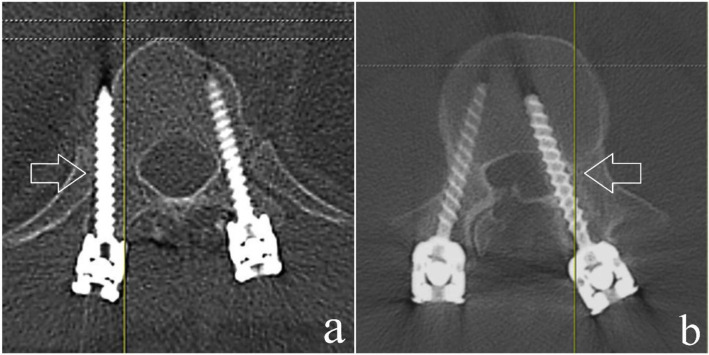
Results: A total of 61 patients with 513 thoracolumbar pedicle screws were available for analysis. Based on our postoperative CT assessment, 28 screws (5.5%; 18 thoracic screws; 10 lumbar screws) had breached the pedicle. There were 14 minor (<3 mm) and 14 major (≥3 mm) breaches. The minimally invasive technique had a significantly lower breach rate compared to open surgery (1.9% vs. 7.9%). By retrospectively analysing the intra-operative fluoroscopic images, we determined certain parameters that could predict a breach during surgery.
Conclusion: Pedicle breaches can still be present despite satisfactory placement of screws visualized intra-operatively. A medial breach is most likely when the length of the pedicle screw spans only up to 50% of the vertebral body as seen on the lateral view but the pedicle screw tip has already transgressed the midline as seen on an AP view. A lateral breach is likely when the tip of the pedicle screw is overlapped by the screw head or is only minimally visualized on an AP view.
Keywords: Fluoroscopy; Pedicle Screws; Spinal Fractures; Spine; Vertebral column.
© the Author(s).
Figures


References
-
- Boucher HH. A method of spinal fusion. J Bone Joint Surg Br. 1959;41-B(2):248–59. - PubMed
-
- Roy-Camille R, Saillant G, Berteaux D, Salgado V. Osteo-synthesis of thoracolumbar spine fractures with metal plates screwed through the vertebral pedicles. Reconstr Surg Traumatol. 1976;15:2–16. - PubMed
-
- Fourney DR, Abi-Said D, Lang FF, McCutcheon IE, Gokaslan ZL. Use of pedicle screw fixation in the management of malignant spinal disease: experience in 100 consecutive procedures. J Neurosurg. 2001;94(1 Suppl):25–37. - PubMed
-
- Masferrer R, Gomez CH, Karahalios DG, Sonntag VK. Efficacy of pedicle screw fixation in the treatment of spinal instability and failed back surgery: a 5-year review. J Neurosurg. 1998;89(3):371–7. - PubMed
LinkOut - more resources
Full Text Sources
Research Materials