Case Reports
. 2021 Mar 17:38:101613.
doi: 10.1016/j.eucr.2021.101613. eCollection 2021 Sep.

Robotic assisted laparoscopic prostatectomy in a patient with prostate cancer and complex urinary tract malformation

Affiliations
Case Reports

Robotic assisted laparoscopic prostatectomy in a patient with prostate cancer and complex urinary tract malformation

Mohamed Abd-Alazeez et al. Urol Case Rep. .

Abstract

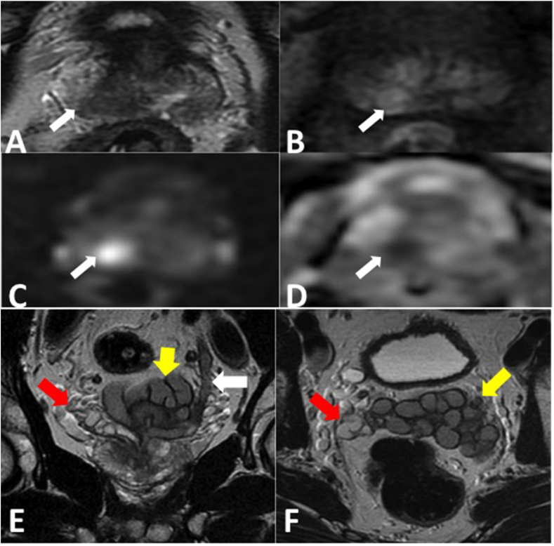
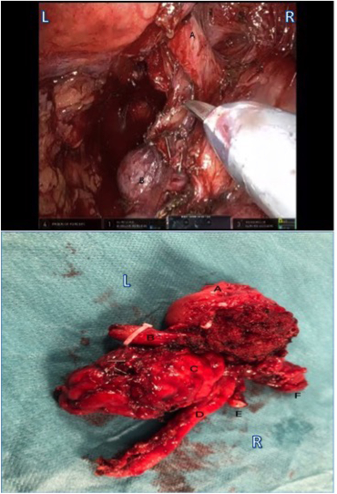
We present a case of prostate cancer with abnormal renal and ureteric anatomy who underwent robotic assisted laparoscopic prostatectomy. This is a 59-year-old European patient who presented with lower urinary tract symptoms (LUTS) and pelvic pain. Investigations revealed prostate cancer as well as a supernumerary right kidney and an atrophic horseshoe left kidney draining into the left seminal vesicle. He was managed with robotic assisted laparoscopic prostatectomy (RALP) using a modified technique. Selective pre-operative investigations and patient counselling led to proper operative planning and good surgical technique and outcome.

Keywords: Ectopic ureter; Prostate cancer; Robot; Supernumerary kidney.


Figures

Fig. 1
Fig. 1
(A) Coronal oblique contrast enhanced CT reconstruction in the excretory phase shows two kidneys in the right lumbar region. The upper kidney (red arrow) has an anterior orientation whilst the lower partially fused kidney (yellow arrow) has an anterolateral orientation. The atrophic left kidney with poor cortical enhancement and no contrast excretion is seen on the left (green arrow) and is joined to the supernumerary right kidney by a fibrous parenchymal bridge (asterisk). (B) 3 D reconstruction of contrast enhanced CT excretory phase showing right supernumerary kidney with lower kidney renal pelvis rotated anterolateral and both ureters joining together and taking a normal course to insert into the urinary bladder. The atrophic left horseshoe kidney shows poor parenchymal enhancement and no contrast excretion and therefore is not seen on this image. (C) DMSA scan shows all the activity in the right supernumerary kidney with no activity in the atrophic horseshoe left kidney. (For interpretation of the references to colour in this figure legend, the reader is referred to the Web version of this article.)
Fig. 2
Fig. 2
A-D: Multi-parametric MRI. Axial T2w (A), dynamic contrast enhanced (B) DWI (C) and ADC map (D) show a hypointense circumscribed area of low T2 signal intensity in the right medial basal peripheral zone (arrow) which displays focal early asymmetrical enhancement and is high signal on the DWI with corresponding low signal intensity on the ADC. This is scored as a PI-RADS 4/5 lesion as the size of the abnormality is less than 1.5 cm, stage T2. Targeted biopsy revealed Gleason 3 + 4. E& F: Coronal T2w(E) and axial T2w(F) of the prostate and seminal vesicles show the ectopic insertion of the left ureter (white arrow) into the left seminal vesicle. The left seminal vesicle (yellow arrow) is asymmetrically enlarged compared to the normal sized right seminal vesicle (red arrow). (For interpretation of the references to colour in this figure legend, the reader is referred to the Web version of this article.)
Fig. 3
Fig. 3
Left: Intraoperative screenshot during posterior approach part of the procedure. A; left ureter, B; left seminal vesicle. Right: Postoperative prostatectomy specimen. A; prostate, B; left ureter, C; left seminal vesicle, D; left vas deferens, E; right vas deferens, F; right seminal vesicle.

References

    1. N'Guessan G., Stephens F.D. Supernumerary kidney. J Urol. 1983;130:649–653. - PubMed
    1. Kumar M., Kumar G., Barwal K., Raina P. Right supernumerary kidney: a rare entity. Urol Case Rep. 2019;23:97–98. - PMC - PubMed
    1. Gonzalvo Perez V., Ramada Benlloch F., Blasco Alfonso J.E., Donderis Guastavino C., Navalon Verdejo P., Zaragoza Orts J. [Supernumerary kidney with ectopic ureteral opening to the vagina associated with horseshoe kidney] Actas Urol Esp. 1992;16:796–798. - PubMed
    1. Unal M., Erem C., Serce K., Tuncer C., Bostan M., Gokce M. The presence of both horseshoe and a supernumerary kidney associated with coarctation of aorta. Acta Cardiol. 1995;50:155–160. - PubMed
    1. Matsumoto T., Koie T., Soma O. [Management of high-risk prostate cancer and left ectopic ureter inserting into seminal vesicle with ipsilateral hypoplastic kidney of a young patient : a case report] Hinyokika Kiyo. 2016;62:329–333. - PubMed

Publication types

LinkOut - more resources