. 2021 Mar 29:11:664221.
doi: 10.3389/fcimb.2021.664221. eCollection 2021.

The Active Subunit of the Cytolethal Distending Toxin, CdtB, Derived From Both Haemophilus ducreyi and Campylobacter jejuni Exhibits Potent Phosphatidylinositol-3,4,5-Triphosphate Phosphatase Activity

Affiliations

The Active Subunit of the Cytolethal Distending Toxin, CdtB, Derived From Both Haemophilus ducreyi and Campylobacter jejuni Exhibits Potent Phosphatidylinositol-3,4,5-Triphosphate Phosphatase Activity

Grace Huang et al. Front Cell Infect Microbiol. .

Abstract

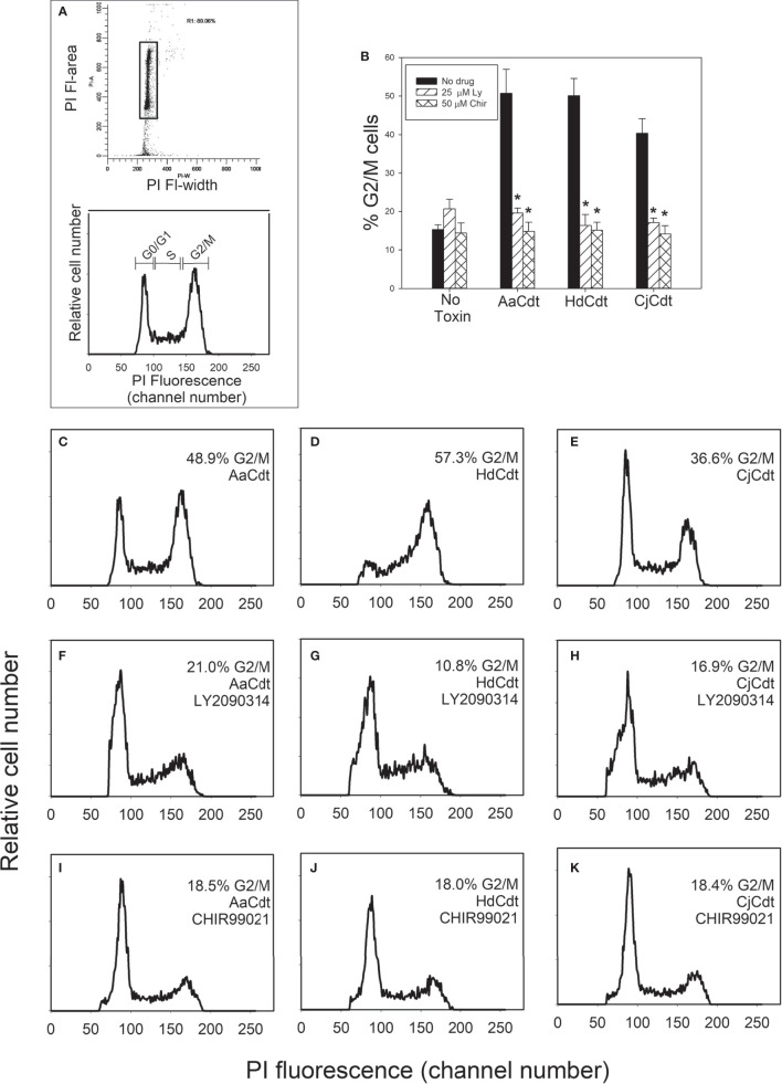
Human lymphocytes exposed to Aggregatibacter actinomycetemcomitans (Aa) cytolethal distending toxin (Cdt) undergo cell cycle arrest and apoptosis. In previous studies, we demonstrated that the active Cdt subunit, CdtB, is a potent phosphatidylinositol (PI) 3,4,5-triphosphate phosphatase. Moreover, AaCdt-treated cells exhibit evidence of PI-3-kinase (PI-3K) signaling blockade characterized by reduced levels of PIP3, pAkt, and pGSK3β. We have also demonstrated that PI-3K blockade is a requisite of AaCdt-induced toxicity in lymphocytes. In this study, we extended our observations to include assessment of Cdts from Haemophilus ducreyi (HdCdt) and Campylobacter jejuni (CjCdt). We now report that the CdtB subunit from HdCdt and CjCdt, similar to that of AaCdt, exhibit potent PIP3 phosphatase activity and that Jurkat cells treated with these Cdts exhibit PI-3K signaling blockade: reduced levels of pAkt and pGSK3β. Since non-phosphorylated GSK3β is the active form of this kinase, we compared Cdts for dependence on GSK3β activity. Two GSK3β inhibitors were employed, LY2090314 and CHIR99021; both inhibitors blocked the ability of Cdts to induce cell cycle arrest. We have previously demonstrated that AaCdt induces increases in the CDK inhibitor, p21CIP1/WAF1, and, further, that this was a requisite for toxin-induced cell death via apoptosis. We now demonstrate that HdCdt and CjCdt also share this requirement. It is also noteworthy that p21CIP1/WAF1 was not involved in the ability of the three Cdts to induce cell cycle arrest. Finally, we demonstrate that, like AaCdt, HdCdt is dependent upon the host cell protein, cellugyrin, for its toxicity (and presumably internalization of CdtB); CjCdt was not dependent upon this protein. The implications of these findings as they relate to Cdt's molecular mode of action are discussed.

Keywords: apoptosis; cell cycle arrest; cytolethal distending toxin; host-parasite interactions; lymphocytes; pathogenesis; toxins.


Conflict of interest statement

The authors declare that the research was conducted in the absence of any commercial or financial relationships that could be construed as a potential conflict of interest.

Figures

Figure 1
Figure 1
Assessment of the HdCdtB and CjCdtB subunits for PIP3 phosphatase activity. Varying amounts of CdtB derived from HdCdt and CjCdt were assessed and compared to AaCdtB for their ability to hydrolyze PIP3 as described in Materials and Methods. The amount of phosphate release was measured using a malachite green binding assay. Data are plotted as phosphate release (nM/30min;mean ± SEM) vs protein concentration. Results are derived from three experiments each performed in triplicate; *indicates statistical significance (p < 0.01) relative to background control samples (0.05 nM/30 min) not receiving protein samples.
Figure 2
Figure 2
Comparison of the effect of HdCdt and CjCdt with AaCdt on phosphorylation of downstream components of the PI-3K signaling pathway. Jurkat cells were treated with AaCdt (AaCdtA/C: 0.5 nM; AaCdtB: 5.7 nM), HdCdt (HdCdtA/C: 0.2 nM; HdCdtB: 5.7 nM), and CjCdt (CjCdtA/C: 1.0 nM; CdtB: 5.7 nM) for 2 hr and then assessed for levels of Akt, pAkt(S473), GSK3ß and pGSK3ß(S9) by Western blot. (A) shows a representative Western blot; (B) shows the compiled results of four experiments (mean ± SEM); *indicates statistical significance p < 0.05 when compared to untreated control cells.
Figure 3
Figure 3
Assessment of GSK3β inhibitors for ability to block Cdt-induced cell cycle arrest. Jurkat cells were pre-treated with medium, 25 µM LY2090314, or 50 µM CHIR99021 for 1 hr followed by the addition of AaCdt (130 pM holotoxin), HdCdt (HdCdtA/C: 0.2 nM; HdCdtB: 0.3 nM) or CjCdt (CjCdtA/C: 1.0 nM; CjCdtB: 1.2 nM). Cells were harvested 24 hr later and analyzed for cell cycle distribution using propidium iodide and flow cytometry as described in Materials and Methods. (A) shows the gating strategy to identify cell cycle distribution. Debris and cell doublets were gated out using plots of propidium iodide fluorescence area versus width (top panel; rectangle); analytical gates were set to identify cells in the G0/G1, S and G2/M phases of the cell cycle based upon propidium iodide fluorescence (bottom panel). The percentage (mean ± SEM) of G2/M cells from three experiments, each performed in duplicate is shown in panel (B); *indicates statistical significance (p<0.01) when compared to cells treated with toxin alone. Representative histograms are presented from a single experiment: panel (C–E) show the results from cells pre-treated with medium only, panel (F–H) show results from cells pre-treated with LY2090314, and panel (I–K) show results from cells pre-treated with CHIR99021. Cells exposed to medium alone exhibited 15.3 ± 2.1% G2/M cells. Panels (C, F, I) were treated with AaCdt, panels (D, G, J) were treated with HdCdt and panels (E, J, K) were treated with CjCdt.
Figure 4
Figure 4
Assessment of the requirement for the CDK inhibitor p21CIP1/WAF1 in Cdt-induced toxicity. (A) JurkatWT cells and Jurkatp21CIP1/WAF1- were treated with AaCdt, HdCdt and CjCdt (same concentrations as above) for 48hr and then analyzed for apoptotic cells by determining the percentage of TUNEL positive cells as detailed in Materials and Methods. The percentage of apoptotic cells is plotted for both JurkatWT (solid bars) and Jurkatp21CIP1/WAF1- (cross hatched bars) cells and represent the mean ± SEM of three experiments. *indicates statistical significance (p < 0.01) when compared to JurkatWT. (B) JurkatWT cells and Jurkatp21CIP1/WAF1- were treated with AaCdt, HdCdt and CjCdt for 16hr and assessed for the percentage of cells in the G2/M phase of the cell cycle as described in Materials and Methods. The percentage of G2/M cells is plotted for both JurkatWT (solid bars) and Jurkatp21CIP1/WAF1- (cross hatched bars) cells and represent the mean ± SEM of three experiments. (C) Jurkat cells were pre-treated with medium (solid bars) or Geldanamycin A [GA; (cross-hatched bars)] for one hr before the addition of Cdt. Cells were analyzed for apoptosis using the TUNEL assay 48 hr later; results represent the mean ± SEM of three experiments. **indicates statistical significance (p < 0.05).
Figure 5
Figure 5
Comparison of the effects of AaCdt, HdCdt, and CjCdt on phosphorylation of H2AX. JurkatWT cells were incubated in the presence AaCdt, HdCdt, and CjCdt (same concentrations as above) for one and four hrs. Cells were then fractionated and analyzed by Western blot for the presence of pH2AX. Etoposide-treated cells were used as a positive control. A representative blot of three experiments is shown.
Figure 6
Figure 6
Comparison of HdCdt and CjCdt for the requirement of the host cell protein cellugyrin to elicit toxicity. (A) JurkatWT (solid bars) and JurkatCg- (cross-hatched bars) cells were treated with AaCdt, HdCdt, and CjCdt as described above for 16 hr and then assessed for cell cycle distribution. Results show the percentage of cells in the G2/M phases of the cell cycle and are plotted as mean ± SEM of three experiments. (B) JurkatWT (solid bars) and JurkatCg- (cross-hatched bars) cells were treated with AaCdt, HdCdt, and CjCdt for 48 hr and analyzed to determine the percentage of apoptotic cells using the TUNEL assay; results are plotted as the mean ± SEM of three experiments. *indicates statistical significance (p < 0.01) when compared to JurkatWT cells.
Figure 7
Figure 7
Schematic model showing proposed mechanism for CdtB internalization and toxicity in human lymphocytes. AaCdt, HdCdt, and CjCdt bind to cells via cholesterol in the context of membrane lipid rafts. As a result of exposure to Cdt, cellugyrin-containing SLMVs translocate from cytosol to membrane lipid rafts. We propose that this translocation leads to the association of AaCdtB and HdCdtB with the cellugyrin-containing SLMVs. This interaction may involve direct binding to cellugyrin either on extra- or intra-vesicular loops or indirect association via an un-identified binding partner. We further propose that CdtB is transported via SLMVs to intracellular target sites, such as locations containing PIP3 pools where the enzymatically active CdtB subunit is released from SLMVs and is then able to dephosphorylate the signaling lipid, resulting in PI-3K blockade and toxicity. Based upon CjCdt toxicity independence of cellugyrin for toxicity (and presumably internalization and trafficking), we propose that CjCdtB enters through a cellugyrin- independent mechanism, hijacks retrograde transport mechanisms, and, in lymphocytes, accumulates in sites of PIP3 pools similar to AaCdtB and HdCdtB.

Similar articles

Cited by

References

    1. Abbas T., Dutta A. (2009). P21 in Cancer: Intricate Networks and Multiple Activities. Nat. Rev. Cancer 9 (6), 400–414. 10.1038/nrc2657 - DOI - PMC - PubMed
    1. Bezine E., Vignard J., Mirey G. (2014). The cytolethal distending toxin effects on Mammalian cells: a DNA damage perspective. Cells 3 (2), 592–615. 10.3390/cells3020592 - DOI - PMC - PubMed
    1. Boesze-Battaglia K., Besack D., McKay T., Zekavat A., Otis L., Jordan-Sciutto K., et al. . (2006). Cholesterol-rich membrane microdomains mediate cell cycle arrest induced by Actinobacillus actinomycetemcomitans cytolethal-distending toxin. Cell Microbiol. 8 (5), 823–836. 10.1111/j.1462-5822.2005.00669.x - DOI - PMC - PubMed
    1. Boesze-Battaglia K., Brown A., Walker L., Besack D., Zekavat A., Wrenn S., et al. . (2009). Cytolethal distending toxin-induced cell cycle arrest of lymphocytes is dependent upon recognition and binding to cholesterol. J. Biol. Chem. 284 (16), 10650–10658. 10.1074/jbc.M809094200 - DOI - PMC - PubMed
    1. Boesze-Battaglia K., Walker L. P., Zekavat A., Dlakic M., Scuron M. D., Nygren P., et al. . (2015). The Aggregatibacter actinomycetemcomitans Cytolethal Distending Toxin Active Subunit CdtB Contains a Cholesterol Recognition Sequence Required for Toxin Binding and Subunit Internalization. Infect. Immun. 83 (10), 4042–4055. 10.1128/IAI.00788-15 - DOI - PMC - PubMed

Publication types