

A Spectrofluorophotometrical Method Based on Fura-2-AM Probe to Determine Cytosolic Ca2+ Level in Pseudomonas syringae Complex Bacterial Cells
- PMID: 33855111
- PMCID: PMC8032496
- DOI: 10.21769/BioProtoc.3949
A Spectrofluorophotometrical Method Based on Fura-2-AM Probe to Determine Cytosolic Ca2+ Level in Pseudomonas syringae Complex Bacterial Cells
Abstract
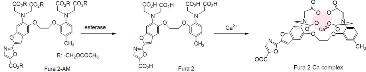
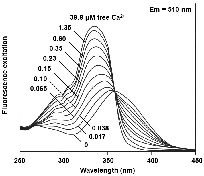
Calcium signaling is an emerging mechanism by which bacteria respond to environmental cues. To measure the intracellular free-calcium concentration in bacterial cells, [Ca2+]i, a simple spectrofluorometric method based on the chemical probe Fura 2-acetoxy methyl ester (Fura 2-AM) is here presented using Pseudomonad bacterial cells. This is an alternative and quantitative method that can be completed in a short period of time with low costs, and it does not require the induction of heterologously expressed protein-based probes like Aequorin. Furthermore, it is possible to verify the properties of membrane channels involved in Ca2+ entry from the extracellular matrix. This method is in particular valuable for measuring [Ca2+]i in the range of 0.1-39.8 µM in small cells like those of prokaryotes.
Keywords: Cytosolic calcium concentration; Fura 2-AM; Live cell signaling; Pseudomonad; Spectrophotometer.
Copyright © 2021 The Authors; exclusive licensee Bio-protocol LLC.
Conflict of interest statement
Competing interestsThe authors declare no conflict-of-interest and have no competing financial interests. Informed consent was obtained from all individual participants included in the study.
Figures

Similar articles
-
Intracellular ionized calcium changes in squid giant axons monitored by Fura-2 and aequorin.Ann N Y Acad Sci. 1991;639:112-25. doi: 10.1111/j.1749-6632.1991.tb17295.x. Ann N Y Acad Sci. 1991. PMID: 1785835
-
Fura-2 antagonises calcium-induced calcium release.Cell Calcium. 2003 Jan;33(1):27-35. doi: 10.1016/s0143-4160(02)00179-3. Cell Calcium. 2003. PMID: 12526885
-
Redox properties of the calcium chelator Fura-2 in mimetic biomembranes.Cell Calcium. 2008 Jun;43(6):615-21. doi: 10.1016/j.ceca.2007.10.002. Epub 2007 Nov 14. Cell Calcium. 2008. PMID: 18001832
-
Intracellular compartmentalization of fura-2 dye demonstrated by laser-excitation fluorescence microscopy: a problem in measuring cytosolic free calcium concentration using fura-2 fluorescence in vascular smooth muscle cells.Tohoku J Exp Med. 1989 Sep;159(1):23-35. doi: 10.1620/tjem.159.23. Tohoku J Exp Med. 1989. PMID: 2815073
-
Assessment of Fura-2 for measurements of cytosolic free calcium.Cell Calcium. 1990 Feb-Mar;11(2-3):63-73. doi: 10.1016/0143-4160(90)90060-8. Cell Calcium. 1990. PMID: 2191782 Review.
References
LinkOut - more resources
Full Text Sources
Other Literature Sources
Miscellaneous
