. 2021 Mar 15;24(4):102312.
doi: 10.1016/j.isci.2021.102312. eCollection 2021 Apr 23.

MSC-like cells increase ability of monocyte-derived dendritic cells to polarize IL-17-/IL-10-producing T cells via CTLA-4

Affiliations

MSC-like cells increase ability of monocyte-derived dendritic cells to polarize IL-17-/IL-10-producing T cells via CTLA-4

Anett Mázló et al. iScience. .

Abstract

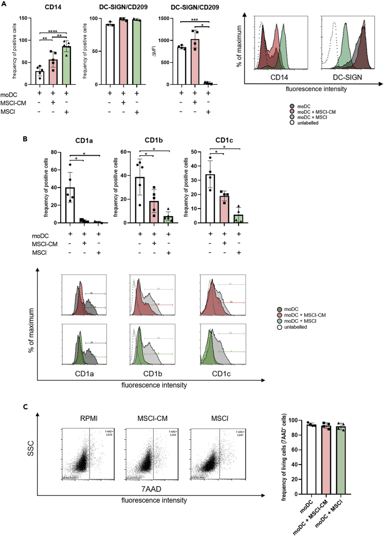
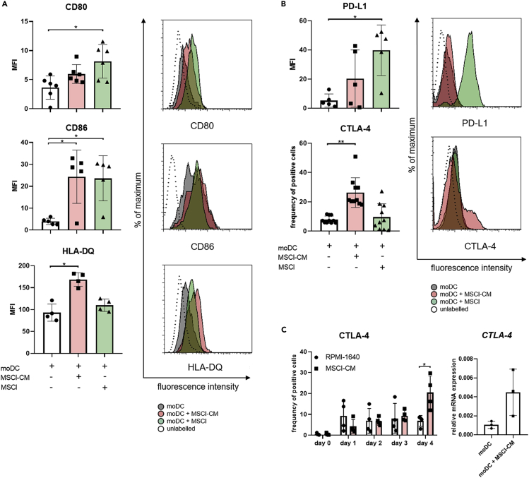
Mesenchymal stromal cell-like (MSCl) cells generated from human embryonic stem cells are considered to be an eligible cell line to model the immunomodulatory behavior of mesenchymal stromal cells (MSCs) in vitro. Dendritic cells (DCs) are essential players in the maintenance and restoration of the sensitive balance between tolerance and immunity. Here, the effects of MSCl cells on the in vitro differentiation of human monocytes into DCs were investigated. MSCl cells promote the differentiation of CTLA-4 expressing DCs via the production of all-trans retinoic acid (ATRA) functioning as a ligand of RARα, a key nuclear receptor in DC development. These semi-matured DCs exhibit an ability to activate allogeneic, naive T cells and polarize them into IL-10 + IL-17 + double-positive T helper cells in a CTLA-4-dependent manner. Mapping the molecular mechanisms of MSC-mediated indirect modulation of DC differentiation may help to expand MSCs' clinical application in cell-free therapies.

Keywords: Components of the Immune System; Immunology; Molecular Biology; Stem Cells Research.


Conflict of interest statement

The authors declare no conflicts of interest related to this research.

Figures

None
Graphical abstract
Figure 1
Figure 1
The cytokine and chemokine production of moDCs is modulated by MSCl cells To examine the regulatory effects of MSCl cells or the MSCl-CM on the cytokine and chemokine production of moDCs, on day 4 of moDC differentiation the integrated density of soluble mediators was measured by Human XL Cytokine Array Kit (A) and the concentration of secreted cytokine IL-6, IL-10, TGFβ was detected by ELISA (B). In case of co-cultures, the diagrams represent the secretion of cytokines released by moDCs and MSCl cells. Mean values of relative cytokine levels and concentrations were calculated from 4 or more independent experiments. Data are represented as individual data points with the mean ± standard deviation. In the statistical analysis, one-way ANOVA followed by Bonferroni's multiple comparison test was used with significance defined as ∗p < 0.05, ∗∗p < 0.01 and ∗∗∗∗p < 0.0001. See also Figure S1.
Figure 2
Figure 2
MSCl cells and MSCl-CM modify the cell surface expression of CD14, DC-SIGN/CD209 and group1 CD1 family members CD14+ monocytes were cultured with recombinant IL-4 and GM-CSF ± MSCl cells or MSCl-CM for 4 days. On day 4, the cell surface expression of CD14 and DC-SIGN (A), and that of CD1a, CD1b, and CD1c (B) were analyzed by flow cytometry on monocyte-derived cells. To exclude the possibility of unspecific staining because of the presence of dead cells, the viability of cells was measured by 7-aminoactinomycin D (7-AAD) staining using flow cytometry (C). The MFI (median fluorescence intensity) and the mean values of the ratio of cells positive for the measured surface molecules were calculated from at least three independent experiments. Data are represented as individual data points with the mean ± standard deviation. Histograms show one of at least four independent experiments. In the statistical analysis, one-way ANOVA followed by Bonferroni's multiple comparison test was used with significance defined as ∗p < 0.05, ∗∗p < 0.01, ∗∗∗p < 0.001 and ∗∗∗∗p < 0.0001. See also Figure S2.
Figure 3
Figure 3
MSCl cells and MSCl-CM alter the cell surface expression of molecules involved in T cell activation CD14+ monocytes were cultured with recombinant IL-4 and GM-CSF ± MSCl cells or MSCl-CM for 4 days. On day 4, monocyte-derived cells were analyzed for the cell surface expression of the T cell stimulatory CD80, CD86, HLA-DQ (A) and the regulatory CTLA-4 and PD-L1 (B) molecules by flow cytometry. The frequency of CTLA-4-expressing cells was monitored on each day during the differentiation process (C). The gene expression level of CTLA-4 was measured by qPCR on day 4 (C). The MFI and the mean values of the ratio of cells positive for the measured cell surface molecules were calculated from at least four independent experiments. Mean values of relative mRNA levels were calculated from three independent experiments. Data are represented as individual data points with the mean ± standard deviation. Histograms show one of at least four independent experiments. In the statistical analysis, one-way ANOVA followed by Bonferroni's multiple comparison test (A and B), as well as Student's t-test and Mann-Whitney rank-sum test (C) were used with significance defined as ∗p < 0.05, ∗∗p < 0.01. See also Figure S3.
Figure 4
Figure 4
MSCl cells change the immune regulatory potential of moDCs at least partially via nuclear factor RARα and ATRA To investigate how MSCl-CM affects the differentiation of moDC, monocytes were differentiated in the presence or absence of 1 nM RARα activator, ATRA followed by a 75 min incubation period with or without 1 μM BMS614 (BMS) specific RARα-antagonist prior to exchange the cell culture medium to RPMI-1640 or MSCl-CM. Monocytes were differentiated into moDC in the presence or absence of MSCl-CM for 4 days. On day 4, moDCs were analyzed for the cell surface expression of CD1a, the T cell stimulatory HLA-DQ, CD86 and the T cell co-inhibitory CTLA-4 proteins by flow cytometry (A). To test and compare the capability of freshly isolated monocytes, moDCs and MSCl to produce ATRA, mRNA was extracted from the different cell types. The relative mRNA expression level of target genes RDH10, ALDH1A1, ALDH1A2, and ALDH1A3 was measured by qRT-PCR (B). To prove the effect of ATRA to the differentiation process of monocytes into moDCs, the RALDH enzymes essential for the synthesis of ATRA were inhibited specifically by 1μM DEAB in the MSCl cultures for 24 hr before the collection of MSCl-CM. CD14+ monocytes were cultured with recombinant IL-4 and GM-CSF and MSCl-CM with or without ATRA for 4 days. On day 4, moDCs were analyzed for the cell surface expression level of CD1a, HLA-DQ, CD86 and the regulatory CTLA-4 molecules by flow cytometry (C). Mean values of MFI and moDCs positive for the measured cell surface antigen were calculated from at least four independent experiments. Mean values of relative mRNA levels were calculated from three independent experiments. Data are represented as individual data points with the mean ± standard deviation. In the statistical analysis, one-way ANOVA followed by Bonferroni's multiple comparison test was used with significance defined as ∗p < 0.05, ∗∗p < 0.01 and ∗∗∗p < 0.001, ∗∗∗∗p < 0.0001.
Figure 5
Figure 5
CTLA-4 expression on moDCs differentiated in the presence of MSCl-CM is essential to drive the development of IL-10- and IL-17-producing T cells CD14+ monocytes were differentiated into moDC in the presence or absence of MSCl-CM for 4 days. On day 4, the moDCs were treated with anti-CTLA-4 blocking antibody and were co-cultured with allogeneic T lymphocytes for further 3, 5 or 9 days. ELISPOT assays were used to determine the number of IL-17- (A) and IL-10- (B) producing T cells. The average values of spot numbers indicating T-lymphocyte responses were counted from 3 micro-wells. Mean values of spot numbers were calculated from 3 independent experiments. IL-10- and IL-17-producing CD4+ T cells were detected by flow cytometry (C). Mean values of the ratio of T cells positive for the measured cell intracellular cytokines were calculated from four independent experiments. Data are represented as individual data points with the mean ± standard deviation. Contour plots show one of the three independent experiments. In the statistical analysis, one-way ANOVA followed by Bonferroni's multiple comparison test was used with significance defined as ∗p < 0.05. See also Figures S4–S6.

References

    1. Abumaree M.H., Bahattab E., Alsadoun A., Al Dosaimani A., Abomaray F.M., Khatlani T., Kalionis B., El-Muzaini M.F., Alawad A.O., Alaskar A.S. Characterization of the interaction between human decidua parietalis mesenchymal stem/stromal cells and natural killer cells. Stem Cell Res. Ther. 2018;9:102. - PMC - PubMed
    1. Bacskai I., Mazlo A., Kis-Toth K., Szabo A., Panyi G., Sarkadi B., Apati A., Rajnavolgyi E. Mesenchymal stromal cell-like cells set the balance of stimulatory and inhibitory signals in monocyte-derived dendritic cells. Stem Cells Dev. 2015;24:1805–1816. - PubMed
    1. Bene K., Varga Z., Petrov V.O., Boyko N., Rajnavolgyi E. Gut microbiota species can provoke both inflammatory and tolerogenic immune responses in human dendritic cells mediated by retinoic acid receptor alpha ligation. Front. Immunol. 2017;8:427. - PMC - PubMed
    1. Braza F., Dirou S., Forest V., Sauzeau V., Hassoun D., Chesne J., Cheminant-Muller M.A., Sagan C., Magnan A., Lemarchand P. Mesenchymal stem cells induce suppressive macrophages through phagocytosis in a mouse model of asthma. Stem Cells. 2016;34:1836–1845. - PubMed
    1. Chen P.M., Liu K.J., Hsu P.J., Wei C.F., Bai C.H., Ho L.J., Sytwu H.K., Yen B.L. Induction of immunomodulatory monocytes by human mesenchymal stem cell-derived hepatocyte growth factor through ERK1/2. J. Leukoc. Biol. 2014;96:295–303. - PubMed

LinkOut - more resources