. 2021 Mar 10;3(2):fcab022.
doi: 10.1093/braincomms/fcab022. eCollection 2021.

Reward sensitivity and action in Parkinson's disease patients with and without apathy

Affiliations

Reward sensitivity and action in Parkinson's disease patients with and without apathy

Kinan Muhammed et al. Brain Commun. .

Abstract

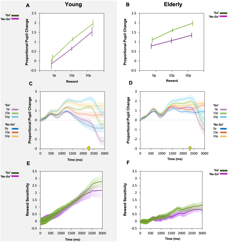
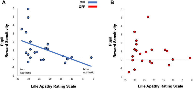
Clinical apathy results in dysfunction of goal directed behaviour, a key component of which is the initiation of action. Previous work has suggested that blunting of reward sensitivity is an important mechanism underlying apathy. However, an additional component might be impoverished initiation of action itself. This study aims to investigate the link between motivation and motor output and its association with apathy and dopamine. An oculomotor task that measures pupillary and saccadic response to monetary incentives was used to assess reward sensitivity, first in 23 young and 18 elderly controls, and then in 22 patients with Parkinson's disease tested ON and OFF dopaminergic medication. To distinguish between pupillary responses to anticipated reward alone versus responses associated with motor preparation, a saccadic 'go/no-go' task was performed. Half of the trials required a saccade to be initiated to receive a reward and in the remaining trials no action was required but reward was still obtained. No significant difference in pupil response was demonstrated between the two conditions in all groups tested, suggesting pupillary responses to rewards are not contingent upon motor preparation in Parkinson's disease. Being ON or OFF dopamine did not influence this response either. Previous work demonstrated associations between apathy and pupillary reward insensitivity in Parkinson's disease. Here we observed this effect only when an action was required to receive a reward, and only in the ON state. These findings suggest that apathy in Parkinson's disease is linked to reduced reward sensitivity and that this is most prominently observed when actions have to be initiated to rewarding goals, with the effect modulated by being ON dopaminergic medication. OFF medication, there was no such strong relationship, and similarly in the 'no-go' conditions, either ON or OFF dopaminergic drugs. The results provide preliminary data which suggest that apathy in Parkinson's disease is associated with a reduction in reward sensitivity and this is most evident when associated with initiation of goal directed actions in the presence of adequate dopamine.

Keywords: Parkinson’s disease; apathy; dopamine; pupillometry; reward sensitivity.


Figures

None
Graphical abstract
Figure 1
Figure 1
Oculomotor experimental paradigm. During the task, 50% of trials required a saccade to be made to obtain a reward (‘go’ trials indicated by a ‘+’ at the start of the trial). In the remaining 50% of trials, participants were required to maintain fixation centrally (‘no-go’ trials indicated by an ‘x’ at the start of the trial). ‘go’ and ‘no-go’ trials were randomly intermixed. Reward obtained was based on reaction time in the ‘go’ trials and reward rates in both arms were matched.
Figure 2
Figure 2
Pupil responses in young (A, C, E) and elderly (B, D, F) participants across ‘go’ and ‘no-go’ trials. (A) Pupil proportional change to reward taken as an average across a 1000 ms epoch from 1400 ms to 2400 ms in young participants. Increased pupil response to reward was present as reward increased but no difference was seen between ‘go’ trials in green and ‘no-go’ trials in violet. (B) Pupil proportional change to reward in elderly participants. Increased pupil response to reward was present as reward level increased but like in the young, no difference was seen between ‘go’ trials (green line) and ‘no-go’ trials (violet line). (C) Average proportional pupil changes over the course of the trials in young participants, broken down into responses to ‘go’ and ‘no-go’ cues. Cue onset was at time 0 ms. Average onset of saccades indicated by yellow arrow (2296 ms). (D) Average proportional pupil changes over the course of the trials in the elderly. Average onset of saccades indicated by yellow arrow (2347 ms). (E) Average reward sensitivity over time in young participants, taken as the difference in the proportional pupil change between the 50p reward condition and the 0p reward. Broken into reward sensitivity for ‘go’ trials—green, versus ‘no-go’ trials—violet. No significant difference in reward sensitivity was present at any time point across the length of the entire trial. (F) Average reward sensitivity over time in the elderly, calculated as the difference in the proportional pupil change between the 50p reward condition and the 0p reward for each time point. Reward sensitivity for ‘go’ trials plotted in green and ‘no-go’ trials in violet. No significant difference in pupil reward sensitivity was present.
Figure 3
Figure 3
Reward sensitivity in ‘no-go’ (A, B) and ‘go’ (C, D) trials compared between young and elderly groups. (A) Average reward sensitivity over time in ‘no-go’ trials, taken as the difference in the proportional pupil change between the 50p reward condition and the 0p reward. Broken into reward sensitivity for young (yellow) and elderly participants (grey). Black bar denotes significant difference between the two groups over a short ∼350 ms period. (B) Pupil reward sensitivity (50p-0p reward) taken as an average across a 1000 ms time epoch from 1400 ms to 2400 ms in the ‘no-go’ trials (Purple background shading in A). Young participants had significantly larger pupil response to reward over the time epoch of interest compared to elderly participants. (C) Average reward sensitivity over time in ‘go’ trials, taken as the difference in the proportional pupil change between the 50p reward condition and the 0p reward. Reward sensitivity in young participants (yellow) and elderly (grey). Black bar denotes significant difference between the two groups over a large period of the trial starting from ∼1000 ms post reward cue onset to the end of the trial. (D) Pupil reward sensitivity (50p-0p reward) taken as an average across a 1000 ms period from 1400 ms to 2400 ms in the ‘go’ trials (Purple background shading in C). Young participants had significantly larger pupil response to reward compared to elderly participants.
Figure 4
Figure 4
Parkinson’s disease pupil response to reward in ‘go’ and ‘no-go’ trials in the ON and OFF state. (A) When ON dopamine, pupil proportional change to reward taken as an average across a 1000 ms period of time from 1400 ms to 2400 ms. Increased pupil response to reward was present as reward magnitude increased but no difference was seen between ‘go’ trials (green) and ‘no-go’ trials (violet). (B) When OFF dopamine, pupil proportional change also increased as reward increased, with no difference in ‘go’ trials (green) and ‘no-go’ trials (violet).
Figure 5
Figure 5
Parkinson’s disease pupil reward sensitivity during ON (A) and OFF (B) dopamine states. (A) Average reward sensitivity over time in Parkinson’s disease when ON in ‘go’ trials (green) compared to ‘no-go’ trials (violet) taken as the difference in the proportional pupil change between the 50p reward condition and the 0p reward. Using permutation testing at each time point, no significant difference was demonstrated between the two conditions over the course of the trial. (B) Average reward sensitivity over time in Parkinson’s disease when OFF in ‘go’ trials (green) compared to ‘no-go’ trials (violet). No significant difference was present between the two conditions either.
Figure 6
Figure 6
Correlation with clinical apathy questionnaire scores (Lille Apathy Rating Scale) and pupil reward sensitivity when a saccade was made to obtain a reward in Parkinson’s disease patients ON and OFF dopamine. (A) A significant correlation between average pupil reward sensitivity in Parkinson’s disease patients ON (blue) and their clinical interview LARS score was present only in the ‘go’ trials. More apathetic patients exhibit less pupillary reward sensitivity compared to more motivated patient. Spearman correlation, rs = −0.472, P < 0.03. Mean apathetic patients reward sensitivity in ‘go’ trials ON dopamine 0.56 (SD 0.76). Non-Apathetic patients mean 1.65 (SD 1.98). No correlation was demonstrated in ‘no-go’ trials when ON or OFF dopamine. (B) The correlation between average pupil reward sensitivity in Parkinson’s disease during ‘go’ trials and LARS score was abolished when OFF dopamine (red). Spearman correlation, rs = −0.091, P = 0.686. Mean apathetic patients reward sensitivity in ‘go’ trials OFF dopamine 0.33 (SD 0.7). Non-Apathetic patients mean 1.68 (SD 2.7).
Figure 7
Figure 7
Parkinson’s disease and elderly pupil response to reward in the ON and OFF state during ‘no-go’ (A) and ‘go’ (B) trials. (A) No differences in the average pupil change between ON, OFF or elderly controls in ‘no-go’ trials were present either, however all groups demonstrated increases in pupil size for larger reward levels. (B) Average pupillary response over the time epoch of interest (1400–2400 ms) in all groups displayed an increase in pupil size for increasing reward level in ‘go’ trials. There were no differences in the average pupil change between ON (blue), OFF (red) or elderly controls (grey).

References

    1. Marin RS. Apathy: a neuropsychiatric syndrome. J Neuropsychiatry Clin Neurosci. 1991;3(3):243–254. - PubMed
    1. Cummings JL, Mega M, Gray K, Rosenberg-Thompson S, Carusi DA, Gornbein J.. The Neuropsychiatric Inventory: comprehensive assessment of psychopathology in dementia. Neurology. 1994;44(12):2308–2314. - PubMed
    1. Stuss D, Van Reekum R, Murphy K.. Differentiation of states and causes of apathy. In: The Neuropsychology of Emotion. 2000:340–363.
    1. Robert PH, Clairet S, Benoit M, et al.The Apathy Inventory: assessment of apathy and awareness in Alzheimer’s disease, Parkinson’s disease and mild cognitive impairment. Int J Geriatr Psychiatry. 2002;17(12):1099–1105. - PubMed
    1. Starkstein SE, Leentjens AFG.. The nosological position of apathy in clinical practice. J Neurol Neurosurg Psychiatry. 2008;79(10):1088–1092. - PubMed

LinkOut - more resources