

Mouse Mandibular Retromolar Taste Buds Associated With a Mucus Salivary Gland
- PMID: 33855345
- PMCID: PMC9034206
- DOI: 10.1093/chemse/bjab019
Mouse Mandibular Retromolar Taste Buds Associated With a Mucus Salivary Gland
Abstract
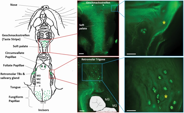
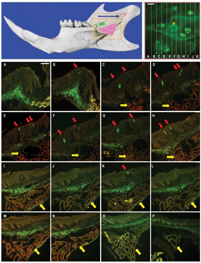
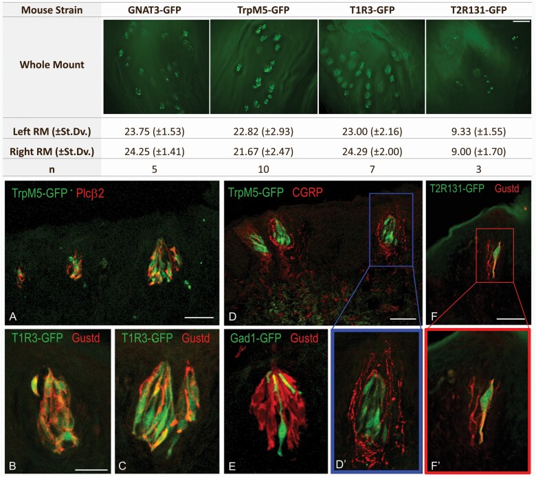
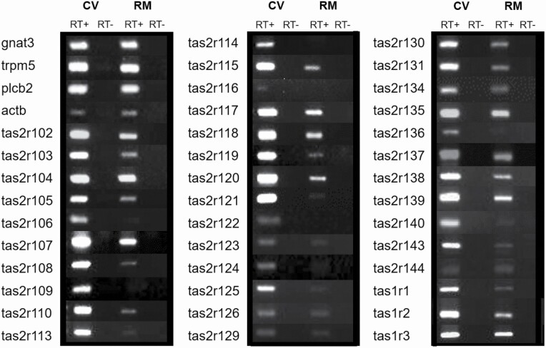
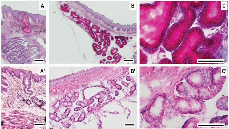
We have characterized a recently rediscovered chemosensory structure at the rear of the mandibular mucosa in the mouse oral cavity originally reported in the 1980s. This consists of unorganized taste buds, not contained within troughs, associated with the ducts of an underlying minor salivary gland. Using whole-mount preparations of transgenic mice expressing green fluorescent protein under the promoter of taste-signaling-specific genes, we determined that the structure contains taste bud clusters and salivary gland orifices at the rear of each mandible, distal to the last molar and anterior to the ascending ramus. Immunohistochemical analysis shows in the retromolar taste buds expression of the taste receptors Tas2R131 and T1R3 and taste cascade molecules TrpM5, PLCβ2, and GNAT3, consistent with type II taste cells, and expression of GAD1, consistent with type III taste cells. Furthermore, the neuronal marker, calcitonin gene-related peptide, in retromolar mucosa tissue wrapping around TrpM5+ taste buds was observed. RT-PCR showed that retromolar taste buds express all 3 mouse tas1r genes, 28 of the 35 tas2r genes, and taste transduction signaling genes gnat3, plcb2, and trpm5, making the retromolar taste buds similar to other lingual and palate taste buds. Finally, histochemistry demonstrated that the mandibular retromolar secretory gland is a minor salivary gland of mucous type. The mandibular retromolar taste structure may thus play a role in taste sensation and represent a potential novel pharmacological target for taste disorders.
Keywords: chemosensory; minor salivary gland; retromolar; taste buds; taste receptors.
© The Author(s) 2021. Published by Oxford University Press. All rights reserved. For permissions, please e-mail: journals.permissions@oup.com.
Figures
Similar articles
-
Calcitonin Gene-Related Peptide Reduces Taste-Evoked ATP Secretion from Mouse Taste Buds.J Neurosci. 2015 Sep 16;35(37):12714-24. doi: 10.1523/JNEUROSCI.0100-15.2015. J Neurosci. 2015. PMID: 26377461 Free PMC article.
-
Taste-signaling proteins are coexpressed in solitary intestinal epithelial cells.Chem Senses. 2007 Jan;32(1):41-9. doi: 10.1093/chemse/bjl034. Epub 2006 Oct 9. Chem Senses. 2007. PMID: 17030556
-
Taste bud papillae on the retromolar mucosa of the rat, mouse and golden hamster.Acta Anat (Basel). 1983;117(4):374-81. doi: 10.1159/000145810. Acta Anat (Basel). 1983. PMID: 6666539
-
Role of saliva in the maintenance of taste sensitivity.Crit Rev Oral Biol Med. 2000;11(2):216-29. doi: 10.1177/10454411000110020501. Crit Rev Oral Biol Med. 2000. PMID: 12002816 Review.
-
Taste buds as peripheral chemosensory processors.Semin Cell Dev Biol. 2013 Jan;24(1):71-9. doi: 10.1016/j.semcdb.2012.12.002. Epub 2012 Dec 20. Semin Cell Dev Biol. 2013. PMID: 23261954 Free PMC article. Review.
Cited by
-
The TRP channels serving as chemical-to-electrical signal converter.Physiol Rev. 2025 Jul 1;105(3):1033-1074. doi: 10.1152/physrev.00012.2024. Epub 2025 Jan 15. Physiol Rev. 2025. PMID: 39813402 Free PMC article. Review.
-
Cellular Diversity and Regeneration in Taste Buds.Curr Opin Physiol. 2021 Apr;20:146-153. doi: 10.1016/j.cophys.2021.01.003. Epub 2021 Jan 12. Curr Opin Physiol. 2021. PMID: 33615087 Free PMC article.
References
-
- Akal UK, Kucukyavuz Z, Nalcaci R, Yilmaz T. 2004. Evaluation of gustatory function after third molar removal. Int J Oral Maxillofac Surg. 33:564–568. - PubMed
-
- Albuquerque AFM, Soares ECS, De barros silva PG, De lima BB. Carvalho FSR, Ribeiro TR, De Sa Cavalcante D, Costa FWG. 2019. Clinical investigation of gustatory and neurosensory alterations following mandibular third molar surgery: an observational prospective study. Clin Oral Investig. 23:2941–2949. - PubMed
-
- Anand R, Shankar DP, Manodh P, Devadoss P, Aparna M, Neelakandan RS. 2018. Short-term evaluation of gustatory changes after surgical removal of mandibular third molar-a prospective randomized control trial. J Oral Maxillofac Surg. 76:258–266. - PubMed
-
- Bartoshuk L. 1991. Losses of taste in regions of the tongue’s surface. Appetite. 17:69. - PubMed
-
- Bromley SM. 2000. Smell and taste disorders: a primary care approach. Am Fam Phys. 61:427–436, 438. - PubMed
Publication types
MeSH terms
Grants and funding
LinkOut - more resources
Full Text Sources
Other Literature Sources
