

Knee articular cartilage injury treatment with matrix-induced autologous chondrocyte implantation (MACI): correlation at 24 and 120 months between clinical and radiological findings using MR arthrography
- PMID: 33855594
- PMCID: PMC8364544
- DOI: 10.1007/s00256-021-03775-y
Knee articular cartilage injury treatment with matrix-induced autologous chondrocyte implantation (MACI): correlation at 24 and 120 months between clinical and radiological findings using MR arthrography
Abstract
Objective: To evaluate the long-term evolution of matrix-induced autologous chondrocyte implantation (MACI) with magnetic resonance (MR) arthrography and verify the correlation between radiological and clinical findings.
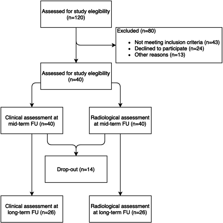
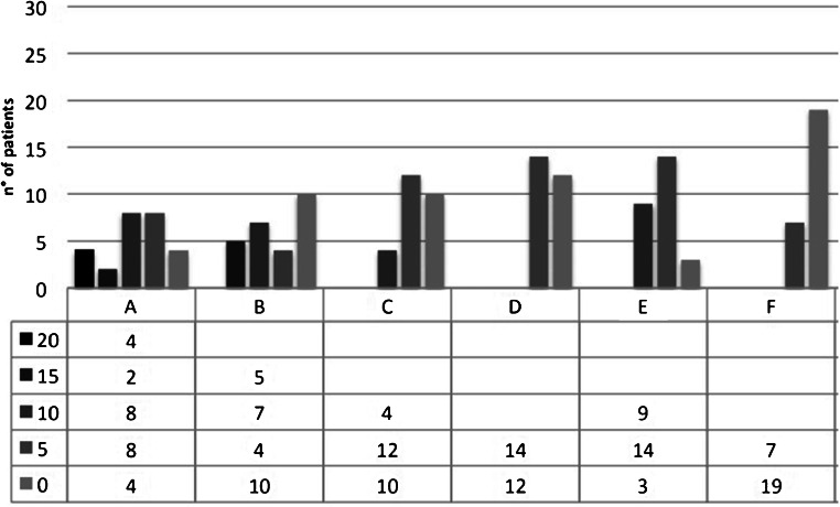
Materials and methods: Twenty-six patients (20 m/6f) were diagnosed with knee chondral injuries and treated with MACI implantation. Each patient received MR arthrography and clinical examination at mid-term (range 22-36 months) and long term (range 96-194 months) after surgery. MR arthrography was performed with dedicated coil and a 1.5-Tesla MR unit. The modified MOCART scale was used to evaluate the status of chondral implants. Implant coating, integration to the border zone, and the surface and structure of the repaired tissue were evaluated. Presence of bone marrow oedema was evaluated. The Cincinnati Knee Rating System (CKRS) was used for clinical assessment.
Results: At long term, 4/26 patients had complete alignment; 5/26 had a complete integration of the margins; in 4/26 cases, the implant surface was undamaged; in 14/26 cases, the reparative tissue was homogeneous. In 9/26 cases, the implant showed isointense signal compared to articular cartilage, while the presence of subchondral bone oedema was documented in 19/26 cases. The average radiological score decreased from 59.2 (mid-term) to 38.6 (long term). The average clinical score decreased from 8.9 to 8.3.
Conclusions: Decrease in clinical results was not significant (0.6 points p = .06), but mMOCART scores decreased significantly (p = .00003). Although imaging studies showed deterioration of the grafts, the patients did not have significant clinical deterioration (231/250).
Keywords: Arthrography; Articular; Cartilage; Chondrocytes; Follow-up studies; Magnetic resonance imaging.
© 2021. The Author(s).
Conflict of interest statement
The authors declare no competing interests.
Figures


Similar articles
-
Matrix-induced autologous chondrocyte implantation of the knee: mid-term and long-term follow-up by MR arthrography.Skeletal Radiol. 2011 Jan;40(1):47-56. doi: 10.1007/s00256-010-0939-8. Epub 2010 May 6. Skeletal Radiol. 2011. PMID: 20446086 Clinical Trial.
-
Cartilage T2 Relaxation Times and Subchondral Trabecular Bone Parameters Predict Morphological Outcome After Matrix-Associated Autologous Chondrocyte Implantation With Autologous Bone Grafting.Am J Sports Med. 2020 Dec;48(14):3573-3585. doi: 10.1177/0363546520965987. Epub 2020 Nov 17. Am J Sports Med. 2020. PMID: 33200942
-
10-Year Prospective Clinical and Radiological Evaluation After Matrix-Induced Autologous Chondrocyte Implantation and Comparison of Tibiofemoral and Patellofemoral Graft Outcomes.Am J Sports Med. 2024 Mar;52(4):977-986. doi: 10.1177/03635465241227969. Epub 2024 Feb 21. Am J Sports Med. 2024. PMID: 38384192 Free PMC article.
-
MR imaging of cartilage repair surgery of the knee.Clin Imaging. 2019 Nov-Dec;58:129-139. doi: 10.1016/j.clinimag.2019.07.004. Epub 2019 Jul 11. Clin Imaging. 2019. PMID: 31325895 Review.
-
Microfracture Versus Autologous Chondrocyte Implantation for Articular Cartilage Lesions in the Knee: A Systematic Review of 5-Year Outcomes.Am J Sports Med. 2018 Mar;46(4):995-999. doi: 10.1177/0363546517701912. Epub 2017 Apr 19. Am J Sports Med. 2018. PMID: 28423287
Cited by
-
The Predictive Value of Early Postoperative MRI-Based Bone Marrow Parameters for Mid-Term Outcome after MACI with Autologous Bone Grafting at the Knee.Cartilage. 2022 Jul-Sep;13(3):19476035221093061. doi: 10.1177/19476035221093061. Cartilage. 2022. PMID: 35993371 Free PMC article.
-
Regenerative Cartilage Treatment for Focal Chondral Defects in the Knee: Focus on Marrow-Stimulating and Cell-Based Scaffold Approaches.Cells. 2025 Aug 7;14(15):1217. doi: 10.3390/cells14151217. Cells. 2025. PMID: 40801648 Free PMC article. Review.
-
A prospective randomized controlled trial comparing biphasic cartilage repair implant with microfracture in small chondral lesions of knee: findings at five-year-follow-up.J Orthop Surg Res. 2025 Jan 20;20(1):73. doi: 10.1186/s13018-024-05392-6. J Orthop Surg Res. 2025. PMID: 39833829 Free PMC article. Clinical Trial.
-
Cell therapies for chondral defects of the talus: a systematic review.J Orthop Surg Res. 2022 Jun 11;17(1):308. doi: 10.1186/s13018-022-03203-4. J Orthop Surg Res. 2022. PMID: 35690865 Free PMC article.
-
Electrical Stimulation of Mesenchymal Stem Cells as a Tool for Proliferation and Differentiation in Cartilage Tissue Engineering: A Scaffold-Based Approach.Bioengineering (Basel). 2024 May 22;11(6):527. doi: 10.3390/bioengineering11060527. Bioengineering (Basel). 2024. PMID: 38927763 Free PMC article.
References
-
- Recht M, Bobic V, Burstein D, Disler D, Gold G, Gray M, et al. Magnetic resonance imaging of articular cartilage. Clin Orthop Relat Res. 2001:S379–96. - PubMed
MeSH terms
LinkOut - more resources
Full Text Sources
Other Literature Sources
