

[Radiographic study of effect of lateral placement of bone graft on shoulder joint degeneration after modified arthroscopic Latarjet surgery with elastic fixation]
- PMID: 33855823
- PMCID: PMC8171623
- DOI: 10.7507/1002-1892.202011089
[Radiographic study of effect of lateral placement of bone graft on shoulder joint degeneration after modified arthroscopic Latarjet surgery with elastic fixation]
Abstract
Objective: To investigate the mid-term effect of lateral placement of bone graft on shoulder joint degeneration after modified arthroscopic Latarjet surgery with elastic fixation for recurrent anterior shoulder dislocation with an anterior glenoid bone defect.
Methods: According to the inclusion and exclusion criteria, 18 patients with recurrent anterior shoulder dislocation and anterior glenoid bone defect who received the modified arthroscopic Latarjet surgery with elastic fixation between January 2015 and November 2016 were enrolled in this study. There were 12 males and 6 females with an average age of 26.2 years (range, 19-37 years). The number of shoulder dislocation ranged from 4 to 30 times (mean, 8.8 times). The disease duration was 8-49 months (mean, 23.8 months). The mean anterior glenoid bone defect was 25.2% of the glenoid surface (range, 20%-29%). The mean preoperative Instability Severity Index Score (ISIS) was 7.6 (range, 7-10). According to Samilson-Prieto classification, the shoulder joint degeneration was rated as grade 0 in 13 cases, grade Ⅰ in 3 cases, and grade Ⅱ in 2 cases. Before and after operation, the visual analogue scale (VAS) score, American Society of Shoulder and Elbow Surgery (ASES) score, Walch-Duplay score, Rowe score, and shoulder mobility were used to evaluate the effectiveness. Imaging examination was performed to observe the shoulder joint degeneration, the position of the bone graft, and the postoperative shaping of the scapular glenoid.
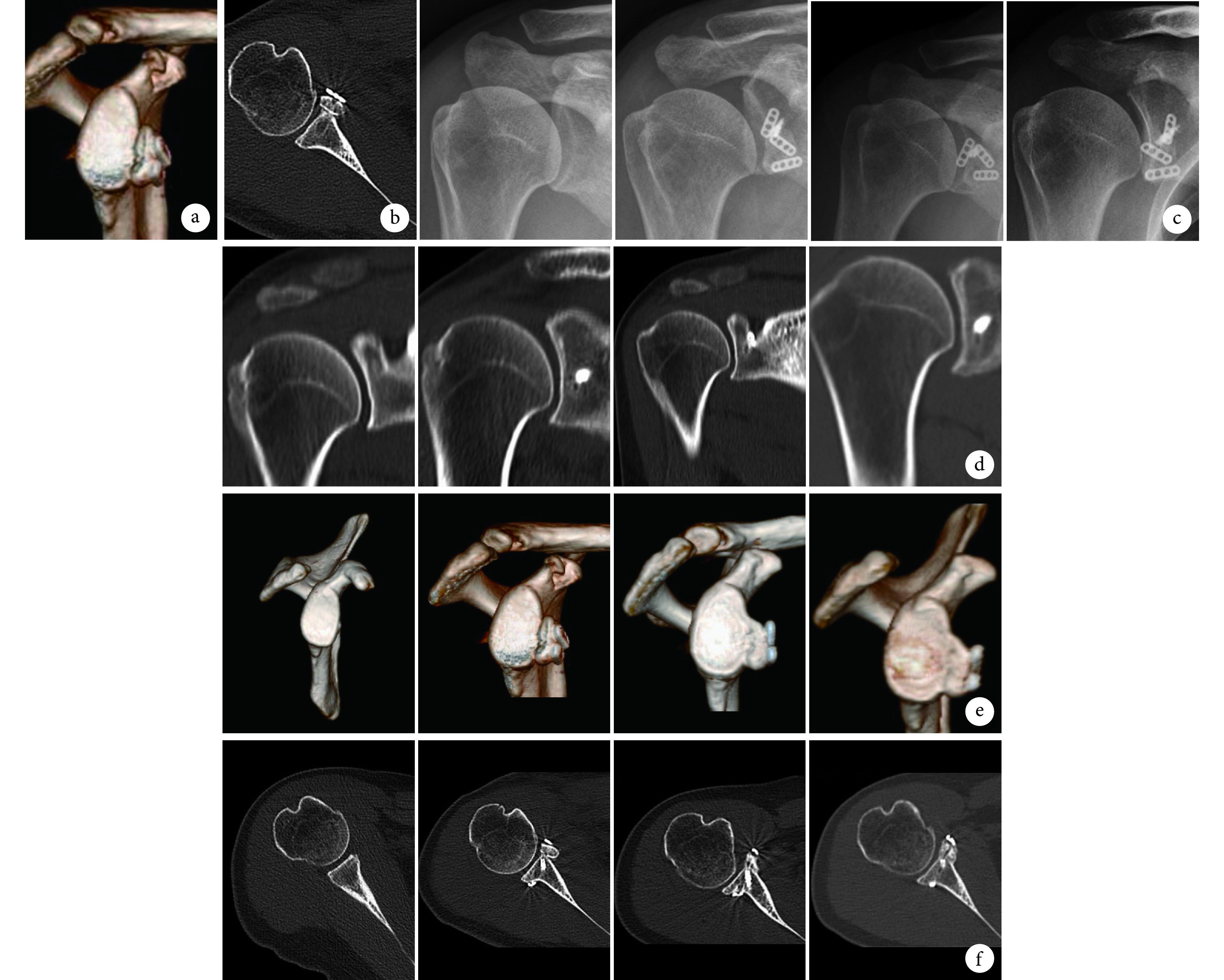
Results: All patients were followed up 55-62 months, with an average of 59.6 months. There was no neurovascular injuries, infections, fixation-related and bone graft-related complications. No re-dislocation and revision occurred. All patients returned to normal life, 17 of whom returned to sport. The VAS score was significantly decreased and ASES, Walch-Duplay, and Rowe scores were significantly improved at last follow-up ( P<0.05). No significant difference was found in range of motion of forward flexion, abduction, lateral rotation at 90° abduction, internal rotation at 90° abduction, or lateral rotation at 0° between pre- and post-operation ( P>0.05). Three-dimensional CT showed that the centers of all bone grafts were between 3∶30 and 4∶30 (right shoulder) or between 7∶40 and 8∶20 (left shoulder) and no bone grafts were positioned superiorly or inferiorly in the glenoid En-face view. All bone grafts were positioned lateral to the scapular glenoid with an average distance of 3.5 mm (range, 2.3-4.6 mm) in cross-sectional imaging by CT. Compared with the preoperative Samilson-Prieto classification results, all cases showed no progression of shoulder joint degeneration at 36, 48 months and last follow-up. All bone grafts remodeled to a steady state within 24 months after operation. The bone graft and glenoid finally remodeled analogous to the shape of the intact glenoid in the En-face view and became flush with the glenoid rim, remodeling to a curved shape congruent to the humeral head in cross-sectional imaging by CT. The shape of the remodeled glenoid at last follow-up was not significantly different from that at 24 months after operation.
Conclusion: The lateral placement of the bone graft during modified arthroscopic Latarjet surgery with elastic fixation do not accelerate the imaging changes of shoulder joint degeneration.
目的: 探讨改良关节镜下弹性固定 Latarjet 手术治疗伴有明显肩胛盂前缘骨缺损的肩关节复发性前脱位时,移植骨块偏外放置在术后中期对肩关节退变的影响。.
方法: 回顾分析 2015 年 1 月—2016 年 11 月收治且符合选择标准的 18 例肩关节复发性前脱位伴肩胛盂前缘骨缺损患者临床资料,均接受改良关节镜下弹性固定 Latarjet 手术。男 12 例,女 6 例;年龄 19~37 岁,平均 26.2 岁。肩关节脱位 4~30 次,平均 8.8 次。病程 8~49 个月,平均 23.8 个月。肩胛盂前缘骨缺损为 20%~29%,平均 25.2%。肩关节不稳定严重指数评分(ISIS)为 7~10 分,平均 7.6 分。肩关节退变 Samilson-Prieto 分级:0 级 13 例,Ⅰ级 3 例,Ⅱ级 2 例。手术前后采用疼痛视觉模拟评分(VAS)、美国肩肘外科协会评分(ASES)、Walch-Duplay 评分、Rowe 评分和肩关节活动度评价临床疗效。影像学观察肩关节退变情况以及移植骨块位置、肩胛盂塑形情况。.
结果: 术后患者均获随访,随访时间 55~62 个月,平均 59.6 个月。无血管神经损伤、感染、固定物和移植骨块相关并发症发生,随访期间无肩关节脱位复发和翻修患者。患者均恢复正常生活,17 例重返运动。末次随访时,VAS 评分较术前明显降低,ASES 评分、Walch-Duplay 评分、Rowe 评分明显提高,差异均有统计学意义( P<0.05);肩关节前屈、外展、外展 90° 外旋、0° 外旋和外展 90° 内旋活动度与术前比较,差异均无统计学意义( P>0.05)。术后即刻三维 CT 显示所有移植骨块中心在肩胛盂 En-face 面上位于 3∶30~4∶30(右肩)或 7∶40~8∶20(左肩)位置,无移植骨块过高或过低;在 CT 横断面上相对于肩胛盂偏外 2.3~4.6 mm,平均 3.5 mm。患者术后 36、48 个月及末次随访时的肩关节退变 Samilson-Prieto 分级与术前一致,均未进展。所有移植骨块在术后 24 个月内塑形达稳定,在三维 CT 肩胛盂 En-face 面显示塑形后的肩胛盂外形为与正常肩胛盂相近的正梨形,在 CT 横断面上为与肩胛盂齐平并形成与肱骨头圆形近似的弧形。末次随访时肩胛盂外形与术后 24 个月比较无明显变化。.
结论: 改良关节镜下弹性固定 Latarjet 手术中移植骨块偏外放置,术后中期未出现肩关节退变进展的影像学改变。.
Keywords: Recurrent anterior shoulder dislocation; arthroscopy; elastic fixation; modified Latarjet surgery; shoulder joint degeneration.
Conflict of interest statement
利益冲突:所有作者声明,在课题研究和文章撰写过程中不存在利益冲突。课题经费支持没有影响文章观点和对研究数据客观结果的统计分析及其报道。
Figures
Similar articles
-
[Mid-term effectiveness of LU-tarjet procedure for recurrent anterior shoulder dislocation].Zhongguo Xiu Fu Chong Jian Wai Ke Za Zhi. 2024 Jun 15;38(6):646-654. doi: 10.7507/1002-1892.202404058. Zhongguo Xiu Fu Chong Jian Wai Ke Za Zhi. 2024. PMID: 38918182 Free PMC article. Chinese.
-
[Mid-term effectiveness of modified arthroscopic suture button fixation Latarjet procedure for treatment of recurrent anterior shoulder dislocations].Zhongguo Xiu Fu Chong Jian Wai Ke Za Zhi. 2024 Jun 15;38(6):679-683. doi: 10.7507/1002-1892.202403125. Zhongguo Xiu Fu Chong Jian Wai Ke Za Zhi. 2024. PMID: 38918187 Free PMC article. Chinese.
-
[Suture button fixation Latarjet procedure under total arthroscopy for treatment of anterior shoulder instability with severe bone defect].Zhongguo Xiu Fu Chong Jian Wai Ke Za Zhi. 2024 Jun 15;38(6):666-671. doi: 10.7507/1002-1892.202403121. Zhongguo Xiu Fu Chong Jian Wai Ke Za Zhi. 2024. PMID: 38918185 Free PMC article. Chinese.
-
Low grade of osteoarthritis development after Latarjet procedure with a minimum 5 years of follow-up: a systematic review and pooled analysis.Knee Surg Sports Traumatol Arthrosc. 2022 Jun;30(6):2074-2083. doi: 10.1007/s00167-021-06771-w. Epub 2021 Oct 22. Knee Surg Sports Traumatol Arthrosc. 2022. PMID: 34677621 Free PMC article.
-
Arthroscopic Latarjet Versus Arthroscopic Free Bone Block Procedures for Anterior Shoulder Instability: A Proportional Meta-analysis Comparing Recurrence, Complication, and Reoperation Rates.Am J Sports Med. 2024 Jun;52(7):1865-1876. doi: 10.1177/03635465231188530. Epub 2024 Jan 19. Am J Sports Med. 2024. PMID: 38240595
References
MeSH terms
LinkOut - more resources
Full Text Sources
Medical
Research Materials