. 2021 Oct 22;144(9):2696-2708.
doi: 10.1093/brain/awab148.

COVID-19 neuropathology at Columbia University Irving Medical Center/New York Presbyterian Hospital

Affiliations

COVID-19 neuropathology at Columbia University Irving Medical Center/New York Presbyterian Hospital

Kiran T Thakur et al. Brain. .

Abstract

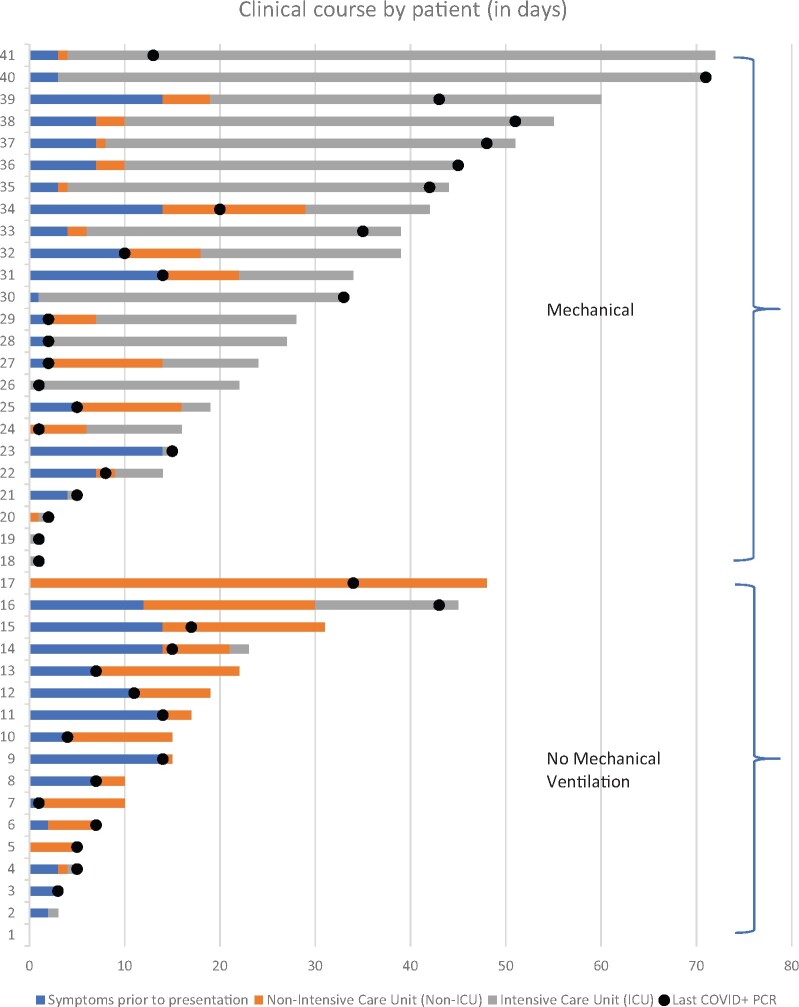
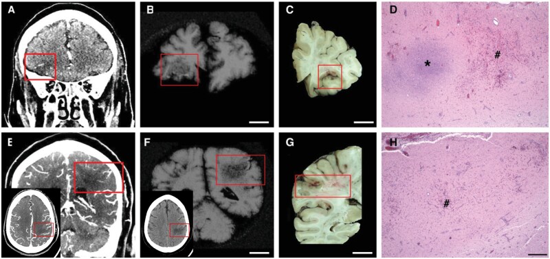
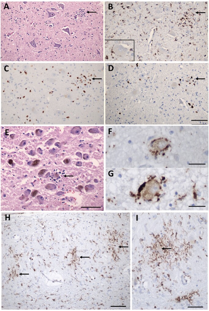
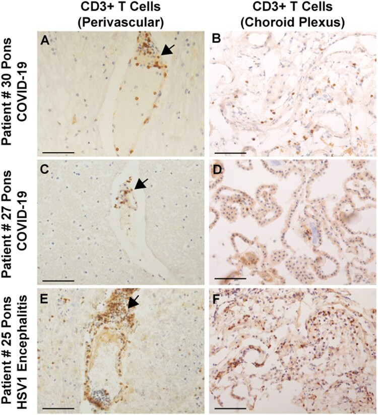
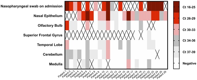
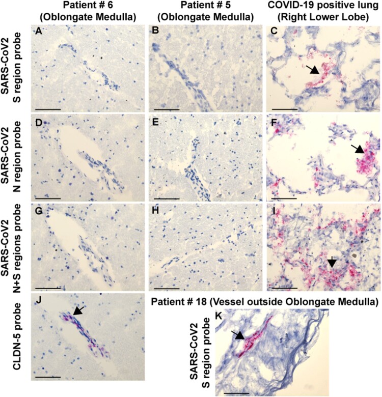
Many patients with SARS-CoV-2 infection develop neurological signs and symptoms; although, to date, little evidence exists that primary infection of the brain is a significant contributing factor. We present the clinical, neuropathological and molecular findings of 41 consecutive patients with SARS-CoV-2 infections who died and underwent autopsy in our medical centre. The mean age was 74 years (38-97 years), 27 patients (66%) were male and 34 (83%) were of Hispanic/Latinx ethnicity. Twenty-four patients (59%) were admitted to the intensive care unit. Hospital-associated complications were common, including eight patients (20%) with deep vein thrombosis/pulmonary embolism, seven (17%) with acute kidney injury requiring dialysis and 10 (24%) with positive blood cultures during admission. Eight (20%) patients died within 24 h of hospital admission, while 11 (27%) died more than 4 weeks after hospital admission. Neuropathological examination of 20-30 areas from each brain revealed hypoxic/ischaemic changes in all brains, both global and focal; large and small infarcts, many of which appeared haemorrhagic; and microglial activation with microglial nodules accompanied by neuronophagia, most prominently in the brainstem. We observed sparse T lymphocyte accumulation in either perivascular regions or in the brain parenchyma. Many brains contained atherosclerosis of large arteries and arteriolosclerosis, although none showed evidence of vasculitis. Eighteen patients (44%) exhibited pathologies of neurodegenerative diseases, which was not unexpected given the age range of our patients. We examined multiple fresh frozen and fixed tissues from 28 brains for the presence of viral RNA and protein, using quantitative reverse-transcriptase PCR, RNAscope® and immunocytochemistry with primers, probes and antibodies directed against the spike and nucleocapsid regions. The PCR analysis revealed low to very low, but detectable, viral RNA levels in the majority of brains, although they were far lower than those in the nasal epithelia. RNAscope® and immunocytochemistry failed to detect viral RNA or protein in brains. Our findings indicate that the levels of detectable virus in coronavirus disease 2019 brains are very low and do not correlate with the histopathological alterations. These findings suggest that microglial activation, microglial nodules and neuronophagia, observed in the majority of brains, do not result from direct viral infection of brain parenchyma, but more likely from systemic inflammation, perhaps with synergistic contribution from hypoxia/ischaemia. Further studies are needed to define whether these pathologies, if present in patients who survive coronavirus disease 2019, might contribute to chronic neurological problems.

Keywords: COVID-19; SARS-CoV-2; microglia activation; microglial nodules; neuropathology.


Figures

Figure 1
Figure 1
Clinical course of COVID-19 patients by days. Bar graph showing the length of the clinical course of our patients, including symptoms prior to presentation (blue) and days in the hospital before death, either in the intensive care unit (grey) or not in the intensive care unit (orange). Dots represent the last positive qRT-PCR result prior to death. Patients are numbered 1–41 and displayed from shortest to longest clinical course.
Figure 2
Figure 2
Acute, focal, haemorrhagic infarcts in COVID-19 patients. Acute, focal, haemorrhagic infarcts of (AD) right inferior frontal and (EH) left lateral parietal lobes. (A and E) Premortem CAT scans; (B and F) post-mortem MRI; (C and D) gross photographs of coronal brain slices through the corresponding infarcts; and (D and H) microscopic images of the corresponding infarcts. The insets in E and F show the parietal infarct in the axial plane, red boxes outline the lesions on the scans and brain slices. (D and H) Areas of fresh haemorrhage (number symbol) and acute necrosis (asterisk) shown with haemoxylin and eosin stain. Scale bar in H = 500 µm for D and H; B, C, F and G = 1 cm.
Figure 3
Figure 3
Inflammatory pathology in COVID-19 brains. (A) Section of the hypoglossal nucleus shows several motor neurons and a microglial module (arrow). (B) An adjacent section stained for CD68, showing clustered microglia in the nodule. Inset: Microglia in close apposition to a hypoglossal neuron (CD68). (C) An adjacent section stained for CD3, showing scattered T cells in the tissue and associated with the microglial nodule. (D) An adjacent section stained for CD8 showing that many of the T cells are CD8+. (E) The locus coeruleus contains a microglial nodule with a degenerating neuron in the centre, identified by its residual neuromelanin (arrow). (F and G) Neurons of the dorsal motor nucleus of the vagus surrounded by CD68+ microglia. (H and I) Microglial nodules in the dentate nucleus (arrows in H), neuron in the middle of a nodule (arrow in I), CD68. Scale bar in D = 200 µm for AD; in E = 10 µm; F and G = 50 µm; H = 100 µm; I = 50 µm; J = 1 mm; and K = 250 µm.
Figure 4
Figure 4
Immunocytochemical staining for CD3+ T cells in COVID-19 brains. (A and C) Sparse perivascular CD3+ T cells in the pons of COVID-19 Patients 27 and 30. (B and D) Sparse CD3+ T cells in the choroid plexus from the lateral ventricle of Patients 27 and 30. (E and F) CD3+ T cell infiltrates around a pontine vessel (E) and in the choroid plexus (F) of Patient 25 with HSV-1 encephalitis; arrows indicate CD3+ T cell infiltrates. All sections are counterstained with haematoxylin. Scale bars = 200 µm.
Figure 5
Figure 5
SARS-CoV-2 qRT-PCR results from the nasal epithelia and brains of COVID-19 patients. Heat map of cycle threshold (Ct) values of brain autopsy sample qRT-PCR for detection of SARS-CoV-2. Nasopharyngeal swab at the time of admission and nasal epithelium are included as samples outside of the CNS. Ct values are presented in quintiles based on the distribution in samples tested. ‘X’ denotes sample not included for that patient.
Figure 6
Figure 6
RNAscope® results from the brains and lungs of COVID-19 patients. RNAscope® with SARS-CoV-2 S region probe on (A) medulla Patient 6, (B) medulla Patient 5, and (C) lung, positive control. SARS-CoV-2 N region probe on (D) medulla Patient 6, (E) medulla Patient 5, and (F) lung, positive control. SARS-CoV-2 N + S region probes on (G) medulla Patient 6, (H) medulla Patient 5, and (I) lung, positive control. (J) CLDN5 probe on medulla Patient 6 showing a positive signal in endothelial cells. (K) SARS-CoV-2 S region probe gives a positive signal in the adventitia of a meningeal vessel outside of medulla Patient 18. All sections were counterstained with haematoxylin. Arrows indicate positive RNAscope® signal. Scale bars = 200 µm.

References

    1. Nath A. Neurologic complications of coronavirus infections. Neurology. 2020;94(19):809–810. - PubMed
    1. Dugue R, Cay-Martínez KC, Thakur K, et al.Neurologic manifestations in an infant with COVID-19. Neurology. 2020;94(24):1100–1102. - PMC - PubMed
    1. Moriguchi T, Harii N, Goto J, et al.A first case of meningitis/encephalitis associated with SARS-Coronavirus-2. Int J Infect Dis. 2020;94:55–58. - PMC - PubMed
    1. Avula A, Nalleballe K, Narula N, et al.COVID-19 presenting as stroke. Brain Behav Immun. 2020;87:115–119. - PMC - PubMed
    1. Beyrouti R, Adams ME, Benjamin L, et al.Characteristics of ischaemic stroke associated with COVID-19. J Neurol Neurosurg Psychiatry. 2020;91(8):889–891. - PMC - PubMed

Publication types

MeSH terms

Substances