. 2021 Jun 9;30(12):1067-1083.
doi: 10.1093/hmg/ddab104.

Functional rescue in an Angelman syndrome model following treatment with lentivector transduced hematopoietic stem cells

Affiliations

Functional rescue in an Angelman syndrome model following treatment with lentivector transduced hematopoietic stem cells

Anna Adhikari et al. Hum Mol Genet. .

Abstract

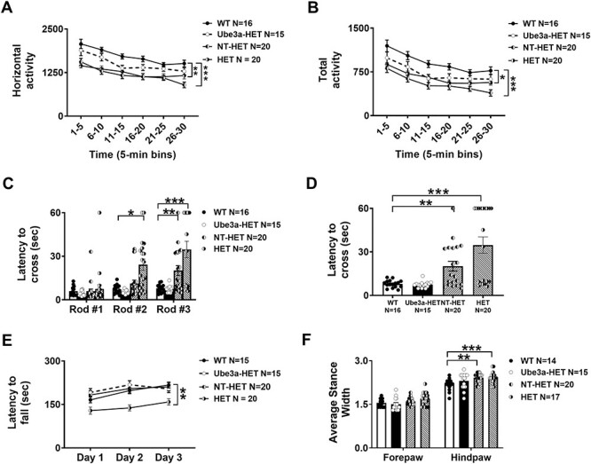
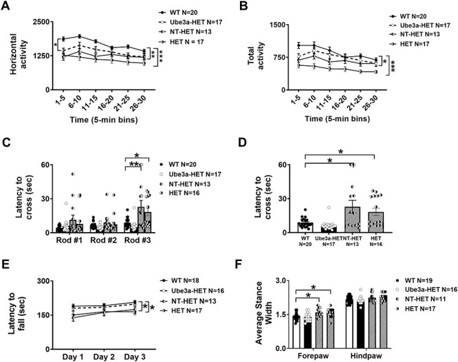
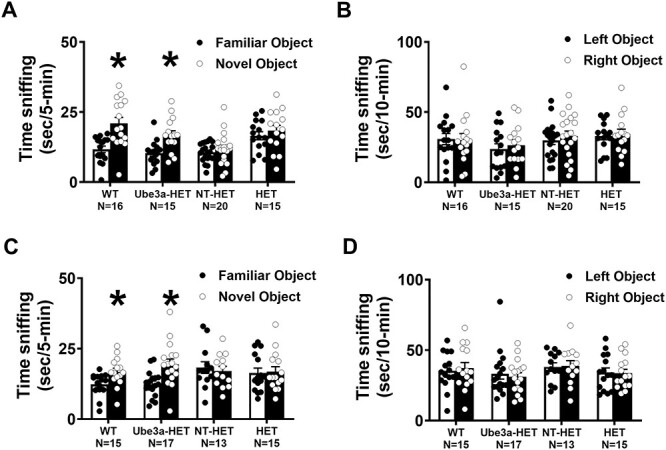
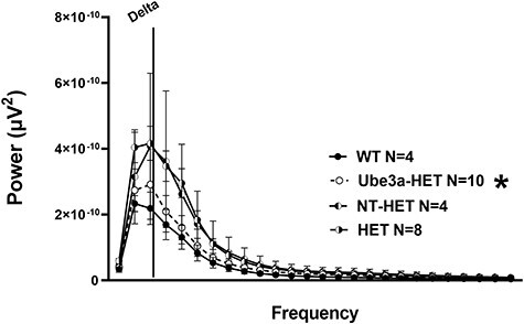
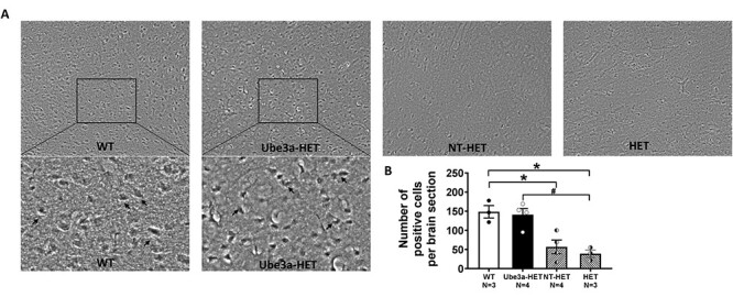
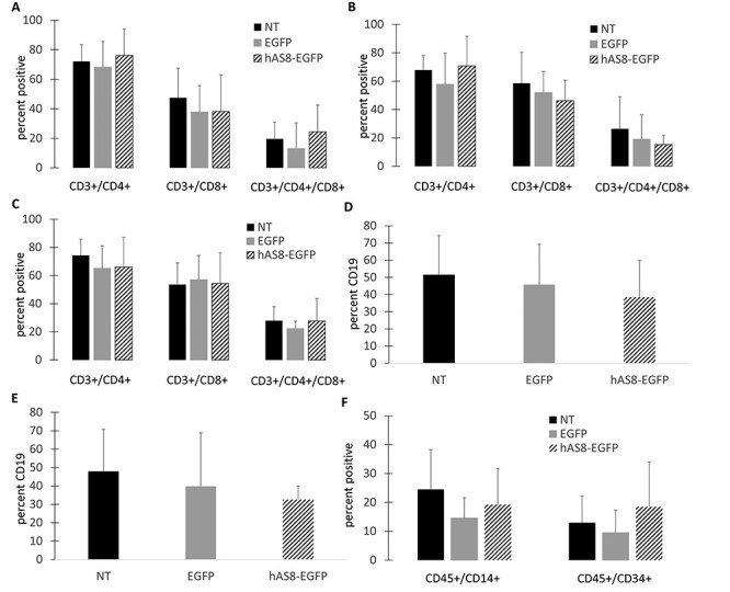
Angelman syndrome (AS) is a rare neurodevelopmental disorder characterized by impaired communication skills, ataxia, motor and balance deficits, intellectual disabilities, and seizures. The genetic cause of AS is the neuronal loss of UBE3A expression in the brain. A novel approach, described here, is a stem cell gene therapy which uses lentivector-transduced hematopoietic stem and progenitor cells to deliver functional UBE3A to affected cells. We have demonstrated both the prevention and reversal of AS phenotypes upon transplantation and engraftment of human CD34+ cells transduced with a Ube3a lentivector in a novel immunodeficient Ube3amat-/pat+ IL2rg-/y mouse model of AS. A significant improvement in motor and cognitive behavioral assays as well as normalized delta power measured by electroencephalogram was observed in neonates and adults transplanted with the gene modified cells. Human hematopoietic profiles observed in the lymphoid organs by detection of human immune cells were normal. Expression of UBE3A was detected in the brains of the adult treatment group following immunohistochemical staining illustrating engraftment of the gene-modified cells expressing UBE3A in the brain. As demonstrated with our data, this stem cell gene therapy approach offers a promising treatment strategy for AS, not requiring a critical treatment window.


Figures

Figure 1
Figure 1
Schematic and functionality of the Ube3a-expressing lentiviral vector: (A) A self-inactivating lentiviral vector backbone, CCLc-MNDU3-X, was used to generate the Ube3a expressing lentiviral vectors. (B) EGFP vector used as an empty vector control. (C) The modified mouse Ube3a isoform 3 (mAS8) was cloned under the control of the MNDU3 promoter. EGFP was cloned downstream under the control of a PGK promoter. (D) The modified human UBE3A isoform 1 was cloned under the control of the MNDU3 promoter. EGFP was cloned downstream under the control of a PGK promoter. (E) Human CD34+ HSPC were transduced with the Ube3a lentivector and derived into mature macrophages. Cell extracts were then generated and evaluated for overexpression of UBE3A by western blot. (F) An S5A ubiquitination assay was performed on the macrophage cell extracts. Control NT, EGFP vector alone (EGFP) transduced cells, and negative and positive controls supplied by the manufacturer were used as controls.
Figure 2
Figure 2
CFU assay and macrophage derivation of Ube3a vector-transduced human CD34+ HSPC: Human CD34+ HPSC were left NT or transduced with either the EGFP alone control vector (EGFP) or the Ube3a vector (hAS8). (A) Cells were cultured in methylcellulose media for 12 days when specific colonies (BFU-E, GM and GEMM) were counted. (B) CFUs were further differentiated into macrophages and analyzed by flow cytometry for the cell surface markers CD4, CD14 and HLADR.
Figure 3
Figure 3
Locomotor ability, balance, motor coordination and gait from Ube3a lentivector transduced HSPC-transplanted neonates: Rigorous assessment of motor translational phenotypes using four standard motor behavioral tests in treated and untreated Ube3amat+/pat+ IL2rg−/y mice that were irradiated and transplanted as neonates with either NT (NT-HET (hatched bar) or the Ube3a lentivector transduced (Ube3a-HET (black bar)) human CD34+ HSPC. Eight weeks post-transplant, mice were subjected to (A, B) open field locomotion, (C, D) balance beam, (E) rotarod and (F) treadmill walking. In all tests, Ube3amat+/pat+ IL2rg−/y deficient mice transplanted with the Ube3a vector transduced human CD34+ HSPC (Ube3a-HET) exhibited wildtype values of performance. (A, B) Open field activity was increased by both the total distance and horizontal activity metrics. (C) Eight weeks post-transplant, mice were also tested on the beam walking assay. Beams decrease in width and are more difficult to cross going from Rod #1 to Rod #2 to Rod #3. (D) Highlight of Rod #3 of C, the most challenging coordination test with remarkable improvements. (E) Latency to fall from the rotarod was significantly improved in the Ube3amat+/pat+ IL2rg−/y mice transplanted with Ube3a vector-transduced human CD34+ HSPC (Ube3a-HET) compared with the NT (NT-HET) cell controls. (F) DigiGait™ analyses showed a narrowing of wide stances in the Ube3a-HET treated group. Data are expressed as ± standard error of mean (SEM). *P < 0.05, **P < 0.001, ***P < 0.0001 indicate when the HET and NT-HET groups differ from the control WT group.
Figure 4
Figure 4
Locomotor ability, balance, motor coordination and gait from Ube3a lentivector transduced HSPC-transplanted adults: Rigorous assessment of motor translational phenotypes using four standard motor behavioral tests in treated and untreated Ube3amat−/pat+ IL2rg−/y mice that were treated with busulfan and transplanted as adults with either NT (NT-HET (hatched bar) or the Ube3a lentivector-transduced (Ube3a-HET (black bar)) human CD34+ HSPC. Four-five weeks old mice were conditioned with busulfan and transplanted via i.v. injection. Six weeks later mice were subjected to (A, B) open field locomotion, (C, D) balance beam, (E) rotarod and (F) treadmill walking. In all tests, Ube3a-deficient mice transplanted with the Ube3a vector-transduced human CD34+ HSPC (Ube3a-HET) exhibited wildtype values of performance. (A, B) Open field activity was increased by both the total distance and horizontal activity metrics. (C) Eight weeks post-transplant, mice were also tested on the beam walking assay. Beams decrease in width and are more difficult to cross going from Rod #1 to Rod #2 to Rod #3. (D) Highlight of Rod #3 of C. (E) Latency to fall from the rotarod was significantly improved in the Ube3a-deficient mice transplanted with Ube3a vector transduced human CD34+ HSPC (Ube3a-HET) compared with the NT (NT-HET) cells controls. (F) DigiGait™ analyses showed a narrowing of wide stances in the Ube3a-HET treated group. Data are expressed as mean ± SEM. *P < 0.05, **P < 0.001, ***P < 0.0001 indicate when the HET and NT-HET groups differ from the control WT group.
Figure 5
Figure 5
Novel object recognition with mice transplanted with Ube3a lentivector transduced HSPC: (A,B) Newborn Ube3a-deficient IL2rg-/y mice were transplanted with either nontransduced (NT-HET) or Ube3a vector-transduced (Ube3a-HET) human CD34+ HSPC. Wild type IL2rg-/y mice (WT) were used as a control. Eight weeks post-transplant, mice were assessed for learning and memory abilities using the novel object recognition test. (A) Ube3a-HET mice exhibited intact object recognition following a one-hour delay, similar to WT while, NT-HET mice did not spend more time with the novel object, exhibiting a lack of recognition memory. (B) All groups explored the two objects similarly during the familiarization phase. (C, D) Ube3a-deficient adult mice were transplanted with either nontransduced (NT-HET) or Ube3a vector-transduced (Ube3a-HET) human CD34+ HSPC. Wild type IL2rg-/y mice (WT) were used as a control. Six weeks post i.v. injection in adult mice, subjects were assessed for learning and memory abilities using the novel object recognition test. (C) Ube3a-HET mice exhibited intact object recognition following a one-hour delay, similar to WT while, NT-HET mice did not spend more time with the novel object, exhibiting a lack of recognition memory. (D) All groups explored the two objects similarly during the familiarization phase. *p < 0.05, novel versus familiar.
Figure 6
Figure 6
Delta power analysis in AS mice transplanted with Ube3a lentivector transduced HSPC: A PSD between using treated and untreated Ube3a-deficient IL2rg−/y mice that were irradiated and transplanted as neonates with either NT HET (semi-filled left) or the Ube3a lentivector transduced Ube3a-HET (black dotted line open circles) human CD34+ HSPC. AS mice on B6 background versus their WT background littermates illustrate observable differences in elevated delta power analogous to clinical phenotypes. Quantification of spectral power bands illustrates that treatment lowers/corrects the elevated delta spectral power toward the levels observed in WT mice.
Figure 7
Figure 7
Immunohistochemical analysis of UBE3A expression in cortex of the mouse brain. Six weeks post-transplant in adult mice, subjects were assessed for UBE3A expression using DAB as a chromogen. (A) Immunohistochemical images showing expression of UBE3A in the mouse cortex of non-transplanted Ube3a wild type IL2rg−/y (WT) mice, those transplanted with Ube3a vector transduced (Ube3a-HET) human CD34+ HSPC, those transplanted with NT human CD34+ HSPC (NT-HET), and non-transplanted HET mice (HET). (B) A significant increase in the UBE3A positive cells, similar to the WT level, was observed in transplanted Ube3a-HET compared with NT-HET and HET when treated as adults. Data are expressed as mean ± SEM. *P < 0.05 indicates when the HET and NT-HET groups differ from the control WT group. #P < 0.05 indicates when the Ube3a-HET group differs from HET group.
Figure 8
Figure 8
Engraftment and development of human immune cells in NRG mice: Human CD34+ HSPC were left NT or transduced with either the EGFP control (EGFP) or Ube3a-expressing (hAS8) lentiviral vector. Cells were transplanted into 2–5-day-old NRG mice. At 16 weeks post-transplant, mice were euthanized, and human T cells were analyzed for CD3, CD4, and CD8 expression in the (A) peripheral blood, (B) spleen, and (C) thymus. Human B cells were analyzed for CD45 and CD19 in the (D) spleen and (E) bone marrow. (F) Human macrophages (CD45/CD14) and human CD34+ HSPC (CD45/CD34) were analyzed in the bone marrow.

References

    1. Williams, C.A. (2005) Neurological aspects of the Angelman syndrome. Brain Dev., 27, 88–94. - PubMed
    1. Buiting, K., Williams, C. and Horsthemke, B. (2016) Angelman syndrome-insights into a rare neurogenetic disorder. Nat. Rev. Neurol., 12, 584–593. - PubMed
    1. Williams, C.A., Driscoll, D.J. and Dagli, A.I. (2010) Clinical and genetic aspects of Angelman syndrome. Genet. Med., 12, 385–395. - PubMed
    1. Chamberlain, S.J. and Lalande, M. (2010) Neurodevelopmental disorders involving genomic imprinting at human chromosome 15q11-q13. Neurobiol. Dis., 39, 13–20. - PubMed
    1. Matsuura, T., Sutcliffe, J.S., Fang, P., Galjaard, R.J., Jiang, Y.H., Benton, C.S., Rommens, J.M. and Beaudet, A.L. (1997) De novo truncating mutations in E6-AP ubiquiting-protein ligase gene (UBE3A) in Angelman syndrome. Nat. Genet., 15, 74–77. - PubMed

Publication types

MeSH terms