. 2021 May 24;6(10):e141159.
doi: 10.1172/jci.insight.141159.

Direct imaging of glymphatic transport using H217O MRI

Affiliations

Direct imaging of glymphatic transport using H217O MRI

Mohammed S Alshuhri et al. JCI Insight. .

Abstract

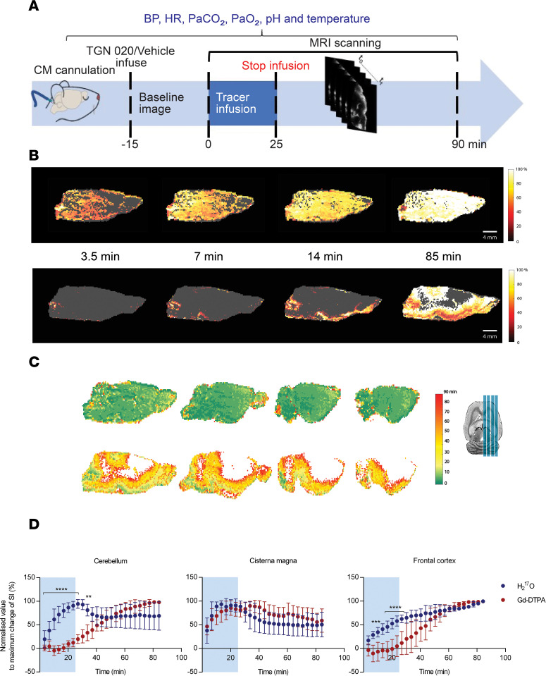
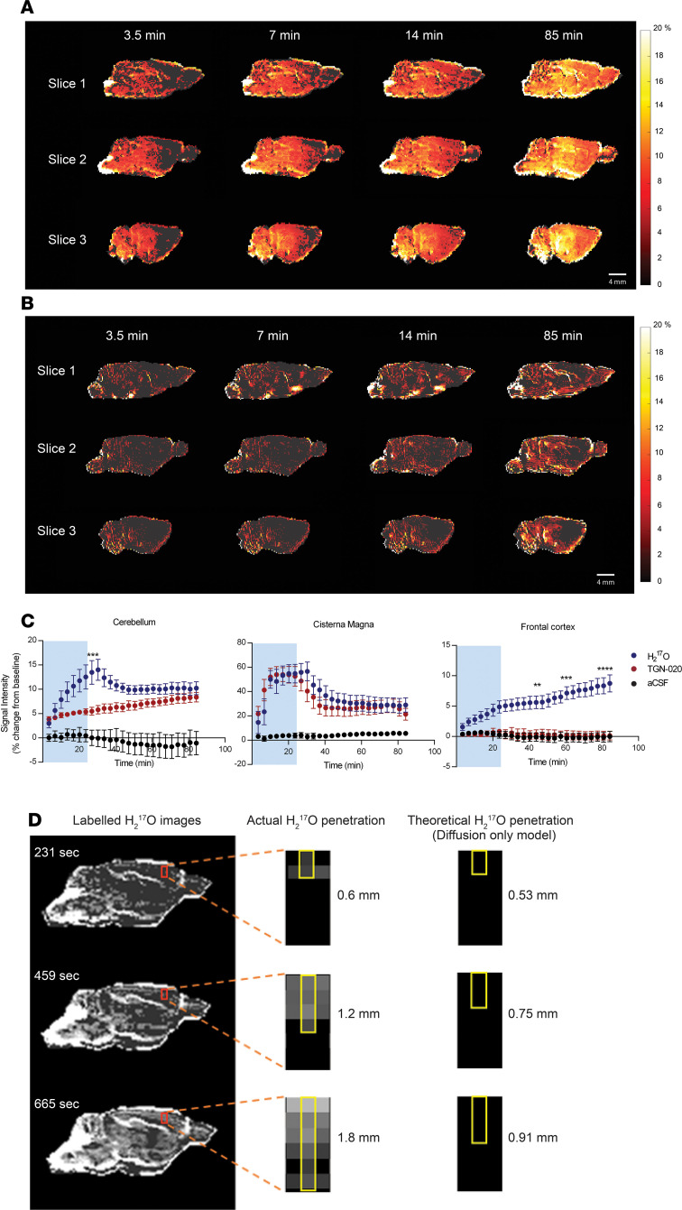
The recently proposed glymphatic pathway for solute transport and waste clearance from the brain has been the focus of intense debate. By exploiting an isotopically enriched MRI tracer, H217O, we directly imaged glymphatic water transport in the rat brain in vivo. Our results reveal glymphatic transport that is dramatically faster and more extensive than previously thought and unlikely to be explained by diffusion alone. Moreover, we confirm the critical role of aquaporin-4 channels in glymphatic transport.

Keywords: Development; Neuroimaging; Neurological disorders; Neuroscience; Transport.


Conflict of interest statement

Conflict of interest: The authors have declared that no conflict of interest exists.

Figures

Figure 1
Figure 1. Serial MRI using H217O tracer reveals rapid glymphatic flow over whole brain.
(A) Study design. Rats received infusion of tracer via cisterna magna. Baseline MRI was acquired, followed by infusion of tracer and continuous MRI. (B) Representative sagittal MRI demonstrating the temporal evolution of tracer over 85 minutes of recording. Normalized pseudocolor scaling illustrates tracer distribution of the 90% 17O-enriched water (MW, 19 Da) (upper panel) and the paramagnetic tracer Gd-DTPA (Magnevist; MW, 938 Da) (bottom panel), where white in the color bar indicates maximum signal change. Representative arrival time maps. The upper panel images show a rat infused with H217O, and the bottom panel images show 1 rat infused with Gd-DTPA. (C) Corresponding tracer arrival time maps for 4 sagittal slices. White color indicates that the tracer did not arrive within the 85-minute recording. (D) Summary data showing the normalized percentage signal change as a function of time for H217O (n = 6, blue circles) and Gd-DTPA (n = 6, red circles) for the cerebellum, cisterna magna and frontal cortex. Blue shading on schematic drawing illustrates the location of ROIs. Blue shading on graphs indicates period of tracer infusion. *P < 0.05, **P < 0.01, ***P < 0.001, ****P < 0.0001. Data are presented as mean ± SD.
Figure 2
Figure 2. Effect of inhibiting AQP4 channels on glymphatic water flow.
(A and B) Serial sagittal MRI revealing the temporal evolution of H217O tracer for vehicle-treated rats (upper panel, n = 6) (A) and rats treated with AQP4 inhibitor (lower panel, TGN 020, n = 6) (B). Pseudocolor scaling illustrates the distribution of H217O throughout the brain over 80 minutes of recording, with AQP4 inhibition resulting in substantially reduced H217O transport compared with the vehicle. (C) Summary data showing the percent signal change as a function of time for the vehicle (blue circles) and AQP4 inhibitor–treated (red circles) groups in the frontal cortex, cerebellum, and cisterna magna. Artificial CSF (aCSF; i.e., without 17O-enriched H217O) was used as a negative control. Blue shading on graphs indicates period of contrast agent infusion. (D) Representative images of H217O transport at different time points. The red rectangle has been magnified to better show the actual penetration of H217O, with the top of the red rectangle position on the cerebral cortex. The theoretically calculated displacements of H217O due to diffusion only are 0.53 mm, 0.75 mm, and 0.91 mm at times 231, 459, and 665 seconds, respectively. It is clear that the rapid H217O penetration of the parenchyma cannot be explained by diffusion alone, indicating the presence of convective ISF flow. *P < 0.05, **P < 0.01, ***P < 0.001, ****P < 0.0001. Data are presented as mean ± SEM.

References

    1. Iliff JJ, et al. A paravascular pathway facilitates CSF flow through the brain parenchyma and the clearance of interstitial solutes, including amyloid β. Sci Transl Med. 2012;4(147):147ra111. - PMC - PubMed
    1. Ding FF, et al. Changes in the composition of brain interstitial ions control the sleep-wake cycle. Science. 2016;352(6285):550–555. doi: 10.1126/science.aad4821. - DOI - PMC - PubMed
    1. Mestre H, et al. Cerebrospinal fluid influx drives acute ischemic tissue swelling. Science. 2020;367(6483):eaax7171. doi: 10.1126/science.aax7171. - DOI - PMC - PubMed
    1. Xu ZQ, et al. Deletion of aquaporin-4 in APP/PS1 mice exacerbates brain A beta accumulation and memory deficits. Mol Neurodegener. 2015;10:58. - PMC - PubMed
    1. Venkat P, et al. White matter damage and glymphatic dysfunction in a model of vascular dementia in rats with no prior vascular pathologies. Neurobiol Aging. 2017;50:96–106. doi: 10.1016/j.neurobiolaging.2016.11.002. - DOI - PMC - PubMed